
Recomendados
Recomendados
Mais conteúdo relacionado
Semelhante a David Baxter - Biosap
Semelhante a David Baxter - Biosap (13)
Mais de COT SSNP
Mais de COT SSNP (18)
Último
Último (20)
David Baxter - Biosap
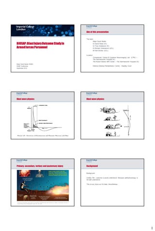
- 1. Aim of this presentation The team: Major David Baxter BIOSAP: Blast Injury Outcome Study in Dr David Sharp (IC) Armed forces Personnel Dr Tony Goldstone (IC) Dr Richard Greenwood (UCL) Mr Neil Kitchen (UCL) Location: Computional, Clinical & Cognitive Neuroimaging Lab (C3NL) – The Hammersmith Hospital (IC) The Robert Steiner MRI Center – The Hammersmith Hospital (IC) Major David Baxter RAMC SSNP Conference Defence Medical Rehabilitation Centre - Headley Court September 2012 Blast wave physics Blast wave physics Primary, secondary, tertiary and quaternary injury Background Background: Civilian TBI – outcome is poorly understood. Because pathophysiology is not well understood. This is true (more so) for blast. Nevertheless… Cernak et al Traumatic brain injury: an overview of pathobiology with emphasis on military populations. Journal of Cerebral Blood Flow & Metabolism (2010) 30, 255–266 1
- 2. Improvements in diagnosis of non-penetrating brain injury Focal injury and white matter damage Contusion location Diffuse axonal injury Conventional MRI; T1, T2, Flair DTI, SWI, Gradient Echo imaging i.e. xray and CT Grade 2: lesions in corpus callosum Grade 3: lesions also in brainstem Adams et al ‘85 Hypothesis and Impact What is the BIOSAP project and what are its aims Aim: 1. To characterize the neuropsychological and endocrine consequences of blast traumatic brain injury. ADMEM database 2. To compare this to civilian traumatic brain injury. Imaging Imaging DMRC Headley Court studies studies Hypothesis: Endocrine Endocrine 1. Blast causes a specific pattern of white matter damage, B’ham hospitals assessment assessment that can be assessed using MRI. Blas t TBI Psychologica Psychologica l assessment l assessment Clinician referral Impact: 1. Gives the capability of screening blast injured soldiers 2. Provide evidence about the mechanism of blast injury. MRI assessment Criteria Inclusion Exclusion Structural MRI • Moderate to severe traumatic brain injury • Cognitive impairment such that the subject will • GCS <14 be unable to cooperate. • LOC >30mins T1 • PTA >24hrs • Significant language or visuo-spatial T2 Flair impairments.. T2 FFE – Gradient Echo • History of Blast injury. • Penetrating head injury or neurosurgery. Diffusion tensor imaging - DTI • Persistent cognitive impairment. Susceptibility Weighted Imaging – SWI • Overt bleeding visible on CT (excluding the presence of microbleeds – a marker of diffuse • No significant premorbid neurological or axonal injury). psychiatric illness. Functional MRI • Able to give informed consent. • Ability to perform the scanning paradigm. • Clinically stable. 2
- 3. Neuropsychological & Psychiatric Test Battery Endocrine Assessment Cognitive: (developed with Professor Jane Powell - Goldsmiths) • Traumatic brain injury produces significant dysfunction in the hypothalamic-pituitary axis in a significant number of patients Intellectual function - WASI similarities, WASI Matrix reasoning [Schneider et al., 2007] Executive function - Trail Making Test, Inhibition/switching, Letter fluency • Impairments persisting in 15-50% of patients. E.g. severe growth hormone deficiency is seen in 10-20% of patients following traumatic Memory function and learning - People Test immediate recall brain injury. Information processing speed - Colour naming (s), Word reading (s) • Limits brain recovery. Psychiatric: • Important therapeutic opportunity. Including: AGHDA, Beck Depression Inventory, Epworth Sleepiness, Nottingham Health Profile, SF-36 PTSD Mood disturbance Anxiety Case 1: History Case 1: Structural imaging findings 28yo male T2Flair Gradient Echo Top cover Wearing Helmet and eye protection 50 Kg IED. Multiple fractures. Superficial lacerations. Left sided pneumothorax. Initial GCS 12/15 2 weeks of retrograde amnesia 6 weeks of post-traumatic amnesia On neuropsychological assessment; Impaired executive function, memory and processing speed Case 1: Outcome Case 2: History 27yo. Male Returned to work in a limited capacity but struggling… Passenger in vehicle Wearing body armour and Helmet IED of unknown size Multiple fractures and lacerations. Left sided pneumothorax. Initial GCS 13/15 1 day retrograde amnesia 4 days post traumatic amnesia On neuropsychological assessment; Impaired memory 3
- 4. Case 2: Outcome Case 2: Structural imaging findings T2Flair Gradient Echo Currently studying for Msc. Case summary Results Conventional imaging • 20 blast injured soldiers 1. Gradient echo imaging is more sensitive than standard T1 and Flair to the changes associated with diffuse axonal injury • 40 age matched civilians with moderate to severe TBI 2. But…the presence of microbleeds can still be associated with very variable clinical outcome. • 40 uninjured civilian controls Study participant demograhics Diffusion tensor imaging Controls Military Civilian Difference Cortex Age 30.6 yrs (SD 29.4 yrs (SD 30.3 yrs (SD No 6.7) 5.8) 7.6) difference Time since n/a 14.6 mnths 12 mnths No injury (SD 5.9) (SD 12.7) difference White matter - TBI Contusion n/a 9 (45%) 10 (50%) No difference Microbleeds n/a 5 (25%) 5 (25%) No difference Low fractional anisotropy Mori et al Neur & High mean diffusivity 4
- 5. DTI Results TBSS Pattern analysis Pattern analysis results Now focus on the bTBI group Endocrine assessment results Region of Interest analysis 5
- 6. Neuropsychological and QoL assessment results Summary 1. MRI changes following blast traumatic brain injury are associated with, neuropsychological, neuropsychiatric and endocrine changes. 2. Blast traumatic brain injury produces a hetreogenous white matter injury pattern, however some regions appear to particularly vulnerable to injury from blast. 3. We will attempt to use this research to identify patients who will go on to have a poor clinical outcome, and provide information about the mechanism of blast. BIOSAP Thanks to; DPMD Prof M Midwinter Dr A K Samra Mjr C Lethbridge DMRC Col Etherington Wng Cmdr A Bennett Kit Malia Doreen Rowland Ronel Terblanche Alison Lutte-Elliott Goldsmiths Prof Jane Powell 6