Resuscitation of the newborn
•Download as PPT, PDF•
3 likes•1,670 views

Power point presentation on the topic of New born resuscitation.
![OBJECTIVES
Have an overview of the Basic Physiologic Changes At Birth
Understand the Resuscitation Flow Diagram/Strategy;
Know the equipment & Personnel Needed;
Be Able To Decide When To Resuscitate [High-Risk]
Understand the Sequelae of birth asphyxia](data:image/gif;base64,R0lGODlhAQABAIAAAAAAAP///yH5BAEAAAAALAAAAAABAAEAAAIBRAA7)
Recommended
More Related Content
What's hot
What's hot (20)
Viewers also liked
Viewers also liked (17)
Similar to Resuscitation of the newborn
Similar to Resuscitation of the newborn (20)
Recently uploaded
Recently uploaded (20)
Resuscitation of the newborn
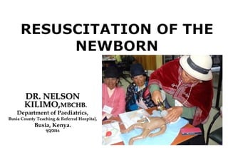
- 1. DR. NELSONDR. NELSON KILIMO,KILIMO,MBCHB.MBCHB. Department of Paediatrics, Busia County Teaching & Referral Hospital, Busia, Kenya. 9/2/20169/2/2016
- 2. OBJECTIVES Have an overview of the Basic Physiologic Changes At Birth Understand the Resuscitation Flow Diagram/Strategy; Know the equipment & Personnel Needed; Be Able To Decide When To Resuscitate [High-Risk] Understand the Sequelae of birth asphyxia
- 3. Prologue • Birth asphyxia kills 0.7 to 1.6 million newborns a year globally • 99% of deaths in developing countries. • Effective newborn resuscitation could reduce this burden of disease • But the training of health-care providers in low income settings is often outdated. (Opiyo, Were et al,2008)
- 4. FETAL CIRCULATION IN THE FETUS: Placenta; lowest vascular resistance – 40% fetal cardiac output Fetal lungs are filled with fluid - resulting in a high vascular resistance – 10% cardiac output 2 right-to-left shunts occur in the fetus 1. Foramen ovale 2. Ductus arteriosus
- 5. TRANSITION AT DELIVERY When the umbilical cord is clamped at birth, the neonate must rapidly make physiologic changes in cardiopulmonary function: Alveolar fluid clearance Lung expansion Circulatory changes with increases in pulmonary perfusion and systemic pressure, and closure of the right-to-left shunts of the fetal circulation
- 6. TRANSITION AT DELIVERY Alveolar fluid clearance: * Labor — increased catecholamine and oxygen tension= active resorption of sodium and liquid * Initial breaths —high trans-pulmonary pressures; drives alveolar fluid from the air spaces into the interstitium and subsequently the pulmonary vasculature. * Thoracic squeeze —pressure upon the chest wall Lung expansion — 1st effective breath, intrathoracic pressure falls, air movement begins Increasing inspiratory pressure expands the alveolar air spaces and establishes functional residual capacity. Surfactant release stimulated, reduces alveolar surface tension, increases compliance, and stabilizes the FRC. Circulatory changes — With the clamping of the umbilical cord, the placenta with its low vascular resistance is removed from the neonatal circulation, resulting in a rise in neonatal systemic blood pressure. * closure of the ductus arteriosus. * closure of the foramen ovale
- 7. DIFFICULTIES IN TRANSITION Lack of respiratory effort: - suggests that the infant is neurologically depressed (usually brain asphyxia) or has impaired muscular function Blockage of the airways: - congenital airway malformation , presence of meconium or mucus in the airway Impaired lung function: - External causes —pneumothorax , pleural effusions - Pulmonary hypoplasia — congenital diaphragmatic hernia, oligohydramnios - Intrinsic lung disease —hyaline membrane disease, acquired pneumonia, transient tachypnea of the newborn Persistent increased pulmonary vascular resistance (also referred to as persistent pulmonary hypertension or persistent fetal circulation) Abnormal cardiac structure and/or function
- 8. SIGNS OF THE COMPROMISED NEWBORN Poor Muscle Tone/HypotoniaPoor Muscle Tone/Hypotonia Depressed Respiratory DriveDepressed Respiratory Drive BradycardiaBradycardia Vascular Collapse/HypotensionVascular Collapse/Hypotension TachypneaTachypnea Color Change/CyanosisColor Change/Cyanosis Poor Response To Stimulation; Depressed Reflexes Seizures & Other Neurological Problems Occur within 1st 12hrs Of Significant Asphyxia Overall Clinical Manifestations & Course Vary, Depending On Occurrence Of/Severity Of Hypoxic- Ischemic Encephalopathy (HIE)
- 9. IN UTERO OR PERINATAL COMPROMISE Primary Apnea When Fetus/Newborn 1st Becomes Deprived Of O2 , An Initial Period Of Attempted Rapid Breathing Is Followed By Primary Apnea; PLUS Falling Heart Rate That Would Improve With Tactile Stimulation Secondary Apnea If O2 Deprivation continues, Secondary Apnea Ensues, Accompanied By Continued Fall In Heart Rate & BP *Secondary Apnea Cannot Be Reversed With Stimulation; Assisted Ventilation Is A Must.
- 11. 0_________ 1__________ 2_____0_________ 1__________ 2_____ AAppearanceppearance Blue/Blue/PalePale Body PinkBody Pink,,Limbs BlueLimbs Blue All pinkAll pink Skin ColorSkin Color PPulse Rateulse Rate 0 <100 >1000 <100 >100 Pulse RatePulse Rate GGrimacerimace 0 Slight Good0 Slight Good Reflex IrritabilityReflex Irritability AActivityctivity Limp Some Movement Active Movements/Limp Some Movement Active Movements/ Limbs Well FlexedLimbs Well Flexed MToneMTone RRespirationespiration 0 Weak, Irregular Good Reg0 Weak, Irregular Good Reg BreathingBreathing RespiratnRespiratn Apgar scores are not used to guide resuscitation but are useful as a measure of the newborn's overall status and response to resuscitation. When the five-minute Apgar score is less than seven, additional scores should be assigned every five minutes for up to 20 minutes. Apgar scores are not good predictors of outcome. Virginia Apgar - 1953 OVERVIEW OF RESUSCITATIVE STEPS
- 12. THE WHO Guidelines ANTICIPATE • Be Prepared For Every Birth By Having Skill To Resuscitate • Review The Risk Factors, If Any, For Perinatal Asphyxia • Clearly Decide On The Responsibilities Of Each Hlth Care Provider During NR • Remember That The Mother Is Also At Risk Of Complications The Following Questions Should Be Answered After Every Birth: • Is The Amniotic Fluid Clear Of Meconium? • Is The Newborn Baby Breathing Or Crying? • Is There A Good Muscle Tone? • Is The Color Pink? • Is The Newborn Baby Born At Term? If Answer=No To Any Of These, Then Consider Resuscitation Immediately
- 13. ANTICIPATION OF NEED • Training: Neonatal resuscitation program; all healthcare providers who care for newborn infants • High risk delivery: Maternal conditions Fetal conditions Ante-partum complications Delivery complications .
- 14. PREPARATION Necessary equipment should be assembled prior to the birth of at-risk newborns: ●The radiant warmer is turned on and is heating. ●The oxygen source is open with adequate flow through the tubing. ●The suctioning apparatus is tested and is functioning properly. ●The laryngoscope is functional with a bright light. ●Testing of resuscitation bag and mask demonstrates an adequate seal and generation of pressure. * In high-risk deliveries of multiple gestations, each infant will require a full complement of personnel and equipment. .
- 15. PREPARATION Preterm infants: greater challenge than term infants • Hypothermia • Inadequate ventilation • Infection • Organ damage • Reduced antioxidant function .
- 16. PREPARATION Additional resources and personnel should be present when a preterm birth is anticipated ●Equipment to keep the infant warm ●Personnel skilled in intubation ●Equipment and personnel should be available to deliver positive pressure and to consider administering surfactant. ●Compressed air sources, oxygen blenders, and pulse oximeters ●Pre-warmed transport incubator .
- 17. PREPARATION ANTENATAL COUNSELING — Each birth institution should have a consistent approach . Counseling should include information regarding prognosis. American Academy of Pediatrics (AAP) guidelines: ●If there is no chance of survival, resuscitation should not be initiated. ●When a good outcome is considered very unlikely, the parents should be given the choice of whether resuscitation should be initiated, and clinicians should respect their preference. ●If a good outcome is considered reasonably likely, clinicians should initiate resuscitation and, together with the parents, continually reevaluate whether intensive care should be continued. .
- 19. OVERVIEW OF RESUSCITATIVE STEPS Basic("ABCDs") in resuscitation still apply in the newborn period. Unique and lead to differences in the initial resuscitative steps. The 2010 AHA/AAP/International Liaison Committee on Resuscitation (ILCOR) guidelines recommend the following approach: ●Initial steps (provide warmth, clear Airway if necessary, dry, and stimulate) ●Breathing (ventilation) ●Chest compressions ●Administration of Drugs, such as epinephrine and/or volume expansion
- 20. INITIAL STEPS Started within a few seconds of birth and should be applied throughout resuscitation. Provide warmth — Prevent hypothermia; warm towel or blanket and pre-warmed radiant heat source maintain the infant's temperature at 36.5ºC ●Swaddling the infant after drying ●"Skin to skin" contact with mother and covering the infant with a blanket ●Use of polyurethane bags or wraps in infants with birth weights less than 1500 g ●Raise the environmental (room) temperature to 26˚C (78.8˚F) ●Warming pads o In infants who require respiratory support, the use of humidified and heated air versus nonheated air decreases the rate of both mild (36 to 36.4ºC) and moderate hypothermia (<36ºC)
- 21. INITIAL STEPS • Airway — back positioned on a flat radiant warmer bed with the neck in a neutral to slightly extended position • neck should not be hyperextended or flexed • The proper position aligns the posterior pharynx, larynx, and trachea, and facilitates air entry. • If needed, a rolled blanket or towel may be placed under the infant's shoulder to slightly extend the neck to maintain an open airway. • Suctioning immediately after birth is reserved for babies with obvious obstruction due to secretions or who require positive pressure ventilation • Use bulb syringe or mechanical suction device
- 22. INITIAL STEPS • Mouth and nose suctioned. Mouth is suctioned first and then the nares to decrease the risk for aspiration. • Suctioning of either the esophagus or stomach should be avoided • Wiping the mouth and nose may be an alternative to suctioning for removal of secretions in infants who are greater than 35 weeks gestation. • Meconium stained amniotic fluid (msaf)— aspiration of upper airway demonstrated no benefit • No longer recommend routine intrapartum suctioning for meconium- stained infants • However endotracheal suctioning of non-vigorous babies with MSAF still recommended.
- 23. INITIAL STEPS Stimulation - after birth, except in "nonvigorous" infant born with MSAF who first requires endotracheal intubation Pulse oximetry — determine oxygen saturation (SpO2) in the following settings because oxyhemoglobin saturation may normally remain in the 70 to 80 percent range for several minutes following birth, which may result in the appearance of cyanosis, and the assessment of skin color is a poor indicator of oxyhemoglobin saturation during the immediate neonatal period: ●When resuscitation is anticipated ●Positive pressure ventilation is used for more than a few breaths ●Persistent cyanosis ●Use of supplementary oxygen Placement at preductal location on the right upper extremity, usually the wrist or medial surface of the palm, as soon as possible.
- 24. NEXT STEPS • Supplemental Oxygen – improved survival with resuscitation in room air rather than 100% oxygen • Positive pressure ventilation – Bag-mask Ventilation (BMV): Self-inflating bag – resource limited settings Flow-inflating bag T-piece resuscitator Laryngeal mask airway • Position • Suction • Air-tight seal: E-C technique • Initial breaths - Adequacy of ventilation is demonstrated by improvement in heart rate
- 25. USE OF ROOM AIRUSE OF ROOM AIR VsVs 100%100% OO22 IN P-P VENTILATIONIN P-P VENTILATION Saugstad, Rootwelt, Aalen on behalf of the Resair 2 Study Group et al Pediatrics,Saugstad, Rootwelt, Aalen on behalf of the Resair 2 Study Group et al Pediatrics,
- 26. Bag & Mask Are The Most Vital Tool In Newborn ResuscitationBag & Mask Are The Most Vital Tool In Newborn Resuscitation
- 27. NEXT STEPS Further resuscitative efforts are based upon the heart rate response of the infant after the initial 30 seconds of BMV. If >100 beats per minute (bpm) and spontaneous effective respiration has begun, BMV can be discontinued and free- flowing oxygen administered as needed, based on the target oxygen saturations for minutes after birth. If between 60 to 100 bpm, continue BMV ventilation and reevaluate after 30 seconds. Reevaluation includes the following sequence of M-Mask readjustment, R-Reposition the airway, S- Suction the mouth and nose, and O- Open the mouth slightly. If <60 bpm, immediately begin chest compression and reassess that adequate positive pressure ventilation is being delivered.
- 28. NEXT STEPS CPAP or PEEP — continuous positive airway (CPAP) or end-expiratory pressure (PEEP) may be beneficial for adequate lung recruitment and reduce subsequent lung injury Data from observational studies and a single clinical trial appear to support the use of CPAP versus BMV in the initial resuscitation of preterm infants Infants treated with single inflation/CPAP, when compared with those who received conventional BMV, were less likely to be intubated, receive more than one dose of surfactant, or develop bronchopulmonary dysplasia (BPD). However, further studies to confirm these findings are needed before CPAP versus BMV can be recommended for neonatal resuscitation. After BMV ventilation as the initial resuscitative intervention, CPAP rather than intubation and mechanical ventilation may be beneficial in the spontaneously vigorous preterm infants who require continued respiratory support or at risk for respiratory distress syndrome.
- 29. NEXT STEPS Chest compressions are initiated if the infant's heart rate remains <60 beats per minute despite adequate ventilation for 30 seconds Thumb technique – In this method, both hands encircle the infant's chest with the thumbs on the sternum and the fingers under the infant. This is the preferred method. Two-finger technique – In this method, the tips of the first two fingers, or the middle and ring finger, are placed in a perpendicular position over the sternum pressure is applied downward perpendicular to the chest wall sufficient to depress the sternum about one-third of the anteroposterior diameter of the chest, and then pressure is released to allow the heart to refill. Avoid applying pressure directly over the xiphoid, as this may cause hepatic injury.
- 30. NEXT STEPS Chest compressions must always be accompanied by positive pressure ventilation (PPV). rate is 90 per minute accompanied by 30 ventilations per minute with one ventilation interposed after every third compression. ventilation rate is reduced from the 40 to 60 breaths per minute used in the absence of chest compression to 30 breaths in the presence of chest compression. After 30 seconds of chest compression and PPV, reassessment of the infant's heart rate, color, and respiratory rate should determine whether further interventions are required (eg, intubation or administration of medications).
- 31. .
- 32. NEXT STEPS Endotracheal intubation; Two care providers are required, time needed for intubation should be limited to 20 seconds, and free flowing oxygen is administered during the procedure. indicated in: ●Tracheal suctioning for meconium is required ●BMV is ineffective or prolonged ●Chest compressions are being performed congenital diaphragmatic hernia, airway stabilization of the extremely low birth weight infant, and for administration of surfactant. Initial stabilization – by BMV Insertion of the laryngoscope Assessment of successful intubation Securing ETT
- 33. NEXT STEPS DRUGS —rarely required in neonatal resuscitation. Delivering adequate ventilation is the most important resuscitative step because the most common cause of bradycardia is inadequate lung inflation or profound hypoxemia. However, if the heart rate remains <60 beats per minute despite adequate ventilation and chest compressions, administration of epinephrine is indicated. Rarely, volume expansion (normal saline, ringers lactate or O-ve blood) or a narcotic antagonist (eg, naloxone) may be useful.
- 34. . • .
- 35. Potentially Hazardous Forms Of Stimulation Slapping Back Or ButtocksSlapping Back Or Buttocks Squeezing Rib CageSqueezing Rib Cage Forcing Thighs Onto AbdomenForcing Thighs Onto Abdomen Dilating Anal SphincterDilating Anal Sphincter Hot Or Cold Compresses Or BathsHot Or Cold Compresses Or Baths ShakingShaking DRUGS, e.g. Hydrocortisone, NaHCODRUGS, e.g. Hydrocortisone, NaHCO33 - Especially With Apnea- Especially With Apnea
- 36. FAILURE OF RESUSCITATION Rarely, infants will not respond to the initial resuscitative efforts. Ensure all the resuscitative steps were fully and properly administered. If the infant fails to respond despite properly executed resuscitation, the following clinical approach may help ascertain the cause: Resuscitation efforts may be discontinued if the neonate has demonstrated no signs of life (no heart beat or no respiratory effort for greater than 10 minutes) after 10 minutes of resuscitation As previously discussed, if additional data obtained after resuscitation is started demonstrates that neonatal outcome is almost certain early death or unacceptably high morbidity, support can be discontinued if agreed upon by the parents and healthcare team.
- 37. WITHOLDING RESUSCITATION With antenatal screening, it is now possible to identify conditions associated with high neonatal mortality or poor outcome. ●The decision not to initiate intensive therapy is made together by the parents and the healthcare team. Discussion, if possible, should occur prior to the birth of the infant. ●Non-initiation of resuscitation may be considered if early death is very likely and survival would be accompanied by unacceptably high morbidity. infants with gestational age <23 weeks or birth weight <400 g, anencephaly, or chromosomal abnormalities incompatible with life (eg, trisomy 13 or 18) ●Intensive care including neonatal resuscitation is always indicated when there is a high likelihood of survival and acceptable morbidity. ●In settings in which the prognosis of the infant is unclear but likely poor, and survival may be associated with a diminished quality of life, parental wishes should determine management decisions.
- 38. WITHOLDING RESUSCITATION At delivery, if the appropriate course is uncertain, it is preferable to initiate resuscitation. If additional data demonstrate that the outcome is almost certain early death or unacceptably high morbidity, support can be discontinued if agreed upon by the parents and healthcare team. Basic care that provides comfort to the infant must be given at all times, even when intensive therapy is not initiated. When there is disagreement between the parents and healthcare team, continued discussion is recommended. Other resources in resolving disagreement include consultation with the hospital's ethics committee or finding healthcare providers that will provide care for the infant in the manner desired by the parents. At times, unresolved disagreement may result in the involvement of the court system. At all times, the clinician must serve as an advocate of the infant and what he/she judges to be in the infant's best interest. The clinician needs to know the relevant laws in his/her local area of practice.
- 39. POSTRESUSCITATION Infants who required resuscitation are at risk of developing postresuscitative complications: ●Hypo- or hyperthermia ●Hypoglycemia (see "Neonatal hypoglycemia") ●Central nervous system (CNS) complications: apnea, seizures, or hypoxic ischemic encephalopathy ●Pulmonary complications: Pulmonary hypertension, pneumonia, pulmonary air leaks, or transient tachypnea of the newborn ●Hypotension ●Electrolyte abnormalities: Hyponatremia or hypocalcemia ●Feeding difficulties: Ileus, gastrointestinal bleeding, or dysfunctional sucking or swallowing The longer and the greater the extent of resuscitation, the more likely that there will be subsequent and serious complications.
- 40. SUMMARY POINTS Preparation & Teaching Is the bedrock of Successful NR Ventilation Is The Primary Goal Oxygenation can be achieved by Room Air Chest Compression & Drugs Are Rarely Needed Ethics Should Carefully Be Considered In Our Circumstances Each Strategy/Step Should Be Assessed Scientifically - More Research Is Required
- 41. SUMMARY POINTS The Most Important & Effective Action In NR Is To Ventilate Baby’s Lungs Effective P-PV In Secondary Apnea Usually Results In Rapid HR Improvement If HR Does Not Increase, Ventilation Could Be Inadequate And/Or Chest Compressions & Epinephrine May Be Needed HR <60 bpm → Additional Steps Needed HR >60 bpm → Chest Compressions Can Be Stopped HR >100 bpm & Breathing → P-PV Can Be Stopped Time Line: If No Improvement After 30 Seconds, Proceed To Next Strategy/Step
- 42. References 1. Opiyo E, English M . Newborn resuscitation: defining best practice for low- income settings. Trans R Soc Trop Med Hyg. 2006 October ; 100(10): 899–908. 2. English M, Esamai F, Wasunna A, Were F, Ogutu B, Wamae A, Snow RW, Peshu N. Delivery of Paediatric Care at the first-referral level in Kenya. Lancet 2004;364:1622–1629 3. 2015 Guidelines for Cardiopulmonary Resuscitation and Emergency Cardiovascular Care of the Neonate, AHA,AAP,ILCOR 4. Guidelines on basic newborn resuscitation. WHO,2012 at http://www.who.int/maternal_child_adolescent/documents/basic_newborn_resuscitation Accessed on 8th February 2016,9:40PM
- 43. END. Now let us watch a short video on resuscitation of a new born. THANK YOU.