SlideShare uma empresa Scribd logo
1 de 47
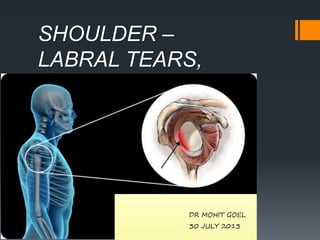
SHOULDER –
LABRAL TEARS,
VARAINTS.
The glenohumeral joint has the
following supporting structures:
Superiorly
coracoacromial arch and
coracoacromial ligament
long head of the biceps tendon
tendon of the supraspinatus muscle
Anteriorly
anterior labrum
glenohumeral ligaments - SGHL, MGHL, IGHL (anterior band)
subscapularis tendon
Posteriorly
posterior labrum
posterior band of the IGHL
infraspinatus and teres minor tendon
Glenoid Labrum
The glenoid labrum is a fibrocartilaginous structure that
attaches to the glenoid rim and is about 4 mm wide.
The labrum may show considerable variation in shape and in
mechanism of attachment to the glenoid. It is usually rounded
or triangular on cross-sectional images.
Biceps Tendon
The tendon of the long head of the biceps muscle attaches to the
anterosuperior aspect of the glenoid rim.
The attachment of the biceps tendon may demonstrate four components,
including fibers that attach to the anterosuperior labrum, the
posterosuperior labrum, the supraglenoid tubercle, and the base of the
coracoid process.
From its site of attachment, the biceps tendon courses laterally and exits
the glenohumeral joint through the intertubercular groove, where it is
secured by the transverse ligament.
coronal section obtained at the level of the labral-bicipital complex illustrates the
biceps tendon (B), superior labrum (L), and glenoid cartilage (C), all of which are
intimately related in this region.
biceps tendon attachment at the level of the superior labrum and glenoid illustrates
attachments to the superior glenoid rim (1), the posterior (2) and anterior (3) labrum, and
the base of the coracoid process (4).
Glenohumeral Ligaments
Superior Glenohumeral Ligament.—
The glenohumeral ligaments play a role as shoulder stabilizers and consist of
thickened bands of the joint capsule.
The superior glenohumeral ligament is the most consistently identified capsular
ligament. It can arise from the anterosuperior labrum, the attachment of the
tendon of the long head of the biceps
muscle, or the middle glenohumeral
ligament.
Middle Glenohumeral Ligament.—
The middle glenohumeral ligament varies most in size and site of attachment to
the glenoid.
It may attach to the superior portion of the anterior glenoid labrum but more
frequently attaches medially on the glenoid neck.
The middle glenohumeral ligament may be absent or may appear thick and
cordlike (as, for example, in Buford complex).
CT arthrogram (2-mm section thickness) shows the middle glenohumeral ligament
(arrowhead) attached to the anterior labrum (arrow).
Transverse fat-saturated MR arthrogram (560/14) demonstrates the middle
glenohumeral ligament attaching medially on the glenoid neck (arrow).
Absent middle glenohumeral ligament in a 40-year-old woman. CT arthrogram (2-
mm section thickness) demonstrates absence of the middle glenohumeral
ligament (*) and a wide anterior joint recess (arrowheads).
Inferior Glenohumeral Ligament.—
The inferior glenohumeral ligament is an important stabilizer of the anterior
shoulder joint and consists of the axillary pouch and anterior and posterior
bands.
The anterior band inserts along the inferior two-thirds of the anterior glenoid
labrum. When redundant, it may overlap the anterior edge of the glenoid
cartilage.
The anterior band is usually quite prominent, although in approximately 25%
of cases it is very thin.
The posterior band is usually thinner than the anterior band.
Sagittal fat-saturated T1-weighted MR
arthrogram (750/15) demonstrates the biceps
tendon (t), subscapularis tendon (S), and anterior
and posterior bands of the inferior glenohumeral
ligament (arrows).
CORONAL
Labral variants
There are many labral variants.
These normal variants are all located in the 11-3 o'clock position.
It is important to recognize these variants, because they can mimick a
SLAP tear.
These normal variants will usually not mimick a Bankart-lesion, since it is
located at the 3-6 o'clock position, where these normal variants do not
occur.
However labral tears may originate at the 3-6 o'clock position and
subsequently extend superiorly.
Sublabral recess
There are 3 types of attachment of the superior labrum at the 12 o'clock
position where the biceps tendon inserts.
In type I there is no recess between the glenoid cartilage and the labrum.
In type II there is a small recess.
In type III there is a large sublabral recess.
This sublabral recess can be difficult to distinguish from a SLAP-tear or a
sublabral foramen.
These images illustrate the differences between an sublabral recess and a SLAP-tear.
Type I labral ---On a coronal MR arthrogram , the
labrum (black arrow) is tightly attached to the
glenoid cartilage and biceps tendon (white arrow)
Type II labral attachment. Coronal fat-saturated T1-
weighted MR arthrogram shows a small recess
between the labrum and the glenoid cartilage (arrow).
Type III labral attachment ----coronal CT
arthrogram shows a large recess between
the labrum and the glenoid (arrow).
Sublabral Foramen
A sublabral foramen or sublabral hole is an unattached anterosuperior
labrum at the 1-3 o'clock position.
It is seen in 11% of individuals.
On a MR-arthtrogram a sublabral
foramen should not be confused
with a sublabral recess or SLAP-tear,
which are also located in this region.
A sublabral recess however is located
at the site of the attachment of the
biceps tendon at 12 o'clock and does
not extend to the 1-3 o?lock position.
A SLAP tear may extend to the 1-3 o'
clock position, but the attachment of
the biceps tendon to the superior
labrum should always be involved.
notice the unattached labrum at the 12-3 o'clock position at the site of the sublabral
foramen. Notice the smooth borders unlike the margins of a SLAP-tear.
Buford complex
A Buford complex is a congenital labral variant.
The anterosuperior labrum is absent in the 1-3 o'clock position and the
middle glenohumeral ligament is usually thickened.
It is present in approximately 1.5% of individuals.
On these axial images a Buford complex can be identified.
The anterior labrum is absent in the 1-3 o'clock position and there is a thickened
middle GHL.
The thickened middle GHL should not be confused with a displaced labrum.
It should always be possible to trace the middle GHL upwards to the glenoid rim and
downwards to the humerus.
Variation in the shapes of labrum
Labral pathology
• BANKART LESION
• BONY BANKART
• REVERSE BANKART
• PERTHES
• REVERSE PERTHES
• APLPSA
• POLPSA
• ALIPSA
• POLIPSA
• GLAD
• GARD
• GAGL
• HAGL
• BHAGL
• DOUBLE LESION
• TRIPLE LESION
Following are various labral pathologies :
Labral pathology
A Clockwise approach to the labrum is the easiest way to diagnose labral
tears and to differentiate them from normal labral variants.
There are two types of labral tears: SLAP tears and Bankart lesions.
SLAP is an acronym that stands for 'Superior Labral tear from Anterior to Posterior'.
SLAP tears start at the 12 o'clock position where the biceps anchor is located, which
tears the labrum off the glenoid.
SLAP tears typically extend from the 10 to the 2 o'clock position, but can extend
more posteriorly or anteriorly and even extend into the biceps tendon.
Bankart lesions are typically located in the 3-6 o'clock position because that's where
the humeral head dislocates.
Dislocation
Anterior dislocation
The shoulder is a very mobile and therefore unstable joint.
The humeral head is almost always displaced anteriorly, inferiorly and medially
below the coracoid process (95% of cases).
Motion to superior is limited by the acromion, coracoid process and rotator cuff
(figure).
Motion in a posterior direction is limited by the posterior rim of the glenoid which is
in an anteverted position.
The dislocation of the humeral head to antero-inferior causes damage to the antero-
inferior rim of the glenoid in the 3 - 6 o'clock position (marked in red).
Especially in younger patients this results in a Bankart fracture or a Bankart lesion
which is a tear of the anteroinferior labrum.
This results in instability and recurrent dislocations.
The images show a subtle Bankart fracture (arrows).
Hill-Sachs
On MR a Hill-Sachs defect is seen at or above the level of the coracoid
process.
Hill-Sachs is a posterolateral depression of the humeral head.
It is above or at the level of the coracoid in the first 18 mm of the proximal
humeral head.
It is seen in 75-100% of patients with anterior instability.
It is chondral or osteochondral.
Bankart and variants
Bankart-lesions and variants like Perthes and ALPSA are injuries to the anteroinferior
labrum.
These injuries are always located in the 3-6 o'clock position because they are caused by
an anterior-inferior dislocation.
Bankart lesion
Bankart lesions are labral tears without an osseus fragment.
MR arthrography or arthroscopy are optimal to diagnose Bankart or Bankart-like
lesions.
There is a detachment of the anteroinferior labrum (3-6 o'clock) with complete
tearing of the anterior scapular periosteum.
The arrow points to the disrupted periosteum.
On MR-athrography the labrum is missing on the anterior glenoid and the labral
fragment is displaced anteriorly (arrow).
Osseus Bankart
Bankart lesions with an osseus fragment are common findings in patients with an
anterior dislocation and are frequently seen on the x-rays or CT-scan.
On MR-arthrography it may be difficult to depict the osseus fragment.
On CT it is easy to appreciate the osseus fragment of the anterior glenoid (arrow).
Reverse Bankart
CT-images in another patient show a reversed osseus Bankart in a patient with
posterior dislocation.
Axial MR-arthrogram of a reverse Bankart.
Perthes lesion
A Perthes lesion is a labroligamentous avulsion like a Bankart, but with a medially
stripped intact periosteum.
On images of the shoulder with the arm in a neutral position, the torn labrum may
be held in its normal anatomic position by the intact scapular periosteum, which
thereby prevents contrast media from entering the tear.
This means that MR-arthrography
with the arm in the neutral position
may fail to detect the labral tear.
In the ABER-position it is obvious that there is a Perthes lesion (black arrow).
Due to the ABER-position the anterior band of the inferior GHL creates tension
on the anteroinferior labrum and contrast fills the tear.
In the ABER position however there is tension on the antero-inferior labrum by
the stretched anterior band of the inferior glenohumeral ligament and you have
more chance to detect the tear.
The arrow points to the intact periosteum.
ALPSA
An ALPSA-lesion is an Anterior Labral Periosteal Sleeve Avulsion.
The anterior labrum is absent on the glenoid rim.
The arrow points to the medially displaced labroligamentous
complex.
Images of a patient with an ALPSA-lesion.
Notice the medially displaced labrum.
POLPSA
Posterior labral periosteal sleeve avulsion.
It is reverse of ALPSA.
GLAD
A GLAD-lesion is a GlenoLabral Articular Disruption.
It represents a patial tear of the anteroinferior labrum with adjacent
cartilage damage.
The arrow points to the cartilage defect.
The images show a partial tear of the anteroinferior labrum
with adjacent cartilage damage at the 4-6 o 'clock position
(arrows).
GLAD-lesion
Defect is at the base of the labrum, predominantly in the glenoid articular
hyaline cartilage.
HAGL is a Humeral Avulsion of the inferior Glenohumeral Ligament.
There is discontinuity of the IGHL attachment on the humerus with
leakage of contrast.
HAGL
Glenoid Avulsion of the inferior Glenohumeral Ligament.
THANKYOU

Mais conteúdo relacionado

Mais procurados

Presentation1.pptx, radiological anatomy of the knee joint.
Presentation1.pptx, radiological anatomy of the knee joint.Presentation1.pptx, radiological anatomy of the knee joint.
Presentation1.pptx, radiological anatomy of the knee joint.Abdellah Nazeer
 
Patello femoral joint - MRI
Patello femoral joint - MRIPatello femoral joint - MRI
Patello femoral joint - MRIDr. Mohit Goel
 
Presentation1.pptx, radiological anatomy of the shoulder joint.
Presentation1.pptx, radiological anatomy of the shoulder joint.Presentation1.pptx, radiological anatomy of the shoulder joint.
Presentation1.pptx, radiological anatomy of the shoulder joint.Abdellah Nazeer
 
Imaging of shoulder - Dr. Vishal Sankpal
Imaging of shoulder - Dr. Vishal SankpalImaging of shoulder - Dr. Vishal Sankpal
Imaging of shoulder - Dr. Vishal SankpalVishal Sankpal
 
Presentation2.pptx wrist joint.
Presentation2.pptx wrist joint.Presentation2.pptx wrist joint.
Presentation2.pptx wrist joint.Abdellah Nazeer
 
Presentation1, radiological imaging of adhesive capsulitis(frozen shoulder).
Presentation1, radiological imaging of adhesive capsulitis(frozen shoulder).Presentation1, radiological imaging of adhesive capsulitis(frozen shoulder).
Presentation1, radiological imaging of adhesive capsulitis(frozen shoulder).Abdellah Nazeer
 
HOW TO READ SHOULDER MRI
HOW TO READ SHOULDER MRI HOW TO READ SHOULDER MRI
HOW TO READ SHOULDER MRI Arjun Kouloth
 
Femoroacetabular%20 impingement[1]
Femoroacetabular%20 impingement[1]Femoroacetabular%20 impingement[1]
Femoroacetabular%20 impingement[1]orthoprince
 
Presentation1.pptx mri of elbow joint
Presentation1.pptx mri of elbow jointPresentation1.pptx mri of elbow joint
Presentation1.pptx mri of elbow jointAbdellah Nazeer
 
Presentation1, radiological imaging of anterior knee pain.
Presentation1, radiological imaging of anterior knee pain.Presentation1, radiological imaging of anterior knee pain.
Presentation1, radiological imaging of anterior knee pain.Abdellah Nazeer
 
Radiology of the Elbow Joint. Dr. Sumit Sharma
Radiology of the Elbow Joint. Dr. Sumit SharmaRadiology of the Elbow Joint. Dr. Sumit Sharma
Radiology of the Elbow Joint. Dr. Sumit SharmaSumit Sharma
 
Presentation1, radiological film reading of the sholder joint.
Presentation1,  radiological film reading of the sholder joint.Presentation1,  radiological film reading of the sholder joint.
Presentation1, radiological film reading of the sholder joint.Abdellah Nazeer
 
Radiographic evaluation of shoulder
Radiographic evaluation of shoulderRadiographic evaluation of shoulder
Radiographic evaluation of shouldermrinal joshi
 
MRI of Knee joint-- hossam massoud
MRI of Knee joint-- hossam massoudMRI of Knee joint-- hossam massoud
MRI of Knee joint-- hossam massoudHossam Massoud
 
Presentation1, radiological film reading of the hip joint.
Presentation1, radiological film reading of the hip joint.Presentation1, radiological film reading of the hip joint.
Presentation1, radiological film reading of the hip joint.Abdellah Nazeer
 
MRI KNEE JOINT ANATOMY
MRI KNEE JOINT ANATOMYMRI KNEE JOINT ANATOMY
MRI KNEE JOINT ANATOMYNikhil Bansal
 
Larynx anatomy ct and mri
Larynx anatomy ct and mriLarynx anatomy ct and mri
Larynx anatomy ct and mriAnish Choudhary
 

Mais procurados (20)

Presentation1.pptx, radiological anatomy of the knee joint.
Presentation1.pptx, radiological anatomy of the knee joint.Presentation1.pptx, radiological anatomy of the knee joint.
Presentation1.pptx, radiological anatomy of the knee joint.
 
MRI of Shoulder anatomy
MRI of Shoulder anatomyMRI of Shoulder anatomy
MRI of Shoulder anatomy
 
Patello femoral joint - MRI
Patello femoral joint - MRIPatello femoral joint - MRI
Patello femoral joint - MRI
 
Presentation1.pptx, radiological anatomy of the shoulder joint.
Presentation1.pptx, radiological anatomy of the shoulder joint.Presentation1.pptx, radiological anatomy of the shoulder joint.
Presentation1.pptx, radiological anatomy of the shoulder joint.
 
Imaging of shoulder - Dr. Vishal Sankpal
Imaging of shoulder - Dr. Vishal SankpalImaging of shoulder - Dr. Vishal Sankpal
Imaging of shoulder - Dr. Vishal Sankpal
 
MRI SPINE ANATOMY
MRI SPINE ANATOMYMRI SPINE ANATOMY
MRI SPINE ANATOMY
 
Presentation2.pptx wrist joint.
Presentation2.pptx wrist joint.Presentation2.pptx wrist joint.
Presentation2.pptx wrist joint.
 
Presentation1, radiological imaging of adhesive capsulitis(frozen shoulder).
Presentation1, radiological imaging of adhesive capsulitis(frozen shoulder).Presentation1, radiological imaging of adhesive capsulitis(frozen shoulder).
Presentation1, radiological imaging of adhesive capsulitis(frozen shoulder).
 
MRI Knee trauma
MRI Knee traumaMRI Knee trauma
MRI Knee trauma
 
HOW TO READ SHOULDER MRI
HOW TO READ SHOULDER MRI HOW TO READ SHOULDER MRI
HOW TO READ SHOULDER MRI
 
Femoroacetabular%20 impingement[1]
Femoroacetabular%20 impingement[1]Femoroacetabular%20 impingement[1]
Femoroacetabular%20 impingement[1]
 
Presentation1.pptx mri of elbow joint
Presentation1.pptx mri of elbow jointPresentation1.pptx mri of elbow joint
Presentation1.pptx mri of elbow joint
 
Presentation1, radiological imaging of anterior knee pain.
Presentation1, radiological imaging of anterior knee pain.Presentation1, radiological imaging of anterior knee pain.
Presentation1, radiological imaging of anterior knee pain.
 
Radiology of the Elbow Joint. Dr. Sumit Sharma
Radiology of the Elbow Joint. Dr. Sumit SharmaRadiology of the Elbow Joint. Dr. Sumit Sharma
Radiology of the Elbow Joint. Dr. Sumit Sharma
 
Presentation1, radiological film reading of the sholder joint.
Presentation1,  radiological film reading of the sholder joint.Presentation1,  radiological film reading of the sholder joint.
Presentation1, radiological film reading of the sholder joint.
 
Radiographic evaluation of shoulder
Radiographic evaluation of shoulderRadiographic evaluation of shoulder
Radiographic evaluation of shoulder
 
MRI of Knee joint-- hossam massoud
MRI of Knee joint-- hossam massoudMRI of Knee joint-- hossam massoud
MRI of Knee joint-- hossam massoud
 
Presentation1, radiological film reading of the hip joint.
Presentation1, radiological film reading of the hip joint.Presentation1, radiological film reading of the hip joint.
Presentation1, radiological film reading of the hip joint.
 
MRI KNEE JOINT ANATOMY
MRI KNEE JOINT ANATOMYMRI KNEE JOINT ANATOMY
MRI KNEE JOINT ANATOMY
 
Larynx anatomy ct and mri
Larynx anatomy ct and mriLarynx anatomy ct and mri
Larynx anatomy ct and mri
 

Destaque

Practical approach to lung cancer
Practical approach to lung cancerPractical approach to lung cancer
Practical approach to lung cancerGamal Agmy
 
Presentation1.pptx. shoulder joint.
Presentation1.pptx. shoulder joint.Presentation1.pptx. shoulder joint.
Presentation1.pptx. shoulder joint.Abdellah Nazeer
 
Shoulder Mri Scan in Delhi by Dr Shekhar Shrivastav
Shoulder Mri Scan in Delhi by Dr Shekhar ShrivastavShoulder Mri Scan in Delhi by Dr Shekhar Shrivastav
Shoulder Mri Scan in Delhi by Dr Shekhar ShrivastavDelhiArthroscopy
 
8th Edition of the TNM Classification for Lung Cancer
8th Edition of the TNM Classification for Lung Cancer8th Edition of the TNM Classification for Lung Cancer
8th Edition of the TNM Classification for Lung CancerMauricio Lema
 

Destaque (7)

Practical approach to lung cancer
Practical approach to lung cancerPractical approach to lung cancer
Practical approach to lung cancer
 
MRI sholdure
MRI sholdureMRI sholdure
MRI sholdure
 
Presentation1.pptx. shoulder joint.
Presentation1.pptx. shoulder joint.Presentation1.pptx. shoulder joint.
Presentation1.pptx. shoulder joint.
 
Abdomin Liver CT
Abdomin Liver CT Abdomin Liver CT
Abdomin Liver CT
 
Shoulder Mri Scan in Delhi by Dr Shekhar Shrivastav
Shoulder Mri Scan in Delhi by Dr Shekhar ShrivastavShoulder Mri Scan in Delhi by Dr Shekhar Shrivastav
Shoulder Mri Scan in Delhi by Dr Shekhar Shrivastav
 
Cáncer de mama
Cáncer de mamaCáncer de mama
Cáncer de mama
 
8th Edition of the TNM Classification for Lung Cancer
8th Edition of the TNM Classification for Lung Cancer8th Edition of the TNM Classification for Lung Cancer
8th Edition of the TNM Classification for Lung Cancer
 

Semelhante a Shoulder labral tears MRI

shoulder radioanatomy.pptx
shoulder radioanatomy.pptxshoulder radioanatomy.pptx
shoulder radioanatomy.pptxIslah Raoof
 
Normal Labral Variants - Walif Chbeir
Normal Labral Variants - Walif ChbeirNormal Labral Variants - Walif Chbeir
Normal Labral Variants - Walif ChbeirWalif Chbeir
 
Examination of shoulder
Examination of shoulderExamination of shoulder
Examination of shoulderArya Anish
 
Sprengle shoulder (congenital elevation of scapula)
Sprengle shoulder (congenital elevation of scapula)Sprengle shoulder (congenital elevation of scapula)
Sprengle shoulder (congenital elevation of scapula)Gaurav Singh
 
appendicular trauma in radiology. .pptx
appendicular trauma in radiology.  .pptxappendicular trauma in radiology.  .pptx
appendicular trauma in radiology. .pptxyashovrattiwari1
 
Osteology upper limb by Dr G Kamau
Osteology upper limb by Dr G KamauOsteology upper limb by Dr G Kamau
Osteology upper limb by Dr G KamauMathewJude
 
Congenital skeletal dysplasia
Congenital skeletal dysplasiaCongenital skeletal dysplasia
Congenital skeletal dysplasiasami123123
 
Do it-yourself-paeds-ortho (Paediatric Orthopaedics for beginners)
Do it-yourself-paeds-ortho (Paediatric Orthopaedics for beginners)Do it-yourself-paeds-ortho (Paediatric Orthopaedics for beginners)
Do it-yourself-paeds-ortho (Paediatric Orthopaedics for beginners)Jonathan Cheah
 
Presentation1, radiological imaging of shoulder dislocation.
Presentation1, radiological imaging of shoulder dislocation.Presentation1, radiological imaging of shoulder dislocation.
Presentation1, radiological imaging of shoulder dislocation.Abdellah Nazeer
 
RADIOLOGICAL ANATOMY OF UPPER LIMB(SHOULDER@ELBOW)
RADIOLOGICAL ANATOMY OF UPPER LIMB(SHOULDER@ELBOW)RADIOLOGICAL ANATOMY OF UPPER LIMB(SHOULDER@ELBOW)
RADIOLOGICAL ANATOMY OF UPPER LIMB(SHOULDER@ELBOW)Bhaskar Sangamreddy
 
usg shoulder by dr. kanishka.pptx
usg shoulder by dr. kanishka.pptxusg shoulder by dr. kanishka.pptx
usg shoulder by dr. kanishka.pptxRajVaghasia
 
Shoulder joint xray & usg by Dr Soumitra Halder
Shoulder joint xray & usg by Dr Soumitra HalderShoulder joint xray & usg by Dr Soumitra Halder
Shoulder joint xray & usg by Dr Soumitra HalderSoumitra Halder
 
Presentation1.pptx, radiological anatomy of the upper limb joint.
Presentation1.pptx, radiological anatomy of the upper limb joint.Presentation1.pptx, radiological anatomy of the upper limb joint.
Presentation1.pptx, radiological anatomy of the upper limb joint.Abdellah Nazeer
 
Recurrent Shoulder Dislocation
Recurrent Shoulder DislocationRecurrent Shoulder Dislocation
Recurrent Shoulder DislocationSidheshwar Thosar
 
Fractures of the distal humerus ppt
Fractures of the distal humerus pptFractures of the distal humerus ppt
Fractures of the distal humerus pptKunal Arora
 
SPRENGEL SHOULDER.pptx
SPRENGEL SHOULDER.pptxSPRENGEL SHOULDER.pptx
SPRENGEL SHOULDER.pptxSalman Syed
 
Supra condylar humerus fracture in children
Supra condylar humerus fracture in childrenSupra condylar humerus fracture in children
Supra condylar humerus fracture in childrenSubodh Pathak
 
Can I Offer More 4
Can I Offer More 4Can I Offer More 4
Can I Offer More 4guest609645b
 
shoulder assessment (full)
shoulder assessment (full)shoulder assessment (full)
shoulder assessment (full)SYED MASOOD
 

Semelhante a Shoulder labral tears MRI (20)

shoulder radioanatomy.pptx
shoulder radioanatomy.pptxshoulder radioanatomy.pptx
shoulder radioanatomy.pptx
 
Normal Labral Variants - Walif Chbeir
Normal Labral Variants - Walif ChbeirNormal Labral Variants - Walif Chbeir
Normal Labral Variants - Walif Chbeir
 
Examination of shoulder
Examination of shoulderExamination of shoulder
Examination of shoulder
 
Sprengle shoulder (congenital elevation of scapula)
Sprengle shoulder (congenital elevation of scapula)Sprengle shoulder (congenital elevation of scapula)
Sprengle shoulder (congenital elevation of scapula)
 
appendicular trauma in radiology. .pptx
appendicular trauma in radiology.  .pptxappendicular trauma in radiology.  .pptx
appendicular trauma in radiology. .pptx
 
Osteology upper limb by Dr G Kamau
Osteology upper limb by Dr G KamauOsteology upper limb by Dr G Kamau
Osteology upper limb by Dr G Kamau
 
DDH
DDHDDH
DDH
 
Congenital skeletal dysplasia
Congenital skeletal dysplasiaCongenital skeletal dysplasia
Congenital skeletal dysplasia
 
Do it-yourself-paeds-ortho (Paediatric Orthopaedics for beginners)
Do it-yourself-paeds-ortho (Paediatric Orthopaedics for beginners)Do it-yourself-paeds-ortho (Paediatric Orthopaedics for beginners)
Do it-yourself-paeds-ortho (Paediatric Orthopaedics for beginners)
 
Presentation1, radiological imaging of shoulder dislocation.
Presentation1, radiological imaging of shoulder dislocation.Presentation1, radiological imaging of shoulder dislocation.
Presentation1, radiological imaging of shoulder dislocation.
 
RADIOLOGICAL ANATOMY OF UPPER LIMB(SHOULDER@ELBOW)
RADIOLOGICAL ANATOMY OF UPPER LIMB(SHOULDER@ELBOW)RADIOLOGICAL ANATOMY OF UPPER LIMB(SHOULDER@ELBOW)
RADIOLOGICAL ANATOMY OF UPPER LIMB(SHOULDER@ELBOW)
 
usg shoulder by dr. kanishka.pptx
usg shoulder by dr. kanishka.pptxusg shoulder by dr. kanishka.pptx
usg shoulder by dr. kanishka.pptx
 
Shoulder joint xray & usg by Dr Soumitra Halder
Shoulder joint xray & usg by Dr Soumitra HalderShoulder joint xray & usg by Dr Soumitra Halder
Shoulder joint xray & usg by Dr Soumitra Halder
 
Presentation1.pptx, radiological anatomy of the upper limb joint.
Presentation1.pptx, radiological anatomy of the upper limb joint.Presentation1.pptx, radiological anatomy of the upper limb joint.
Presentation1.pptx, radiological anatomy of the upper limb joint.
 
Recurrent Shoulder Dislocation
Recurrent Shoulder DislocationRecurrent Shoulder Dislocation
Recurrent Shoulder Dislocation
 
Fractures of the distal humerus ppt
Fractures of the distal humerus pptFractures of the distal humerus ppt
Fractures of the distal humerus ppt
 
SPRENGEL SHOULDER.pptx
SPRENGEL SHOULDER.pptxSPRENGEL SHOULDER.pptx
SPRENGEL SHOULDER.pptx
 
Supra condylar humerus fracture in children
Supra condylar humerus fracture in childrenSupra condylar humerus fracture in children
Supra condylar humerus fracture in children
 
Can I Offer More 4
Can I Offer More 4Can I Offer More 4
Can I Offer More 4
 
shoulder assessment (full)
shoulder assessment (full)shoulder assessment (full)
shoulder assessment (full)
 

Mais de Dr. Mohit Goel

Utrasound Gall-bladder & biliary tract
Utrasound Gall-bladder & biliary tractUtrasound Gall-bladder & biliary tract
Utrasound Gall-bladder & biliary tractDr. Mohit Goel
 
Sectional radio-anatomy of abdomen
Sectional radio-anatomy of abdomenSectional radio-anatomy of abdomen
Sectional radio-anatomy of abdomenDr. Mohit Goel
 
Retroperitoneal masses radiology
Retroperitoneal masses radiologyRetroperitoneal masses radiology
Retroperitoneal masses radiologyDr. Mohit Goel
 
Prostate carcinoma raiology
Prostate carcinoma raiologyProstate carcinoma raiology
Prostate carcinoma raiologyDr. Mohit Goel
 
Precocious puberty - Imaging
Precocious puberty - ImagingPrecocious puberty - Imaging
Precocious puberty - ImagingDr. Mohit Goel
 
PNS (Para-nasal-sinuses) anatomy and variants
PNS (Para-nasal-sinuses) anatomy and variantsPNS (Para-nasal-sinuses) anatomy and variants
PNS (Para-nasal-sinuses) anatomy and variantsDr. Mohit Goel
 
Pineal region masses - radiology
Pineal region masses - radiologyPineal region masses - radiology
Pineal region masses - radiologyDr. Mohit Goel
 
Peripheral arterial doppler
Peripheral  arterial dopplerPeripheral  arterial doppler
Peripheral arterial dopplerDr. Mohit Goel
 
Pediatric stroke radiology
Pediatric stroke radiologyPediatric stroke radiology
Pediatric stroke radiologyDr. Mohit Goel
 
Pediatric chest (part 2)
Pediatric chest (part 2)Pediatric chest (part 2)
Pediatric chest (part 2)Dr. Mohit Goel
 
Orbital pathologies radiology
Orbital pathologies radiologyOrbital pathologies radiology
Orbital pathologies radiologyDr. Mohit Goel
 
Orbital pathologies.pptx (part 1)
Orbital pathologies.pptx (part 1)Orbital pathologies.pptx (part 1)
Orbital pathologies.pptx (part 1)Dr. Mohit Goel
 

Mais de Dr. Mohit Goel (20)

Utrasound Gall-bladder & biliary tract
Utrasound Gall-bladder & biliary tractUtrasound Gall-bladder & biliary tract
Utrasound Gall-bladder & biliary tract
 
Ultrasoud hernia
Ultrasoud herniaUltrasoud hernia
Ultrasoud hernia
 
TVS image gallery
TVS image galleryTVS image gallery
TVS image gallery
 
Shoulder ultrasound
Shoulder ultrasoundShoulder ultrasound
Shoulder ultrasound
 
Sectional radio-anatomy of abdomen
Sectional radio-anatomy of abdomenSectional radio-anatomy of abdomen
Sectional radio-anatomy of abdomen
 
Renal doppler usg
Renal doppler usgRenal doppler usg
Renal doppler usg
 
Retroperitoneal masses radiology
Retroperitoneal masses radiologyRetroperitoneal masses radiology
Retroperitoneal masses radiology
 
Prostate carcinoma raiology
Prostate carcinoma raiologyProstate carcinoma raiology
Prostate carcinoma raiology
 
Precocious puberty - Imaging
Precocious puberty - ImagingPrecocious puberty - Imaging
Precocious puberty - Imaging
 
Pre-FESS PNS CT
Pre-FESS PNS CTPre-FESS PNS CT
Pre-FESS PNS CT
 
PNS (Para-nasal-sinuses) anatomy and variants
PNS (Para-nasal-sinuses) anatomy and variantsPNS (Para-nasal-sinuses) anatomy and variants
PNS (Para-nasal-sinuses) anatomy and variants
 
Pineal region masses - radiology
Pineal region masses - radiologyPineal region masses - radiology
Pineal region masses - radiology
 
Peripheral arterial doppler
Peripheral  arterial dopplerPeripheral  arterial doppler
Peripheral arterial doppler
 
Pediatric stroke radiology
Pediatric stroke radiologyPediatric stroke radiology
Pediatric stroke radiology
 
Pediatric chest (part 2)
Pediatric chest (part 2)Pediatric chest (part 2)
Pediatric chest (part 2)
 
Pediatric chest
Pediatric chestPediatric chest
Pediatric chest
 
Orbital pathologies radiology
Orbital pathologies radiologyOrbital pathologies radiology
Orbital pathologies radiology
 
Osteopetrosis
OsteopetrosisOsteopetrosis
Osteopetrosis
 
Orbital pathologies.pptx (part 1)
Orbital pathologies.pptx (part 1)Orbital pathologies.pptx (part 1)
Orbital pathologies.pptx (part 1)
 
Orbit imaging anatomy
Orbit imaging anatomyOrbit imaging anatomy
Orbit imaging anatomy
 

Último

Oppenheimer Film Discussion for Philosophy and Film
Oppenheimer Film Discussion for Philosophy and FilmOppenheimer Film Discussion for Philosophy and Film
Oppenheimer Film Discussion for Philosophy and FilmStan Meyer
 
MS4 level being good citizen -imperative- (1) (1).pdf
MS4 level   being good citizen -imperative- (1) (1).pdfMS4 level   being good citizen -imperative- (1) (1).pdf
MS4 level being good citizen -imperative- (1) (1).pdfMr Bounab Samir
 
Using Grammatical Signals Suitable to Patterns of Idea Development
Using Grammatical Signals Suitable to Patterns of Idea DevelopmentUsing Grammatical Signals Suitable to Patterns of Idea Development
Using Grammatical Signals Suitable to Patterns of Idea Developmentchesterberbo7
 
31 ĐỀ THI THỬ VÀO LỚP 10 - TIẾNG ANH - FORM MỚI 2025 - 40 CÂU HỎI - BÙI VĂN V...
31 ĐỀ THI THỬ VÀO LỚP 10 - TIẾNG ANH - FORM MỚI 2025 - 40 CÂU HỎI - BÙI VĂN V...31 ĐỀ THI THỬ VÀO LỚP 10 - TIẾNG ANH - FORM MỚI 2025 - 40 CÂU HỎI - BÙI VĂN V...
31 ĐỀ THI THỬ VÀO LỚP 10 - TIẾNG ANH - FORM MỚI 2025 - 40 CÂU HỎI - BÙI VĂN V...Nguyen Thanh Tu Collection
 
Mythology Quiz-4th April 2024, Quiz Club NITW
Mythology Quiz-4th April 2024, Quiz Club NITWMythology Quiz-4th April 2024, Quiz Club NITW
Mythology Quiz-4th April 2024, Quiz Club NITWQuiz Club NITW
 
Blowin' in the Wind of Caste_ Bob Dylan's Song as a Catalyst for Social Justi...
Blowin' in the Wind of Caste_ Bob Dylan's Song as a Catalyst for Social Justi...Blowin' in the Wind of Caste_ Bob Dylan's Song as a Catalyst for Social Justi...
Blowin' in the Wind of Caste_ Bob Dylan's Song as a Catalyst for Social Justi...DhatriParmar
 
Congestive Cardiac Failure..presentation
Congestive Cardiac Failure..presentationCongestive Cardiac Failure..presentation
Congestive Cardiac Failure..presentationdeepaannamalai16
 
Reading and Writing Skills 11 quarter 4 melc 1
Reading and Writing Skills 11 quarter 4 melc 1Reading and Writing Skills 11 quarter 4 melc 1
Reading and Writing Skills 11 quarter 4 melc 1GloryAnnCastre1
 
Active Learning Strategies (in short ALS).pdf
Active Learning Strategies (in short ALS).pdfActive Learning Strategies (in short ALS).pdf
Active Learning Strategies (in short ALS).pdfPatidar M
 
Textual Evidence in Reading and Writing of SHS
Textual Evidence in Reading and Writing of SHSTextual Evidence in Reading and Writing of SHS
Textual Evidence in Reading and Writing of SHSMae Pangan
 
Multi Domain Alias In the Odoo 17 ERP Module
Multi Domain Alias In the Odoo 17 ERP ModuleMulti Domain Alias In the Odoo 17 ERP Module
Multi Domain Alias In the Odoo 17 ERP ModuleCeline George
 
Concurrency Control in Database Management system
Concurrency Control in Database Management systemConcurrency Control in Database Management system
Concurrency Control in Database Management systemChristalin Nelson
 
Grade Three -ELLNA-REVIEWER-ENGLISH.pptx
Grade Three -ELLNA-REVIEWER-ENGLISH.pptxGrade Three -ELLNA-REVIEWER-ENGLISH.pptx
Grade Three -ELLNA-REVIEWER-ENGLISH.pptxkarenfajardo43
 
Unraveling Hypertext_ Analyzing Postmodern Elements in Literature.pptx
Unraveling Hypertext_ Analyzing  Postmodern Elements in  Literature.pptxUnraveling Hypertext_ Analyzing  Postmodern Elements in  Literature.pptx
Unraveling Hypertext_ Analyzing Postmodern Elements in Literature.pptxDhatriParmar
 
Daily Lesson Plan in Mathematics Quarter 4
Daily Lesson Plan in Mathematics Quarter 4Daily Lesson Plan in Mathematics Quarter 4
Daily Lesson Plan in Mathematics Quarter 4JOYLYNSAMANIEGO
 
Q-Factor HISPOL Quiz-6th April 2024, Quiz Club NITW
Q-Factor HISPOL Quiz-6th April 2024, Quiz Club NITWQ-Factor HISPOL Quiz-6th April 2024, Quiz Club NITW
Q-Factor HISPOL Quiz-6th April 2024, Quiz Club NITWQuiz Club NITW
 
4.16.24 Poverty and Precarity--Desmond.pptx
4.16.24 Poverty and Precarity--Desmond.pptx4.16.24 Poverty and Precarity--Desmond.pptx
4.16.24 Poverty and Precarity--Desmond.pptxmary850239
 
Beauty Amidst the Bytes_ Unearthing Unexpected Advantages of the Digital Wast...
Beauty Amidst the Bytes_ Unearthing Unexpected Advantages of the Digital Wast...Beauty Amidst the Bytes_ Unearthing Unexpected Advantages of the Digital Wast...
Beauty Amidst the Bytes_ Unearthing Unexpected Advantages of the Digital Wast...DhatriParmar
 

Último (20)

Oppenheimer Film Discussion for Philosophy and Film
Oppenheimer Film Discussion for Philosophy and FilmOppenheimer Film Discussion for Philosophy and Film
Oppenheimer Film Discussion for Philosophy and Film
 
prashanth updated resume 2024 for Teaching Profession
prashanth updated resume 2024 for Teaching Professionprashanth updated resume 2024 for Teaching Profession
prashanth updated resume 2024 for Teaching Profession
 
MS4 level being good citizen -imperative- (1) (1).pdf
MS4 level   being good citizen -imperative- (1) (1).pdfMS4 level   being good citizen -imperative- (1) (1).pdf
MS4 level being good citizen -imperative- (1) (1).pdf
 
Using Grammatical Signals Suitable to Patterns of Idea Development
Using Grammatical Signals Suitable to Patterns of Idea DevelopmentUsing Grammatical Signals Suitable to Patterns of Idea Development
Using Grammatical Signals Suitable to Patterns of Idea Development
 
31 ĐỀ THI THỬ VÀO LỚP 10 - TIẾNG ANH - FORM MỚI 2025 - 40 CÂU HỎI - BÙI VĂN V...
31 ĐỀ THI THỬ VÀO LỚP 10 - TIẾNG ANH - FORM MỚI 2025 - 40 CÂU HỎI - BÙI VĂN V...31 ĐỀ THI THỬ VÀO LỚP 10 - TIẾNG ANH - FORM MỚI 2025 - 40 CÂU HỎI - BÙI VĂN V...
31 ĐỀ THI THỬ VÀO LỚP 10 - TIẾNG ANH - FORM MỚI 2025 - 40 CÂU HỎI - BÙI VĂN V...
 
Mythology Quiz-4th April 2024, Quiz Club NITW
Mythology Quiz-4th April 2024, Quiz Club NITWMythology Quiz-4th April 2024, Quiz Club NITW
Mythology Quiz-4th April 2024, Quiz Club NITW
 
Blowin' in the Wind of Caste_ Bob Dylan's Song as a Catalyst for Social Justi...
Blowin' in the Wind of Caste_ Bob Dylan's Song as a Catalyst for Social Justi...Blowin' in the Wind of Caste_ Bob Dylan's Song as a Catalyst for Social Justi...
Blowin' in the Wind of Caste_ Bob Dylan's Song as a Catalyst for Social Justi...
 
Congestive Cardiac Failure..presentation
Congestive Cardiac Failure..presentationCongestive Cardiac Failure..presentation
Congestive Cardiac Failure..presentation
 
Reading and Writing Skills 11 quarter 4 melc 1
Reading and Writing Skills 11 quarter 4 melc 1Reading and Writing Skills 11 quarter 4 melc 1
Reading and Writing Skills 11 quarter 4 melc 1
 
Active Learning Strategies (in short ALS).pdf
Active Learning Strategies (in short ALS).pdfActive Learning Strategies (in short ALS).pdf
Active Learning Strategies (in short ALS).pdf
 
Paradigm shift in nursing research by RS MEHTA
Paradigm shift in nursing research by RS MEHTAParadigm shift in nursing research by RS MEHTA
Paradigm shift in nursing research by RS MEHTA
 
Textual Evidence in Reading and Writing of SHS
Textual Evidence in Reading and Writing of SHSTextual Evidence in Reading and Writing of SHS
Textual Evidence in Reading and Writing of SHS
 
Multi Domain Alias In the Odoo 17 ERP Module
Multi Domain Alias In the Odoo 17 ERP ModuleMulti Domain Alias In the Odoo 17 ERP Module
Multi Domain Alias In the Odoo 17 ERP Module
 
Concurrency Control in Database Management system
Concurrency Control in Database Management systemConcurrency Control in Database Management system
Concurrency Control in Database Management system
 
Grade Three -ELLNA-REVIEWER-ENGLISH.pptx
Grade Three -ELLNA-REVIEWER-ENGLISH.pptxGrade Three -ELLNA-REVIEWER-ENGLISH.pptx
Grade Three -ELLNA-REVIEWER-ENGLISH.pptx
 
Unraveling Hypertext_ Analyzing Postmodern Elements in Literature.pptx
Unraveling Hypertext_ Analyzing  Postmodern Elements in  Literature.pptxUnraveling Hypertext_ Analyzing  Postmodern Elements in  Literature.pptx
Unraveling Hypertext_ Analyzing Postmodern Elements in Literature.pptx
 
Daily Lesson Plan in Mathematics Quarter 4
Daily Lesson Plan in Mathematics Quarter 4Daily Lesson Plan in Mathematics Quarter 4
Daily Lesson Plan in Mathematics Quarter 4
 
Q-Factor HISPOL Quiz-6th April 2024, Quiz Club NITW
Q-Factor HISPOL Quiz-6th April 2024, Quiz Club NITWQ-Factor HISPOL Quiz-6th April 2024, Quiz Club NITW
Q-Factor HISPOL Quiz-6th April 2024, Quiz Club NITW
 
4.16.24 Poverty and Precarity--Desmond.pptx
4.16.24 Poverty and Precarity--Desmond.pptx4.16.24 Poverty and Precarity--Desmond.pptx
4.16.24 Poverty and Precarity--Desmond.pptx
 
Beauty Amidst the Bytes_ Unearthing Unexpected Advantages of the Digital Wast...
Beauty Amidst the Bytes_ Unearthing Unexpected Advantages of the Digital Wast...Beauty Amidst the Bytes_ Unearthing Unexpected Advantages of the Digital Wast...
Beauty Amidst the Bytes_ Unearthing Unexpected Advantages of the Digital Wast...
 

Shoulder labral tears MRI

  • 2. The glenohumeral joint has the following supporting structures: Superiorly coracoacromial arch and coracoacromial ligament long head of the biceps tendon tendon of the supraspinatus muscle Anteriorly anterior labrum glenohumeral ligaments - SGHL, MGHL, IGHL (anterior band) subscapularis tendon Posteriorly posterior labrum posterior band of the IGHL infraspinatus and teres minor tendon
  • 3. Glenoid Labrum The glenoid labrum is a fibrocartilaginous structure that attaches to the glenoid rim and is about 4 mm wide. The labrum may show considerable variation in shape and in mechanism of attachment to the glenoid. It is usually rounded or triangular on cross-sectional images.
  • 4. Biceps Tendon The tendon of the long head of the biceps muscle attaches to the anterosuperior aspect of the glenoid rim. The attachment of the biceps tendon may demonstrate four components, including fibers that attach to the anterosuperior labrum, the posterosuperior labrum, the supraglenoid tubercle, and the base of the coracoid process. From its site of attachment, the biceps tendon courses laterally and exits the glenohumeral joint through the intertubercular groove, where it is secured by the transverse ligament.
  • 5. coronal section obtained at the level of the labral-bicipital complex illustrates the biceps tendon (B), superior labrum (L), and glenoid cartilage (C), all of which are intimately related in this region. biceps tendon attachment at the level of the superior labrum and glenoid illustrates attachments to the superior glenoid rim (1), the posterior (2) and anterior (3) labrum, and the base of the coracoid process (4).
  • 6. Glenohumeral Ligaments Superior Glenohumeral Ligament.— The glenohumeral ligaments play a role as shoulder stabilizers and consist of thickened bands of the joint capsule. The superior glenohumeral ligament is the most consistently identified capsular ligament. It can arise from the anterosuperior labrum, the attachment of the tendon of the long head of the biceps muscle, or the middle glenohumeral ligament.
  • 7. Middle Glenohumeral Ligament.— The middle glenohumeral ligament varies most in size and site of attachment to the glenoid. It may attach to the superior portion of the anterior glenoid labrum but more frequently attaches medially on the glenoid neck. The middle glenohumeral ligament may be absent or may appear thick and cordlike (as, for example, in Buford complex). CT arthrogram (2-mm section thickness) shows the middle glenohumeral ligament (arrowhead) attached to the anterior labrum (arrow).
  • 8. Transverse fat-saturated MR arthrogram (560/14) demonstrates the middle glenohumeral ligament attaching medially on the glenoid neck (arrow).
  • 9. Absent middle glenohumeral ligament in a 40-year-old woman. CT arthrogram (2- mm section thickness) demonstrates absence of the middle glenohumeral ligament (*) and a wide anterior joint recess (arrowheads).
  • 10. Inferior Glenohumeral Ligament.— The inferior glenohumeral ligament is an important stabilizer of the anterior shoulder joint and consists of the axillary pouch and anterior and posterior bands. The anterior band inserts along the inferior two-thirds of the anterior glenoid labrum. When redundant, it may overlap the anterior edge of the glenoid cartilage. The anterior band is usually quite prominent, although in approximately 25% of cases it is very thin. The posterior band is usually thinner than the anterior band.
  • 11. Sagittal fat-saturated T1-weighted MR arthrogram (750/15) demonstrates the biceps tendon (t), subscapularis tendon (S), and anterior and posterior bands of the inferior glenohumeral ligament (arrows). CORONAL
  • 12. Labral variants There are many labral variants. These normal variants are all located in the 11-3 o'clock position.
  • 13. It is important to recognize these variants, because they can mimick a SLAP tear. These normal variants will usually not mimick a Bankart-lesion, since it is located at the 3-6 o'clock position, where these normal variants do not occur. However labral tears may originate at the 3-6 o'clock position and subsequently extend superiorly.
  • 14. Sublabral recess There are 3 types of attachment of the superior labrum at the 12 o'clock position where the biceps tendon inserts. In type I there is no recess between the glenoid cartilage and the labrum. In type II there is a small recess. In type III there is a large sublabral recess. This sublabral recess can be difficult to distinguish from a SLAP-tear or a sublabral foramen.
  • 15. These images illustrate the differences between an sublabral recess and a SLAP-tear.
  • 16. Type I labral ---On a coronal MR arthrogram , the labrum (black arrow) is tightly attached to the glenoid cartilage and biceps tendon (white arrow) Type II labral attachment. Coronal fat-saturated T1- weighted MR arthrogram shows a small recess between the labrum and the glenoid cartilage (arrow). Type III labral attachment ----coronal CT arthrogram shows a large recess between the labrum and the glenoid (arrow).
  • 17. Sublabral Foramen A sublabral foramen or sublabral hole is an unattached anterosuperior labrum at the 1-3 o'clock position. It is seen in 11% of individuals. On a MR-arthtrogram a sublabral foramen should not be confused with a sublabral recess or SLAP-tear, which are also located in this region. A sublabral recess however is located at the site of the attachment of the biceps tendon at 12 o'clock and does not extend to the 1-3 o?lock position. A SLAP tear may extend to the 1-3 o' clock position, but the attachment of the biceps tendon to the superior labrum should always be involved.
  • 18. notice the unattached labrum at the 12-3 o'clock position at the site of the sublabral foramen. Notice the smooth borders unlike the margins of a SLAP-tear.
  • 19. Buford complex A Buford complex is a congenital labral variant. The anterosuperior labrum is absent in the 1-3 o'clock position and the middle glenohumeral ligament is usually thickened. It is present in approximately 1.5% of individuals.
  • 20. On these axial images a Buford complex can be identified. The anterior labrum is absent in the 1-3 o'clock position and there is a thickened middle GHL. The thickened middle GHL should not be confused with a displaced labrum. It should always be possible to trace the middle GHL upwards to the glenoid rim and downwards to the humerus.
  • 21. Variation in the shapes of labrum
  • 22. Labral pathology • BANKART LESION • BONY BANKART • REVERSE BANKART • PERTHES • REVERSE PERTHES • APLPSA • POLPSA • ALIPSA • POLIPSA • GLAD • GARD • GAGL • HAGL • BHAGL • DOUBLE LESION • TRIPLE LESION Following are various labral pathologies :
  • 23. Labral pathology A Clockwise approach to the labrum is the easiest way to diagnose labral tears and to differentiate them from normal labral variants. There are two types of labral tears: SLAP tears and Bankart lesions. SLAP is an acronym that stands for 'Superior Labral tear from Anterior to Posterior'. SLAP tears start at the 12 o'clock position where the biceps anchor is located, which tears the labrum off the glenoid. SLAP tears typically extend from the 10 to the 2 o'clock position, but can extend more posteriorly or anteriorly and even extend into the biceps tendon. Bankart lesions are typically located in the 3-6 o'clock position because that's where the humeral head dislocates.
  • 24. Dislocation Anterior dislocation The shoulder is a very mobile and therefore unstable joint. The humeral head is almost always displaced anteriorly, inferiorly and medially below the coracoid process (95% of cases). Motion to superior is limited by the acromion, coracoid process and rotator cuff (figure). Motion in a posterior direction is limited by the posterior rim of the glenoid which is in an anteverted position.
  • 25. The dislocation of the humeral head to antero-inferior causes damage to the antero- inferior rim of the glenoid in the 3 - 6 o'clock position (marked in red). Especially in younger patients this results in a Bankart fracture or a Bankart lesion which is a tear of the anteroinferior labrum. This results in instability and recurrent dislocations.
  • 26. The images show a subtle Bankart fracture (arrows).
  • 27. Hill-Sachs On MR a Hill-Sachs defect is seen at or above the level of the coracoid process. Hill-Sachs is a posterolateral depression of the humeral head. It is above or at the level of the coracoid in the first 18 mm of the proximal humeral head. It is seen in 75-100% of patients with anterior instability. It is chondral or osteochondral.
  • 28. Bankart and variants Bankart-lesions and variants like Perthes and ALPSA are injuries to the anteroinferior labrum. These injuries are always located in the 3-6 o'clock position because they are caused by an anterior-inferior dislocation.
  • 29. Bankart lesion Bankart lesions are labral tears without an osseus fragment. MR arthrography or arthroscopy are optimal to diagnose Bankart or Bankart-like lesions. There is a detachment of the anteroinferior labrum (3-6 o'clock) with complete tearing of the anterior scapular periosteum. The arrow points to the disrupted periosteum.
  • 30. On MR-athrography the labrum is missing on the anterior glenoid and the labral fragment is displaced anteriorly (arrow).
  • 31. Osseus Bankart Bankart lesions with an osseus fragment are common findings in patients with an anterior dislocation and are frequently seen on the x-rays or CT-scan.
  • 32. On MR-arthrography it may be difficult to depict the osseus fragment. On CT it is easy to appreciate the osseus fragment of the anterior glenoid (arrow).
  • 33. Reverse Bankart CT-images in another patient show a reversed osseus Bankart in a patient with posterior dislocation. Axial MR-arthrogram of a reverse Bankart.
  • 34. Perthes lesion A Perthes lesion is a labroligamentous avulsion like a Bankart, but with a medially stripped intact periosteum. On images of the shoulder with the arm in a neutral position, the torn labrum may be held in its normal anatomic position by the intact scapular periosteum, which thereby prevents contrast media from entering the tear. This means that MR-arthrography with the arm in the neutral position may fail to detect the labral tear.
  • 35. In the ABER-position it is obvious that there is a Perthes lesion (black arrow). Due to the ABER-position the anterior band of the inferior GHL creates tension on the anteroinferior labrum and contrast fills the tear. In the ABER position however there is tension on the antero-inferior labrum by the stretched anterior band of the inferior glenohumeral ligament and you have more chance to detect the tear. The arrow points to the intact periosteum.
  • 36.
  • 37. ALPSA An ALPSA-lesion is an Anterior Labral Periosteal Sleeve Avulsion. The anterior labrum is absent on the glenoid rim. The arrow points to the medially displaced labroligamentous complex.
  • 38. Images of a patient with an ALPSA-lesion. Notice the medially displaced labrum.
  • 39. POLPSA Posterior labral periosteal sleeve avulsion. It is reverse of ALPSA.
  • 40. GLAD A GLAD-lesion is a GlenoLabral Articular Disruption. It represents a patial tear of the anteroinferior labrum with adjacent cartilage damage. The arrow points to the cartilage defect.
  • 41. The images show a partial tear of the anteroinferior labrum with adjacent cartilage damage at the 4-6 o 'clock position (arrows). GLAD-lesion
  • 42. Defect is at the base of the labrum, predominantly in the glenoid articular hyaline cartilage.
  • 43. HAGL is a Humeral Avulsion of the inferior Glenohumeral Ligament. There is discontinuity of the IGHL attachment on the humerus with leakage of contrast. HAGL
  • 44. Glenoid Avulsion of the inferior Glenohumeral Ligament.
  • 45.
  • 46.