Varicose veins by M.Fathy Zaidan
•
116 gostaram•21,120 visualizações

anatomy of the lower extremity veins, CVI , ambulatory venous hypertension, varicose veins , clinical examination and performance of various tests of the varicose veins

Recomendados
Mais conteúdo relacionado
Mais procurados
Mais procurados (20)
Destaque
Destaque (20)
Semelhante a Varicose veins by M.Fathy Zaidan
Semelhante a Varicose veins by M.Fathy Zaidan (20)
Último
Último (20)
Varicose veins by M.Fathy Zaidan
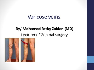
- 1. Varicose veins By/ Mohamad Fathy Zaidan (MD) Lecturer of General surgery
- 2. Venous Anatomy of Lower Limbs
- 3. The Long & Short Saphenous Veins Superficial veins of the leg. • 1-Sapheno-femoral junction (SFJ), • 2-Great saphenous vein (GSV), • 3-Anterior accessory saphenous vein (AASV), • 4-Posterior accessory saphenous vein (PASV), • 5-Anterior thigh circumflex vein (ATCV), • 6- Posterior thigh circumflex vein (PTCV) • 7-Sapheno-popliteal junction (SPJ), • 8-Small saphenous vein (SSV), • 9-Thigh extension of the small saphenous vein (TE SSV), • 10-Intersaphenous vein (Intersaph. V)
- 4. Mainsitesofsuperficialtodeepvenouscommunication Medial malleolus Sapheno-femoral junction Mid thigh perforator (Hunter’s canal) Medial calf perforators Just below Just above 10 cm above Just below the knee The lower perforators are joined to form the Posterior arch vein Thigh perforators connect to the long saphenous main trunk May or Kuster ankle perforators Cockett lower leg perforators(3) Boyd gastrocnemius perforators Dodd perforator
- 5. Valves • Connects the deep and superficial system via the perforators • Divide the veins into segments • Unidirectional (from below upwards ,, or from superficial to deep )
- 6. Venous return • Venomotor tone [Upright position -- dependant pooling – dec. cardiac output - - inc. sympathetic discharge -- inc. venous tone -- inc. venous return.] • Calf muscle contraction Blood is pushed upwards and from superficial to deep sys. prevented from retrograde flow by competent venous valves
- 7. Competent Veno-muscular Pump is composed of: 1. Superficial & deep veins with competent valves. 2. Competent perforating veins communicating the deep & superficial systems 3. Powerful lower limb muscles.
- 8. Ambulatory venous pressure • The ankle venous pressure during walking. • (AVP) is the "gold standard" test of the efficiency of the calf musculovenous pump • by placing a small needle into one of the veins on the back of the foot and connecting the needle to a blood pressure measurement machine. The test has three parts • standing venous pressure is around 90 mmHg , During exercise this should fall to around 30 mmHg.
- 9. "Ankle blow out" syndrome for varicose veins • Perforator vein incompetence in the gaiter areas have been shown to increase ambulatory venous pressures (venous hypertension), a phenomenon which has also been referred to "ankle blow- out" syndrome in the gaiter areas. • The combination of incompetent perforator veins and resultant venous hypertension over time causes damage to capillaries in the skin and subcutaneous capillaries, allowing protein rich fluid and red blood cells to escape into the subcutaneous tissue around the ankle
- 10. CVI (chronic venous insufficiency) • CVI collectively describes the manifestations of impaired venous return due to abnormal venous system function.
- 11. CVI • Whatever the cause of CVI, it will eventually cause venous hypertension of the microcirculation, giving the same symptoms & signs. • The severity of symptoms & signs depend on the degree & duration of venous hypertension When veins fail, the microcirculation suffers.
- 12. CVI(symptoms and signs) • Early 1-Posture related discomfort 2-Lower limb oedema 3-Muscle cramps • Persistent & Sever 4. Ankle brown pigmentation 5. Venous eczema 6.Lipodermatosclerosis 7. Venous ulcer 8- Talipes
- 13. Varicose veins • Tortuous dilated elongated veins. • Defective connective tissue and smooth muscle in the vain wall. • Valve not working. • Gathering of blood within veins.
- 14. CLASSIFICATION • (CEAP) classification from the American Venous Forum, last revised 2004. Clinical •C0 - No visible or palpable signs of venous disease •C1 - Telangiectases or reticular veins •C2 - Varicose veins •C3 - Edema •C4a - Pigmentation or eczema •C4b - Lipodermatosclerosis or atrophie blanche •C5 - Healed venous ulcer •C6 - Active venous ulcer Etiologic •Ec – Congenital •Ep – Primary •Es - Secondary (post- thrombotic) •En - No venous cause identified Anatomic •As - Superficial veins •Ap - Perforator veins •Ad - Deep veins •An - No venous location identified Pathophysiologic •Pr – Reflux •Po – Obstruction •Pr,o – Reflux and obstruction •Pn - No venous pathophysiology identifiable EXAMPLE : C6, Ep, As,p,d, Pr
- 15. Inspection
- 16. 1-Superficial vein affected (long S. , short S., SFJ, SPJ , tributaries, stray V., perforators i.e blow out ). 2-Describe the dilated superficial vein(s). (Telangectasias-Reticular veins- varicose V.) 3-Look for signs of thrombophlebitis. 4-Inspect the ankle and look for signs of CVI. 5-Inspect the groin (static and dynamic) ,searching for masses, dilated V., Impulse, scar . 6-exclude ischemia.
- 17. Site and extent of the affected vein (in the standing position)
- 18. Telangectasias • Small(0.5-1mm) widened blood vessels in skin-small intradermal varicosities “SPIDER VEINS”/”venulectasias" • In anywhere on the body esp-leg • Usually no severe symptoms
- 20. Reticular veins • Subcutaneous dilated veins-enter tributaries of main axial/trunk veins • Size >spider veins <varicose vein
- 21. Blow out • Defect in the deep fascia at the site of incompetent perforator
- 22. Ankle flare
- 23. Pigmentation & eczema • The skin pigmentation of chronic venous insufficiency is associated both with increased melanin production and also with deposition of haemosiderin
- 24. Lipodermatosclerosis • Progressive scaring of skin and subcutaneous fat due to venous insufficiency, area affected becomes thin and hard- area above becomes oedematous (Champagne bottle appearance)
- 25. Venous ulcer • As a complication of CVI and ankle blowout syndrome (due to increase the AVP) • At the gaiter area (site of ankle perforators)
- 27. Clinical features Ischemic ulcer Venous ulcer Gender Men > women Women > men Age Usually presents > 60 years Typically develops 40-60 years Risk factors Smoking, diabetes, hyperlipidemia and hypertension Previous DVT, thrombophilia, varicose vein Symptoms Severe pain unless there is diabetic neuropathy Pain but not severe, relieved by elevation Site Pressure area (heel, metatarsal head and base) Medial and lateral malleoli Edge Regular, punched out Irregular, with neo-epithelium Base Deep, green (sloughy) or black (necrotic) with no granulation tissue, may involve tendon, bone and joint Pink and granulating Surrounding skin Shows signs of ischemia (cold, pale, atrophic….) Varicose eczema, indurations, pigmentation, redness. Veins Empty Full, usually varicosed Swelling Usually absent Often present
- 28. Talipes equinovarus • Progressive scaring around the ulcer leads to permanent deformity
- 29. Scars of previous operations
- 30. Inspection of the groin
- 32. Thrombophlebitis
- 33. Palpation
- 34. Items of palpations • Tenderness • Temperature • Thrombophlebitis (Tenderness-cord like structure) • Defect in the deep fascia (Fegan’s method ) • Ulcer (base) • Calf muscles (DVT) • Groin (LNs.- cough impulse ) • Abdomen (masses) • Special tests
- 35. Comments
- 36. Schwartz’s test
- 37. For DVT • Tender calf • Mose’s sign • Homan’s sign
- 39. Special tests 1.Brodie Trenedlenburg test 2.Multiple Tourniquet test 3.pratt’s test 4.Perthe’s test 5.Modified Perthe’s test
- 43. Do Multiple Tourniquet test pratt’s test only if Brodie Trenedlenburg test positive with maintained pressure
- 45. pratt’s test
- 46. Perthe’s test & Modified Perthe’s test Modified Perthe’s testPerthe’s test
- 51. Doppler
- 52. Doppler ultrasound • The transducer sends and receives sound waves that are amplified through a microphone. • The sound waves bounce off solid objects, including blood cells. • The movement of blood cells causes a change in pitch of the reflected sound waves (called the Doppler effect). • If there is no blood flow, the pitch does not change.
- 53. Duplex • Combines Doppler flow information and conventional imaging information. • Sometimes called B- mode, to allow physicians to see the structure of your blood vessels. • It can also be useful to estimate the diameter of a blood vessel as well as the amount of obstruction, if any, in the blood vessel
- 54. Management • For incompetent perforator 1-Trendelenburg operation (saphenofemoral disconnection) 2- saphenopoplitial disconnection 3-Subfascial ligation (OPEN OR ENDOSCPPIC= SEPS) 4-endovasculer occlusion • For incompetent dilated vein 1-Stripping 2-Endoluminal occlusion of the saphenous vein by radiofrequency (RF) ,laser energy or foam injection. 3-Stab avulsion of varices with or without saphenous vein stripping (phlebectomy) • For valve incompetence ?? • For the ulcer 1-biopsy if suspicious 2-Compression dressings (4 layer , Unna’s boot…atc) 3-coverage
- 56. • Brodie–Trendelenburg percussion test (also accredited to Sir Benjamin Collins Brodie) is a test for incompetent valves in superficial veins • Trendelenburg's cannula: a cannula used during surgery of the larynx to prevent the patient from swallowing blood during surgery involving the head and neck • Trendelenburg gait: an abnormal gait caused by weakness of the abductor muscles of the lower limb, including the gluteus medius and gluteus minimus muscles. • Trendelenburg operation: ligation of the great saphenous vein, for the treatment of varicose veins. This term may also apply to pulmonary thrombectomy • Trendelenburg position, in which the patient is placed on a bed which is put into incline such that the patient's head is lower than his feet • Trendelenburg's sign: a sign of congenital dislocation of the hip • Trendelenburg's test: a test for varicose veins as well as a test to assess hip mobility
- 57. Stripping
- 61. Profore system (four layers compression) • orthopaedic wadding (Velband) was applied over the primary dressing • crepe bandage • Elset bandage • final layer of a cohesive bandage (Coban)
- 62. UNNA boot
- 63. Thank you