12. The Gram stain procedure
Developed in 1884 by the Danish physician
Hans Christian Gram
An important tool in bacterial taxonomy,
distinguishing so-called Gram-positive
bacteria, which remain coloured after the
staining procedure, from Gram-negative
bacteria, which do not retain dye and need to
be counter-stained.
Can be applied to pure cultures of bacteria
or to clinical specimens
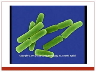
Top: Pure culture of E. coli
(Gram-negative rods)
Bottom: Neisseria gonorrhoeae in a smear of urethral pus
(Gram-negative cocci, with pus cells)
18. will stain will not stain
Gram positive bacteria Gram negative bacteria
Two biochemical groups of bacteria:
peptidoglycan
outer
membrane
19. Bacteria with Chemically Unique Cell Walls
Acid-Fast Cells
Mycobacterium species
Gram + type of cell wall
Unique lipid
Mycolic acid – waxy substance
Does not decolorize
20. Bacterial Growth
Solid media or liquid media
Agar plates, slopes, broth culture
Atmosphere:
Aerobic, anaerobic or microaerophilic
Facultative or obligate anaerobes
Usually at 37 degrees C
Most clinically important bacteria grow overnight, or
within a few days
Mycobacteria can take months
Some can not be grown
21. Capsules or slime layer
E.g., slime layer
allows bacteria to
cling to tooth
enamel or other
substrates
26. Binary fission
Daughter cells are identical copies
(1) (2) (3)
(4) (5) (6)
Chromosome Plasma membrane
Neither mitosis nor meiosis occurs in prokaryotes
27. REPRODUCTION
Asexual, through binary fission
No true sexual reproduction, since neither
mitosis nor meiosis exist in prokaryotes
Horizontal transfer of genetic material
Transformation Uptake of genetic material from the
environment
Transduction Transfer of genetic material between
prokaryotes by viruses
Conjugation Direct transfer of genetic material from one
prokaryote to another
28. Conjugation in E. coli
Sex pilus
Sex pilus connects cells and draws them together
Conjugation tube then forms
29. Bacteria
Surviving harsh conditions
Endospore – forms inside a bacterium and then persists
through inhospitable conditions
endospore
30.
31. The oldest known fossils
First organisms
on Earth
Cyanobacteria
> 3 billion years
old
32. Distributed globally – including many
extremophiles
“Heat-loving”
Archaea
“Salt-loving”
Archaea
33. Methanogens
Methane-generating Archaea
Occur in oxygen-free habitats
E.g., swamp mud, guts of
ruminant animals
Cave Bacteria
Sometimes reaching
acidity of pH 0.5
Distributed globally – including many
extremophiles
34. Ice Bacteria & Archaea
Distributed globally – including many
extremophiles
35. Prokaryote Nutrition – autotrophs & heterotrophs
All organisms require a source of
energy & carbon
Autotrophs can
obtain all their
C from CO2
36. All organisms require a source of
energy & carbon
Heterotrophs
require at least
one organic
nutrient, e.g.,
glucose
Prokaryote Nutrition – autotrophs & heterotrophs
37. All organisms require a source of
energy & carbon
Phototrophs
obtain their
energy from
the sun
Prokaryote Nutrition – autotrophs & heterotrophs
38. All organisms require a source of
energy & carbon
Chemotrophs
obtain their
energy from
chemical
compounds
Prokaryote Nutrition – autotrophs & heterotrophs