
Recommended
More Related Content
What's hot
What's hot (20)
Viewers also liked
Viewers also liked (19)
Similar to Capture and Release Nanopore-Nanofiber Mesh: pH Triggered Nucleic Acid Detection
Similar to Capture and Release Nanopore-Nanofiber Mesh: pH Triggered Nucleic Acid Detection (20)
More from CFTCC
More from CFTCC (20)
Recently uploaded
Recently uploaded (20)
Capture and Release Nanopore-Nanofiber Mesh: pH Triggered Nucleic Acid Detection
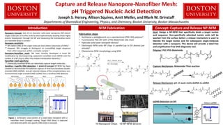
- 1. Capture and Release Nanopore-Nanofiber Mesh: pH Triggered Nucleic Acid Detection Joseph S. Hersey, Allison Squires, Amit Meller, and Mark W. Grinstaff Departments of Biomedical Engineering, Physics, and Chemistry, Boston University, Boston Massachusetts Introduction NFM Fabrication Concept: Capture and Release NP-NFM Nanopore Concept: Sub 10 nm diameter solid-state nanopores (NP) detect single molecules of nucleic acids by electrophoretically drawing these highly anionic biopolymers through the NP and measuring the translocation event as a transient drop in current.1 Nanopore sensitivity: • NPs detect DNA at the single molecule level (detect attomoles of DNA).1 • However, NPs struggle to distinguish an unmodified target sequence from other sequences within a complex sample.2 Nanopore-Nanofiber mesh: We have recently developed a novel NP modification by electrospinning a polymeric nanofiber mesh (NFM) onto the surface of a NP chip to affect the analyte translocation dynamics.3 Nanofiber mesh specificity: • Chemically modified NFMs can selectively capture target molecules. Sensitive + specific DNA detection: A proof-of-concept NP-NFM has been created to demonstrate the selective capture of thiol-functionalized double- stranded DNA (dsDNA) and pH induced release of the complementary non- functionalized single-stranded DNA (ssDNA) into a sensitive DNA detector. Figure 1. Schematic cross-section of a solid-state nanopore with a nanofiber mesh (orange) coating. Target DNA (blue) is captured and detected while non-specific DNA (red) is removed. Fabrication steps • Synthesize a poly(glycerol-co-ε-caprolactone) (PGC-OH) polymer4 • Functionalize PGC-OH with a PEG-Maleimide side chain • Fabricate solid-state nanopore devices5 • Electrospin NFM onto NP chips in parallel (up to 50 devices per minute)3 • Characterize NFM morphology using SEM Goal: Design a NP-NFM that specifically binds a target nucleic acid sequence. Non-specifically adsorbed nucleic acids will be washed from the surface before a release mechanism is used to liberate the target nucleic acid for subsequent single-molecule detection with a nanopore. This device will provide a label-free and amplification-free DNA diagnostic tool. Polymer: PGC-PEG-Maleimide Capture Mechanism: Maleimide-Thiol reaction Release Mechanism: pH 11 wash melts dsDNA to ssDNA ssDNA detection on NP:
- 2. Capture and Release Nanopore-Nanofiber Mesh: pH Triggered Nucleic Acid Detection Joseph S. Hersey, Allison Squires, Amit Meller, and Mark W. Grinstaff Departments of Biomedical Engineering, Physics, and Chemistry, Boston University, Boston Massachusetts Proof of Concept: SYBR I Detection Capture and Release NP-NFM Capture and Release NP-NFM: Results Experimental Design: • Create 500 bp dsDNA with or without a thiol modification on one of the strands. • Electrospin maleimide functionalized NP-NFM. • Apply 4 µL drop of 50 nM dsDNA onto the NFM, wait 18 hours, and image on a gel scanner (excitation 497 nm, emission 520 nm). • Thiol-functionalized DNA that covalently binds to the NP-NFM will not wash away at pH 7 but non-specifically bound DNA will. • A pH 11 wash melts the dsDNA releasing ssDNA causing the SYBR I fluorescence to decrease. Sybr I Fluorescence Low High Experimental Design: • Use the same protocol as the Sybr I experiments except use 2.5 nM dsDNA and wait only 30 minutes. • Apply a voltage (500 mV) across the nanopore and detect DNA as transient drops in current after each wash step. • Wash 1: Remove any unbound material using a pH 7 wash. Wash 2: Confirm unbound DNA was removed. Wash 3: pH 11 buffer melts any remain DNA releasing ssDNA into the nanopore. • dsDNA only: • No dsDNA is captured onto the mesh. • All of the dsDNA is removed during pH 7 wash 1. • Thiol-dsDNA: • No dsDNA is observed in either pH 7 wash 1 or 2 indicating the DNA has been captured. • The pH 11 wash shows signs of translocations; however, this wash also causes the pore to block. Conclusions and Future Work A proof of concept capture and release NP-NFM was developed • Demonstrated the ability to selectively binding a model thiol functionalized dsDNA • Confirmed DNA binding to the NP-NFM optically • Utilized a nanopore system to detect pico-molar double- stranded and single-stranded DNA after each wash step. • pH 11 release washes likely cause the polymer to degrade resulting in nanopore blockage. However, the blockage profile contains translocation events and blockage occurs more quickly if DNA is captured onto the NP-NFM Future work: • Develop a new release mechanism to avoid blockages Literature Cited 1. Wanunu, M. Phys. Life Rev. 2012, 9, 125. 2. Branton, D.; Deamer, D.; et al. Nat. Biotechnol. 2008, 26, 1146. 3. Squires, A. H.; Hersey, J. S.; Grinstaff, M. W.; Meller, A. J. Am. Chem. Soc. 2013, 135, 16304. 4. Wolinsky, J. B.; Ray, W. C.; et al. Macromolecules 2007, 40, 7065. 5. Kim, M. J.; Wanunu, M.; Bell, D. C.; Meller, A. Adv. Mater. 2006, 18, 3149. Acknowledgements The authors gratefully acknowledge the National Institutes of Health for funding support of this project (R21 EB017377) and the Boston University Division of Materials Science and Engineering. For more information visit: http://people.bu.edu/mgrin