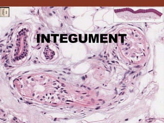
Histology of the Integument
•
9 likes•5,194 views

The skin has two main layers - the epidermis and dermis. The epidermis is made of stratified squamous epithelium and contains melanocytes, Langerhans cells, and keratinocytes. The dermis contains hair follicles, sweat and sebaceous glands, as well as blood vessels and nerves. Skin thickness varies in different regions. Thick skin is found on the palms and soles and contains several epidermal layers, while thin skin covers most of the body and has fewer layers. The skin provides protection, temperature regulation, sensation, and other important functions.

Recommended
More Related Content
What's hot
What's hot (20)
Similar to Histology of the Integument
Similar to Histology of the Integument (20)
More from Garry D. Lasaga
More from Garry D. Lasaga (20)
Recently uploaded
Recently uploaded (20)
Histology of the Integument
- 1. INTEGUMENT
- 2. THE SKIN •The skin or cutis covers the entire outer surface of the body. •Structurally, it has two layers. •The epidermis is formed by an epithelium and is of ectodermal origin. •The dermis, consists of CT and develops from the mesoderm. 31
- 3. • Beneath the 2 layers is a subcutaneous layer of loose connective tissue, the hypodermis or subcutis. • Hair, nails and sweat and sebaceous glands are called the appendages of the skin. • The skin and its appendages together are called the integumentary system. THE SKIN
- 4. Functions of skin Protects against injury and dessication Maintain water balance Excrete various substances Thermoregulation Receive stimuli (temperature, pain, pressure) Fat metabolism in the subcutaneous layer
- 5. CHARACTERISTIC THICK SKIN THIN SKIN Surface Texture Alternating ridges and grooves Smooth Epidermis/Dermis Interface Interdigitating ridges Less prominent ridges Epidermal Strata • S. Basale • S. Spinosum • S. Granulosum • S. Lucidum • S. Corneum • Same as thick skin, except: no S. Lucidum. • The corneum, granulosum, and spinosum layers are reduced in thickness. Hairs and Sebaceous Glands None Regionally variable Sweat Glands Abundant Moderate
- 6. Regional Variation of the Epidermis THICK SKIN – sole of foot (1.4 mm thick) THIN SKIN – eyelid and most of body (0.07 TO 0.12 mm) CORNEA OF EYE – transparent APPENDAGES – hair follicles, follicles, nails, glands
- 7. Regional Variation of the Epidermis The thickness of the skin depends on the epidermis. Once can have a very thick dermis and still be considered thin skin.
- 10. Epidermis The superficial, epithelially-derived region of skin. 1. Stratum basale 2. Stratum spinosum 3. Stratum granulosum 4. Stratum lucidum 5. Stratum corneum
- 11. Thick Skin Papillary layer Reticular layer Dermal papillae Epidermal peg DERMIS Papillary layer • Dermal papillae • Epidermal pegs • Meissner’s corpuscles Reticular layer • Hair follicles • Sebaceous, sweat glands • Krause’s end bulb
- 12. Thick Skin – DERMIS Dermal papillae Meissner’s corpuscles in dermal papillae Epidermal peg Dermis Epidermis Meissner’s corpuscle is a mechanoreceptor nerve ending for sensitivity to light touch. Epidermis
- 13. Skin hand monkey - DERMIS Hypodermis Papillary layer Epidermal pegDermal papillae AdipocytesEccrine sweat glands Pacinian corpuscles
- 14. Epidermal – dermal interphase Epidermal-dermal interphase between the thin skin and thick skin. The thick skin has a more elaborate dermal papillae. The interphase prevent the separation of the 2 layers during mechanical stress.
- 15. Dermal side of the Epidermal – dermal interphase
- 16. Epidermal – dermal interface If the epidermis is pulled off, the dermal papillae can be seen projecting into the holes (cavities) within the epidermis.
- 17. MELANIN is produced by MELANOCYTES 107 Dermal papillae projection
- 18. Epidermal – dermal interface - finger pad
- 19. Thick Skin (Pacinian corpuscle and melanin) Pacinian corpuscle Melanin pigment Melanin capping of nuclei
- 20. Pacinian corpuscles are mechanoreceptors that detect vibration and pressure. 105 monkey finger Pacinian corpuscles
- 21. Epidermis STRATUM CORNEUM • Keratinized flattened, denucleated, dead cells STRATUM GRANULOSUM • Keratohyalin granules STRATUM SPINOSUM • Tonofibrils – desmosomes STRATUM BASALE • Continual renewal of epidermis
- 22. Thick Skin 5. Stratum basale Hemidesmosomes 4. Stratum spinosum 3. Stratum granulosum 2. Stratum lucidum 1. Stratum corneum Desmosomes Keratohyalin granules 1 2 3 4 5 2 3 4 5
- 23. Thick Skin Stratum basale Hemidesmosomes Stratum spinosum Stratum granulosum Stratum lucidum Desmosomes Keratohyalin granules 1 S. granulosum: flattened cells undergoing the terminal differentiation process of keratinization – forming the skin’s barrier against water loss when sealed with contents of membrane coating granules. .
- 24. Thick Skin on finger Stratum basale Hemidesmosomes Keratohyalin granules 1 The epidermis of thick skin is subject to continuous friction and pressure so the abundant desmosomes (and tonofibrils) withstand this and hold the cell layers together. Stratum spinosum Dermis Epidermis Desmosomes
- 25. Cells in EPIDERMIS KERATINOCYTES – main cell type – ectoderm MELANOCYTES – pigmentation – neural crest LANGERHANS CELL – immunologic role MERKEL CELLS – associated with nerve endings
- 26. MELANOCYTE – PIGMENT SYNTHESIS NEURAL CREST ORIGIN (EYE & CNS) The embryonic origin of melanocytes is the neural crest derivatives that migrate into the embryonic epidermis’ stratum basale.
- 27. 111 107 MELANOCYTES Location: stratum basale; they appear as clear cells
- 28. Melanocyte – Pigment Synthesis Melanogenesis : tyrosine – RER – Golgi – production of melanin granules Melanocytes w/ projections – pass melanin granules to keratinocytes through cytocrine secretion
- 29. CYTOCRINE SECRETION - PASS MELANIN GRANULES FROM MELANOCYTES TO KERATINOCYTES
- 30. MELANOCYTE - PIGMENT SYNTHESIS LOCATED IN THE STRATUM BASALE CLEAR CELL – NO DESMOSOMAL CONNECTION
- 31. Melanin granules above nucleus – protects (UV) it as it is the source of cells that are mitotically active. Stratum basale Stratum spinosum Stratum granulosum Stratum corneum Melanin capping of nuclei
- 32. Melanocytes respond to melanocyte stimulating hormone secreted by the pars intermedia. Melanin granule accumulate over the nuclei of mitotic cells of the stratum basale to protect nuclear DNA from UV damage. 107
- 33. LANGERHANS CELLS • BONE MARROW ORIGIN • LOCATED IN STRATUM SPINOSUM – GOLD CHLORIDE STAIN • CLEAR CELL – NO DESMOSOMES • DENDRITIC CELL
- 34. LANGERHANS CELLS • Dendritic cell • Rod or racket-shaped granules • Function – immunologic role as antigen-presenting cell • Contact allergic responses and other cell-mediated reactions of the skin
- 35. HAIR
- 36. External Root Sheath Sebaceous Gland (Holocrine Secretion) Eccrine Sweat Glands Arrector Pili muscle Hair Bulb Connective Tissue (Dermal) Papilla Hair Shaft Canal 107 Hair Follicle ?
- 37. Skin, scalp (Hair follicle)
- 38. Thin Skin (scalp) Hair follicle location Arrector pili muscle Sebaceous glands Arrector Pili Muscle – bundles of smooth m. extending from hair follicle to the papillary dermis. • Elevate the hair, forming “goose bumps”.
- 40. Skin, scalp Mode of secretion of the sebaceous glands is holocrine where by the sebum is released when cells burst. sebaceous glands Eccrine sweat glands Human Skin, scalp 108
- 41. Simple columnar epithelium of rectum with goblet cells Stratified squamous epithelium of anal wall 66
- 42. THREE TYPES OF GRANULES IN KERATINOCYTES MELANIN • SKIN PIGMENT • PRODUCED BY MELANOCYTES AND PASSED BY CYTOCRINE SECRETION TO KERATINOCYTES MEMBRANE COATING GRANULES (LAMELLATED GRANULES) • WATER PROOFING FUNCTION • PRODUCED BY KERATINOCYTES KERATINOHYALIN GRANULES • PRODUCED BY KERATINOCYTES
- 43. THREE TYPES OF GRANULES IN KERATINOCYTES MEMBRANE COATING GRANULES (LAMELLATED GRANULES) • Small, ovoid structures from the Golgi containing various lipids and they undergo exocytosis to produce a lipid-rich impermeable layer around the cells of the s. granulosum – water proofing.
- 44. THREE TYPES OF GRANULES IN KERATINOCYTES KERATINOHYALIN GRANULES • CHEMICAL NATURE NOT CLEARLY ESTABLISHED • MATRIX OF CELLS IN STRATUM CORNEUM, STABILITY DUE TO DISULFIDE BONDS • ABSENT IN HAIR AND NAILS