
Recomendados
Mais conteúdo relacionado
Mais procurados
Mais procurados (20)
Destaque
Destaque (20)
Semelhante a Eosophageal Atresia - Tracheo-esophageal Fistula
Semelhante a Eosophageal Atresia - Tracheo-esophageal Fistula (20)
Último
Último (20)
Eosophageal Atresia - Tracheo-esophageal Fistula
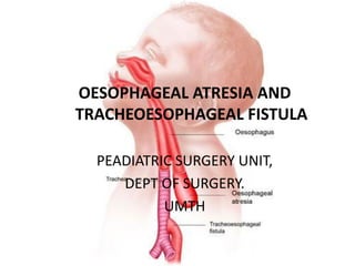
- 1. OESOPHAGEAL ATRESIA AND TRACHEOESOPHAGEAL FISTULA PEADIATRIC SURGERY UNIT, DEPT OF SURGERY. UMTH
- 2. Learning objectives • Discuss predictors of poor outcome and long term morbidity of EA
- 3. Outline • Introduction • Case summary • Embryology/aetiology • Classification • Clinical Features • Investigations • Risk categorisation • Treatment • Complications • Outcome • Recent advances • Summary
- 4. Introduction • Epitome of Paediatric Surgery “To anastomose the ends of an infant’s esophagus the Surgeon must be as delicate and precise as a skilled watchmaker. No operation offers a greater opportunity for pure technical artistry.” Willis Pott, 1950 • Overall Improvement in survival with focus on complications
- 5. Introduction • Significant challenge in modern paediatric surgery (urgency to make timely and appropriate diagnosis) • Survival is likely, unless NB belong to a specific risk group • Improvement in neonatal care, have contributed to the survival of these high risk patients
- 6. History • 1935 pre-survival period • Donovan 1935- first survivor of isolated EA – Initial gastrostomy, esophageal replacement 15yr by Humpries • Haight and Towley 1943 • First successful primary anastomosis
- 7. History • Watersone 1964 – Risk groups based on weight, pneumonia & congenital anomaly • Spitz 1994 – Revised at risk groups for the 1990s currently been used
- 8. Epidemiology • Incidence – 1:3500-4500 live birth • Varies geographically • Incidence high – Finland 1:2440, Europe 2.82/10,000, whites >60%, 1st pregnancy, ↑maternal age 35-45yrs, in vitro fertilization, twin gestation • Slight male preponderance
- 9. CASE PRESENTATION • BHH • 2hr old term male neonate.
- 10. PRESENTING COMPLAINT • Excessive salivation since birth • Absent anal opening since birth
- 11. HxPC • Product of a term pregnancy, delivered to a 28yr old P8 +0 A7 • Noticed to have excessive salivation since birth. • Associated hx of choking on feeding • Cough and difficulty in breathing, • No hx suggestive of cyanotic spell • No vomiting following feeding
- 12. • Noticed absent anal opening since birth • No associated hx of abdominal distention, passage of feacal material in urine, or through other opening around the perineum.
- 13. • Pregnancy was booked at four month gestation. • Regular with follow up visit and antenatal medications • No hx suggestive of illicit drug use during pregnancy
- 14. • There was positive hx of polyhydramnious from mother, which was also picked on abd uss. • No hx of maternal febrile illness, rashes, jaundice or foul smell vaginal discharge • No hx of exposure to radiation
- 15. • Mother not a known diabetic or hypertensive pt • Delivery was via SVD, • Child cried immediately after birth
- 16. SOCIAL HX • 7th child ,in a monogamous non sanguineous setting, • Lost elder sibling in her 4 months, said to be due to congenital hydronephrosis. Nil family hx of similar disease. • Mother is a FTHW with no formal education. Father is a civil servant.
- 17. O/E • A calm neonate, pink, with excessive salivation, no dysmorphic facae, afebrile, not pale, acyanosed, anicteric, not dehydrated, no peripheral lymph node enlargement or pedal oedema. • Birth wt: 2.9kg OFC: 34cm • Lt: 49cm HR: 152bpm • RR:40cpm
- 18. Abdomen • Full, moves with resp. with visible peristalsis. Liver enlarged by 2cm, Spleen not enlarged, Kidneys no ballotable, BS present and hyper active. • Normal male external genitalia • Absent anal opening • Well developed gluteal fold
- 19. • Resp: clear lung fields, BVBS • CVS: HS 1 and 2 no murmur • CNS: conscious, normal skull with hyperpigmented patch on the sacral area, no defect on the spine, Complete moro, good grasp, normal tone. • MSS: Upper and lower limbs appeared grossly normal.
- 20. DIAGNOSIS EOSOPHAGEAL ATRESIA + TRACHEOESOPHAGEAL FISTULA and ANORECTAL AGENESIS WITHOUT FISTULA.
- 21. INVESTIGATIONS • Babygram confirm arrest of nasogastric tube about 10cm from the lower incisor. • Plain Abd X-ray result showed gas in the stomach.
- 22. • PCV: 0.68 • RBC: 6.6mmol/l • E/U/C : azotemia of 8.0. Creatinine not retrieved other parameters essentially within normal limit • Total bilirubin: 20.3mg/dl • Conj. bilirubin: 11.2mg/dl, 56% conjugated.
- 23. • Operations: – Closure of fistula – Esophagoesophagostomy • Intra op findings were: – Proximal blind pouch – Distal tracheoesophageal fistula
- 24. EMBRYOLOGY • The embryology of the foregut is still subject to controversy. • Development of the digestive system begins at 4th week during folding • Endoderm gives rise to most of its epithelium & glands • The cranial end is ectodermal,namely the stomodeum.
- 26. • During the fourth week of gestation the foregut starts to differentiate into a ventral respiratory part and a dorsal esophageal part. • The laryngotracheal diverticulum then evaginates ventrally into the mesenchyme.
- 27. • The ventral respiratory system separates from the esophagus by the formation of lateral tracheoesophageal folds that fuse in the midline and create the tracheoesophageal septum. • At 6 to 7 weeks of gestation, the separation is complete. • Incomplete fusion of the folds would result in a defective tracheoesophageal septum and abnormal connection between the trachea and esophagus.
- 28. • This theory has been challenged. • In chicken embryo studies, these folds could not be demonstrated. • Instead, cranial and caudal folds were found in the region of tracheoesophageal separation. • According to this theory, EA/TEF would then be due to an imbalance in the growth of these folds
- 29. The Adriamycin (doxorubicin) rat model • It has greatly helped with understanding the development of EA/TEF. • From this model, it appears the EA develops first, with the lung bud arising from the atretic foregut dividing into three rather than two branches. • The middle branch would be the distal esophagus eventually connecting with the stomach.
- 30. AETIOLOGY • The pathogenesis of Eosophageal atresial malformations remains unknown. • Many theories proposed to explain EA, EA-TEF, TEF, no single unifying theory has been proposed that addresses all the variations seen with this group of anomalies.
- 31. • Most EA occur sporadically, highly unlikely that simple, inheritable genetic mechanism is responsible. • Most likely heterogenous and multifactorial, and involves multiple genes and complex gene-enviroment interactions
- 32. • Environmental factors implicated: – use of methimazole in early pregnancy – prolonged use of contraceptive pills – maternal diabetes and thalidomide exposure. – EA is occasionally seen in the fetal alcohol syndrome and – in maternal phenylketonuria
- 33. • Also associated with of trisomy 18 and trisomy 21. • Trisomy 18 carries is a greater risk for EA development. • Three separate genes have been associated with EA/TEF: – MYCN haploinsufficiency in Feingold syndrome, – CHD7 in CHARGE syndrome, and – SOX2 in the anophthalmia-esophageal-genital (AEG) syndrome. • EA may occasionally be part of the – Opitz G/BB syndrome, – Fanconi anemia, – oculo-auriculo-vertebral syndrome
- 34. CLASSIFICATION • E.C. Vogt a radiologist in 1932 classified the anomalies anatomically into: • Type 1: absent eosophagus • Type 2: EA without TEF • Type 3: EA with TEF – a: EA with Proximal TEF – b; EA with Distal TEF – c: EA with proximal and distal TEF • Type 4: isolated TEF with intact eosophagus.
- 35. TYPE % GROSS TYPE 1. EA with distal TEF 85.8 C 2. EA without TEF 7.8 A 3. TEF without EA 4.2 E 4. EA with TEF to both pouches 1.4 D 5. EA with proximal TEF 0.8 B
- 36. CLASSICATION: PROGNOSTIC GROUP SURVIVAL WATERSON CLASSIFICATION A 100% Birth weight >2.5kg and otherwise healthy B 85% Birth weight 2-2.5kg and well or higher weight with moderate associated anomalies (non-cardiac plus PDA, VSD or ASD). C 65% Birth weight <2kg or higher with severe assocaited cardiac anomalies. Waterston’s 1962 classification seperated patients into groups based on birth weight, pneumonia and congenital anomalies. Group A were treated with immediate repair Group B “ “ “ delayed repair Group C “ “ “ staged repair
- 37. CLASSIFICATION: PROGNOSTIC GROUP SURVIVAL I. Birth weight >1.5kg without major CHD 97% II. Birthweight <1.5kg or major CHD 59% III. Birth weight <1.5kg and major CHD 22% SPITZ Classification – Most commonly used currently.
- 38. Diagnosis • Prenatal • Not common-18/52 • USS- 20%-40% yield – Small stomach – Absent stomach bubble – ployhydramnios
- 40. Diagnosis • Regurgitation at feed • Choking, coughing • Cyanosis with or without feeding • Abdominal distension-distal fistula – Scaphoid abdomen-isolated EA
- 41. • Inability to pass a NGT • Look for associated anomalies –(20%-70%) • Incidence is high with isolated EA – VACTERL/CHARGE associations – SCHISIS malformations
- 42. Associated malformations • VACTERL—H • V vertebra • A anorectal • C cardiac • T tracheo- • E esophageal • R renal • L limb • H hydrocephalus
- 43. Associated malformations • CHARGE associations • C coloboma of eye • H heart • A anorectal • R renal • G hypoGonadism • E deafness (Ear)
- 44. Associated Malformations • Systems affected are as follows: • Cardiovascular – 29% • Anorectal – 14% • GUS – 14% • GIT – 13% • Vertebral / Skeletal – 10% • Respiratory – 6% • Genetic – 4% • Others - 11% • Ein et al 1989
- 45. Investigations • Oropharyngeal tube met resistance ≈10cm from the lower incisor • Limited contrast study -0.5-1ml of diluted barium into the upper pouch
- 46. Investigations • Air in the stomach or bowel suggestive of distal TEF
- 47. Investigations • Absent gas in the abdomen reflects isolated EA
- 48. Investigations • EA and TEF with duodenal atresia. NGT coiled in upper pouch.
- 49. Investigations • Upper pouch outlined by barium
- 50. Investigations • Broncoscopy-fistula proximal to the carina
- 51. Investigations • Echocardiography- – roll out 1.8-2.5% right sided aortic arch – Cardiac anomalies • Renal USS • Chromosomal analysis
- 52. Investigations • TEF without EA – High index of suspicion – Delayed – Coughing/ choking during feeding – Recurrent aspiration – Use video esophagography – broncoscopy
- 53. Risk Categorisation Waterstone - 1962 Gp A bwt ≥ 2.5 kg and well (95%) Gp B Bwt - 1.8 – 2.5 Kg and well or - ≥2.5 Kg with moderate pneumonia and birth defects (85%) Group C Bwt ≤ 1.5 Kg or Any weight with severe pneumonia and associated malformations (65%) Spitz - 1994 • Gp I -Bwt ≤ 1.5 Kg no major cardiac anomaly (97%) • Gp II - ‹ 1.5kg or major cardiac anomaly (59%) • Gp III - ≤ 1.5Kg and major cardiac anomaly (22%)
- 54. Okamoto modification of the Spitz Classification: Predictors of Survival in Cases of Esophageal Atresia Class Description Risk Survival Class I No major cardiac anomaly, BW ≥2000 g Low 100% Class II No major cardiac anomaly, BW < 2000 g Moderate 81% Class III Major cardiac anomaly,BW ≤2000 g Relatively high 72% Class IV Major cardiac anomaly, BW < 2000 g High 27%
- 55. Pre-operative care • 24-48hrs pre-op stabilization • Pneumonitis – Aspiration from upper pouch secretions – Reflux of gastric acid • Broad spectrum antibiotics • Pulmonary physiotherapy • IVF • Vit k • Parenteral nutrition
- 56. • nurse 30 degrees head up/lateral position • Suction under low pressure with Replogle catheter Pre-op care
- 57. Pre-op care • Avoid routine endotracheal intubation – Risk gastric perforation – Worsening resp.distress as the abdomen becomes distended from ventilation through the TEF
- 58. No Treatment • Potters Syndrome • Severe uncorrectable CHD
- 59. Treatment • Primary • Delayed • Stage
- 60. Treatment • Open thoracotomy – Division of fistula, primary anastomosis of the esophagus • Preliminary broncoscopy
- 61. Treatment • Position – Left lateral decubitus
- 62. Treatment • Access – Curved infrascapula skin incision extending from anterior axillary line to paravertebral region – Right posterolateral thoracotomy – Pleural space entered b/w 4th intercostal space – Extra pleural vs Transpleural
- 63. Treatment • Azygous vein is divided • Distal TEF mobilized and closed leaving 1-2mm of the esophagus on the trachea • Check air tightness of tracheal closure – Instill warm saline in the pleural cavity, look for bubbles with positive pressure ventilation
- 64. Treatment • Trans anastomotic tube – Aspirate air in distended stomach – Patency of distal lumen • Avoid excessive handling of distal esophagus • Proximal pouch identification can be facilitated by the anaesthesiologist
- 65. Treatment • End –end esophagoesophagostomy • Leave a drain-? Transpleural • End-side anastomosis + single suture to ligate the fistula Salama et al – High rate of recurrent fistula – Poenaru et al identified low rate of GER
- 66. Treatment • Minimally invasive (thoracoscopic repair EA - TEF) • Thoracoscopy has been used since 2 decades • Not yet gold standard for OA but widely used
- 67. Treatment – Operative Minimally invasive surgery Advantages • Superior visualisation • Improved cosmesis • Eliminates morbidity • Less chest wall complications- scoliosis,wing scapula,chronic pain,chest wall asymmetry, maldevlopment Disadvantages • Not suitable in unstable pts • Also in preterm and • Pts with severe abdominal distension
- 68. Emergency Treatment • Preterm • Large distal fistula with gross abdominal distension • Transpleural ligation of fistula
- 69. Emergency treatment • Other options • Gastric division • Banding of gastroesophgeal jxn • Positioning of the tip endotracheal tube below fistulous orifice • Fogarty balloon catheter to ablate the flow of ventilator air • Emergency gastrostomy to decompress the air-filled stomach
- 70. Emergency treatment • Cervical esophagostomy • advantages • Early sham feeding – Stimulates neural maturation – Development of learning skills needed for feeding – Speech acquisition • disavantages • Need for esophageal replacement later
- 71. Treatment • Intraoperative maneuvers to decrease gap – Circular (Livaditis) /spiral myotomy – Full thickness anterior/posterior flap esophagoplasty (Gough) – Bagolan traction elongation technique – Gastric transposition/hiatal mebolization
- 73. Post operative care • Broad spectrum antibiotics • ICU • IVF • Trans anastomotic tube feeding 48hrs • Elective paralysis+ ventilator support 3-5days • ?anastomosis under tension
- 74. Post operative care • Ventilator care
- 75. Post operative care • H2 antagonist prophylaxis • ? Anastomotic stricture • Esophagogram 5-7days • Prophylactic dilatation at 3wks
- 76. Treatment (Long gap EA) • Delayed primary repair • Definition varies – >6cm – >6vertebral segments – Cannot be anastomose primarily
- 77. • Prone to – Prolonged hospitalization – Associated significant morbidity – Later swallowing difficulties
- 78. Pre operative delayed procedures • Upper pouch bougienage • Electromagnetic field pull together metallic bullets • Kimura’s multi stage, extrathoracic esophageal elongation technique
- 79. Foker Method • Foker’s elongation traction technique
- 80. Stage procedures-Isolated EA • 50% incidence of prematurity • 11% incidence of Down’s syndrome • 10% incidence of duodenal atresia
- 81. Stage procedure-isolated EA • Gastrostomy, Oesophagostomy and replacement – A gap length of 6 vertebral bodies – Priliminary broncoscopy to eliminate presence of occluded distal TEF • Types of replacement – Colonic – Gastric transposition – Gastric tube – Jejunal interposition
- 82. Isolated (H-type) TEF • Fistula run oblique (N-type) from trachea to esophagus • 4% of esophageal anomalies • Occur3-4 wks of life • Xterized – Choking on feeding – Cyanotic spells – Intermittent abdominal distention on crying or coughing
- 83. Isolated (H-type) TEF • Olderchildren have recurrent bouts right upper lobe of pneumonia • Radiograph – Aspiration pneumonia – Gastric distention – Tube video esophagography – Broncoscopy/esophagoscopy confirm the diagnosis
- 84. Isolated TEF - Treatment • Identify fistula and divide – Use pleural/inter costal muscle flaps to minimize fistula recurrence • Patient should remain intubated – Risk of tracheal oedema and recurrent laryngeal nerve palsy • Use Nd:Yag laser – coagulate fistula
- 85. EA and Right sided Aortic Arch • Incidence – 1.8 – 2.5% • Preop Echo very important • Leak rate very high ≈42% with right thoracotomy • If anastomosis difficult divide fistula and do a left thoracotomy
- 86. Complications Early • Anastomotic leak • Anastomotic stricture • Recurrent TOF Late • Pulmonary dysfxn • GOR • Tracheomalacia • Motility disorders • Chest wall deformities • “Anastomotic” stricture –usually 2o to GORD • Recurrent TOF
- 87. Anastomotic leak • Occur in 13-16% • most are less significant • Major disruption rear • Pt can deteriorate 24-48hrs from tension pneumothorax, mediastinitis
- 88. Anastomotic leak • Can be manage – drainage, nutritional support, antibiotic cover – Early exploration to secure anastomosis and establish satisfactory drainage
- 89. Anastomotic leak • Risk factors – Poor surgical technique – Ischaemia of the esophageal ends – Use of myotomy – Tension at anastonotic site
- 90. Esophageal stricture • Incidence varies 37-55% • In 80% of will require dilatation • Due to – poor anastomotic technique – long gap EA – ischaemia – GERD – anastomotic leak
- 91. • Symptoms – Dysphagia – Recurrent aspiration – Foreign body obstruction • Approach to Rx – Balloon dilation – Ante/retrograde bougienage – Resection and anastomosis
- 92. Recurrent TEF • 54% occur in the first year of repair • 49% occur 15yrs later • Approach to Rx – Pleural flap – Vascularised pericardial flap – Azygous Vn flap – Intercostal muscle flap
- 93. • Other options include: – Diathermy fulguration – Nd:YAG laser – Injection of sclerosants • Recurrence with above options high
- 94. GERD • Occur in 30-70% of EA • Due to – shortening of intra abdominal portion of the esophagus – Esophageal motor dysfunction • Risk of Barrett esophagus-9%
- 95. GERD • Symptoms • Vomiting, dysphagia, recurrent anastomotic stenosis, stridor, cyanotic spells, recurrent pneumonia • Investigation – UPI – 24hr PH probe(multichannel esophageal impidence+24hr PH probe) –promising – Manometry-dysordered peristalsis -↓LOS pressure
- 96. GERD • Higher rates in – Delayed Io repair – Patients on gastrostomy – Patients with anastomosis under tension • Aggressive medical Rx or surgical fundoplication (High failure rate – 15-38%)
- 97. Tracheomalacia • Generalized or localized weakness of the trachea that allow ant and post walls to come together during expiration or coughing • Reported in 75% of post mortem specimen • symptomatic in 10-25%
- 98. Tracheomalacia • Symptoms mimick- recurrent TEF, GERD, anastomotic leak • Brassing or barking cough • Symptoms can improve with time • Rx – CPAP,aortopexy, tracheal stenting, tracheostomy(severe)
- 99. Dysmotility disorders • Dysphagia in the absence of anastomotic stricture • Usually bolus obstruction – esophageal emptying by gravity • Patients take excessive amounts of liquids to push food down
- 100. Dysmotility disorders • May be 2o to intrinsic innervation problems or Vagal n damage at surgery • Manometery
- 101. Risk for death & long term complications • Low birth weight<2500g/prematurity • Twin birth • Long gap EA • Pre operative intubation • Prolong post operative intubation >4days
- 102. • Inability to feed at 1month • Anastomotic leak • Major cardiac anomaly • Severe associated anomalies
- 103. Outcomes • Survival 85-95% compared to 50yrs ago • Respiratory morbidity (pneumonia 15% at 5yrs, 5% at 15yr) • Esophageal motility disorders 20% adolescent, 40% adults • GER 15-50% • Quality of life (behavioral problems)
- 104. Outcomes (UMTH) Year Age Type Other anomalies Rx Outcome 2012 9days C Nil Died (Pneumonia) 2013 5days C ARM Primary repair Died (aspiration) 2013 2days C Nil Gastrostomy+banding survived 2014 14days C Nil Gastrostomy+banding Died(sepsis) 2014 3days C Nil Gastrostomy + banding survived
- 105. Future prospects – Role of Tissue Engineering • Recent advances in biomaterial science and cellular biology • Use of stem cells seeded on synthetic biopolymers
- 106. Summary • EA a challenging condition to manage, but timely resuscitation and appropriate surgical treatment underscore it’s outcome in absence of associated anomalies.