
![Sources ,[object Object],[object Object],[object Object],[object Object],[object Object],[object Object],[object Object],[object Object],[object Object],[object Object],[object Object],[object Object],[object Object],[object Object],[object Object],[object Object],[object Object],[object Object],[object Object],[object Object],[object Object]](data:image/gif;base64,R0lGODlhAQABAIAAAAAAAP///yH5BAEAAAAALAAAAAABAAEAAAIBRAA7)
Recomendados
Mais conteúdo relacionado
Mais procurados
Mais procurados (20)
Destaque
Semelhante a F mri
Semelhante a F mri (20)
Mais de Rad Tech
Mais de Rad Tech (20)
F mri
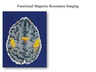
- 1. Functional Magnetic Resonance Imaging
- 10. How does fMRI form an image of neural activity? How to form an image of neural activity using NMR? Need to measure: 1. Spatial Location 2. Neural Activity Magnitude
- 13. Spatial Location in 1-D Frequency Encoding in practice Phase Encoding
- 14. Spatial Location in 2-D Echo Planar Imaging (Mansfield 1977): use frequency encoding to determine the “ x” direction and echo encoding to determine the “ y ” direction. sampling windows
- 22. Issues! There is not a one-to-one correspondence between T2 and the neural activity that we are trying to measure. There are pathways that might decrease the decay rate and hence results in a decreased MR signal!
- 25. This figure relates the temporal and spatial resolution of methods for the study of brain function to the size scale of neuronal features and to the “invasiveness” of the methods. Comparison: temporal and spatial resolution; and invasiveness
Notas do Editor
- NMR- see p1 Noll; hydrogen - p 1 Noll susceptibility - p2 Cohen
- 1 - p1 Noll 2 - p2 Cohen 3 - p2 Cohen and p1 Noll
- 2 - p2 Cohen
- 1 - p2 Noll, Hornak table - Hornak
- p2 Noll in phase - p3 Cohen
- p 4 Cohen p3-4 Noll
- p5 Noll p5 Cohen
- p6 Noll
- p7 Noll, p5-6 Cohen
- p9 Noll, p12 Cohen rapid data acquisition techniques special reception coils, increasing static magnetic field intensity SNR depends on temporal resolution - lower temporal resolution head - head restraints, bite bars, post-processing techniques, different gradient systems, multi-shot techniques, movement correction algorithms, use cortical landmarks
- p13 Cohen: mapping cortical and subcortical function will require methods with an appropriate balance of temporal and spatial resolution.
- invasiveness ==> it is possible to perform longitudinal studies