Callus Induction and Shoot Regeneration in VIGNA RADIATA

Plant Tissue Culture is a practice used to propagate plants under sterile conditions, often to produce clones of a plant. Different techniques in plant tissue culture may offer certain advantages over traditional methods of propagation. We have taken the Vigna radiata seeds as explant for callus induction and shoot regeneration. Because Mungbean is a food grain, legume crop all over the world. This crop is regarded as a quality pulse in India for its excellent protein and high digestibility. Several biotic and abiotic factors as well as low genetic variability are supposed to be responsible for low production of this crop. Explant was sterilized and inoculated on callus induction and shoot regeneration medium separately supplemented with hormones. The medium used for callus induction includes MS medium and other hormones like 2,4-D and Kinetin and medium used for shoot regeneration includes MS medium and other hormones like Kinetin and BAP and the explants were incubated in tissue culture lab under aseptic conditions and light and temperature of 25 ± 20C was provided. After first week, discolorations of explants were observed, after 3 weeks small proliferations appeared on the explant surface. The undifferentiated mass of cells i.e. callus is developed after 5 weeks. In shoot regeneration culture tubes after 2 weeks leaf primordia was observed, and the differentiation and elongation of shoots were observed during 6 weeks.

Recomendados
Mais conteúdo relacionado
Mais procurados
Mais procurados (20)
Semelhante a Callus Induction and Shoot Regeneration in VIGNA RADIATA
Semelhante a Callus Induction and Shoot Regeneration in VIGNA RADIATA (20)
Mais de ijsrd.com
Mais de ijsrd.com (20)
Último
Último (20)
Callus Induction and Shoot Regeneration in VIGNA RADIATA
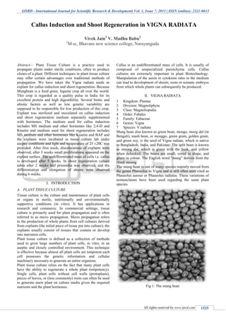
- 1. IJSRD - International Journal for Scientific Research & Development| Vol. 1, Issue 7, 2013 | ISSN (online): 2321-0613 All rights reserved by www.ijsrd.com 1525 Abstract— Plant Tissue Culture is a practice used to propagate plants under sterile conditions, often to produce clones of a plant. Different techniques in plant tissue culture may offer certain advantages over traditional methods of propagation. We have taken the Vigna radiata seeds as explant for callus induction and shoot regeneration. Because Mungbean is a food grain, legume crop all over the world. This crop is regarded as a quality pulse in India for its excellent protein and high digestibility. Several biotic and abiotic factors as well as low genetic variability are supposed to be responsible for low production of this crop. Explant was sterilized and inoculated on callus induction and shoot regeneration medium separately supplemented with hormones. The medium used for callus induction includes MS medium and other hormones like 2,4-D and Kinetin and medium used for shoot regeneration includes MS medium and other hormones like Kinetin and BAP and the explants were incubated in tissue culture lab under aseptic conditions and light and temperature of 25 ±20C was provided. After first week, discolorations of explants were observed, after 3 weeks small proliferations appeared on the explant surface. The undifferentiated mass of cells i.e. callus is developed after 5 weeks. In shoot regeneration culture tubes after 2 weeks leaf primordia was observed, and the differentiation and elongation of shoots were observed during 6 weeks. I. INTRODUCTION A. PLANT TISSUE CULTURE Tissue culture is the culture and maintenance of plant cells or organs in sterile, nutritionally and environmentally supportive conditions (in vitro). It has applications in research and commerce. In commercial settings, tissue culture is primarily used for plant propagation and is often referred to as micro propagation. Micro propagation refers to the production of whole plants from cell cultures derived from explants (the initial piece of tissue put into culture); the explants usually consist of tissues that contain or develop into meristem cells. Plant tissue culture is defined as a collection of methods used to grow large numbers of plant cells, in vitro, in an aseptic and closely controlled environment. This technique is effective because almost all plant cells are totipotent each cell possesses the genetic information and cellular machinery necessary to generate an entire organism. Plant tissue culture relies on the fact that many plant cells have the ability to regenerate a whole plant (totipotency). Single cells, plant cells without cell walls (protoplasts), pieces of leaves, or (less commonly) roots can often be used to generate anew plant on culture media given the required nutrients and the plant hormones. Callus is an undifferentiated mass of cells. It is usually of composed of unspecialized parenchyma cells. Callus cultures are extremely important in plant Biotechnology. Manipulation of the auxin to cytokinin ratio in the medium can lead to development of shoots, roots or somatic embryos from which whole plants can subsequently be produced. II. VIGNA RADIATA 1 Kingdom: Plantae 2 Division: Magnoliphyta 3 Class: Magnoliopsdia 4 Order: Fabales 5 Family: Fabaceae 6 Genus: Vigna 7 Spieces: V.radiata Mung bean also known as green bean, mongo, moog dal (in Bengali), mash bean, or monggo, green gram, golden gram, and green soy, is the seed of Vigna radiate, which is native to Bangladesh, India, and Pakistan. The split bean is known as moong dal, which is green with the husk, and yellow when dehusked. The beans are small, ovoid in shape, and green in colour. The English word "mung" derives from the Hindi moong The mung bean is one of many species recently moved from the genus Phaseolus to Vigna and is still often seen cited as Phaseolus aureus or Phaseolus radiatus. These variations of nomenclature have been used regarding the same plant species. Fig 1: The mung bean Callus Induction and Shoot Regeneration in VIGNA RADIATA Vivek Jain1 V. Madhu Babu2 1 M.sc, Bhavans new science college, Narayanguda
- 2. Callus Induction and Shoot Regeneration in VIGNA RADIATA (IJSRD/Vol. 1/Issue 7/2013/0034) All rights reserved by www.ijsrd.com 1526 III. ORIGIN AND DISTRIBUTION Fig. 2 Mung bean has been considered to have been domesticated in India (Vavilov 1926). His theory has been supported by other authors based on the morphological diversity (Singh et al. 1974), existence of wild and weedy types (Chandel 1984, Paroda and Thomas 1988), and archaeological remains (Jain and Mehra 1980) of mungbean in India. Wild forms of mungbean, V.radiata var.sublobata show a wide area of distribution, stretching from Central and East Africa, Madagascar, through Asia, New Guinea, to North and East Australia (Tateishi 1996). Mung bean is grown throughout the southern Asia including India, Pakistan, Bangladesh, Sri Lanka, Thailand, Cambodia, Vietnam, Indonesia, Malaysia and China, etc. It is also grown in the parts of Africa and U.S.A. and has recently been introduced in Australia. USES IV. BEAN SPROUTS Mung bean sprouts are germinated by leaving them watered with 4 hours of daytime light and spending the rest of the day in the dark. Mung bean sprouts can be grown under artificial light for 4 hours over the period of a week. Fluorescent bulbs or incandescent light bulbs would be the best to use for mung bean sprouts. They are usually sold simply as "bean sprouts”. Mung bean sprouts are stir-fried as a Chinese vegetable accompaniment to a meal, usually with ingredients such as garlic, ginger, spring onions, or pieces of salted dried fish to add flavor. Uncooked bean sprouts are used in filling for Vietnamese spring rolls. V. STARCH Mung bean starch, which is extracted from ground mung beans, is used to make transparent cellophane noodles (also known as bean thread noodles, bean threads, glass noodles. Cellophane noodles become soft and slippery when they are soaked in hot water. A wider variety of cellophane noodles, called mung bean sheets or green bean sheets, are also available. In the South Asia, mung bean is used to make "dhal", which is the most common dish made from various kinds of split legumes with spices. In the Southeast and East Asian countries, mung bean is used to make various kinds of sweet, bean jam, sweetened bean soup, vermicelli, and bean sprout. VI. CALLUS INDUCTION AND SHOOT REGENERATION IN MUNGBEAN Fig 3 The plant, which was selected for callus induction and shoot regeneration, is a plant called Vigna radiata. The explants used in this process were its seeds. The material and glassware used were sterilized in an autoclave at 1210 C at 15lb pressure for 15 min. The entire procedure was done under aseptic conditions in laminar airflow chamber in the tissue culture laboratory. VII. MATERIALS AND METHODS: A. MATERIALS Glassware (conical flasks, hard glass test tubes plugged with sterile cotton plugs, pipettes, glass rods, Petri plates, beakers), laminar hood, micro pipettes, measuring cylinders, micro tips, sterilization materials(tween- 20,HgCl2,70%ethanol, sterile distilled water), forceps, surgical blades, scalpels, tray, explants, spirit lamp, MS medium, hormones (2,4-D, Kinetin, BAP). B. PROTOCOL Collect explant (seeds) ↓ Sterilize the explant (seeds) ↓ Prepare sterile basal medium ↓ Inoculate sterile explants in culture tubes ↓ Incubate the culture tubes in culture room aseptically for 4 day C. PROCEDURE Explant: Seeds of Vigna radiata are taken Washing and sterilization of explants (seeds) Step 1: Explants were washed under running tap water thoroughly for 5 minutes. Then the explants were washed with the Tween-20 for 2 minutes. The explants were washed thoroughly with distilled water to remove the soap solution
- 3. Callus Induction and Shoot Regeneration in VIGNA RADIATA (IJSRD/Vol. 1/Issue 7/2013/0034) All rights reserved by www.ijsrd.com 1527 Fig. 4 Step 2: The explants were then kept under laminar airflow chamber and the explants were sterilized with 70% ethanol solution for 3 minutes. Then the explants were washed with mercuric chloride(HgCl2) solution for 2 minutes followed by thorough washing with sterile double distilled water for 5-6 times. Step 3: MEDIA PREPARATION Tissue culture media used in the present study were based on MS medium. The medium used for seed germination (preconditioning) consisted of MS salts, B5 vitamins, 3.0% (w/v) sucrose. All media were adjusted to pH 5.8 before addition of agar and sterilized at 121_C for 20 min. The sterile medium, which is prepared, was transferred into culture test tubes 15ml into each and the test tubes were plugged tightly with sterile cotton plugs. The medium was allowed to solidify for the explants to be inoculated. Step 4: The sterile explants (seeds) 2 for each test tube were inoculated on the media to get the plant. Fig. 4 1 Callus induction 2 Shoot regeneration D. CALLUS INDUCTION 1) PROTOCOL Excise the leaves from plant ↓ Prepare sterile callus induction medium ↓ Fig. 5 Inoculate the sterile explants in culture tubes ↓ Incubate the culture tubes in culture room aseptically for 5 weeks 2) PROCEDURE Step 1: Excision of explants Leaves were excised aseptically from the 4-day-old in vitro germinated seedlings. Leaves were excised using the sterile blade STEP 2: Preparation of medium The medium used for callus induction includes MS medium and other hormones 2,4-D and Kinetin which induce callus formation. The media prepared was sterilized in an autoclave. After the sterile medium was cooled, hormones (200 micro liters each) were added. The sterile callus induction medium was transferred into test tubes 15 ml into each and the test tubes were plugged tightly with sterile cotton plugs. The medium was allowed to solidify for the explants to be inoculated. Fig. 6 Step 3: The sterile explants were inoculated on callus induction media. Callus induction procedure includes-
- 4. Callus Induction and Shoot Regeneration in VIGNA RADIATA (IJSRD/Vol. 1/Issue 7/2013/0034) All rights reserved by www.ijsrd.com 1528 a) Inoculation of explants, and b) Incubation of the inoculated explants Fig. 7 E. INOCULATION OF EXPLANT 1 Clean the work place and table of laminar air flow chamber with 70% ethanol 2 Put on the switch of UV lamp of chamber for 1 hour before work 3 Put on the power switch and allow the air flow to blow air for at least 15 minutes before work 4 Put off the UV light. 5 Put all the sterilized materials for inoculation on the table of laminar airflow. 6 Wear a clean apron 7 Clean the hands and the work area with alcohol and allow to dry it 8 Pour the alcohol in a clean sterilized beaker and dip all the instruments into it 9 Light the spirit lamp F. INCUBATION OF EXPLANT 1 After inoculating the tissue on the culture media, cultures are incubated on the racks at 25-280 C 2 Culture tubes were placed at 30-450 inclined position 3 Illumination is provided by white florescent light 18 inches above the culture tubes 4 Cultures were incubated for 5 weeks G. SHOOT REGENERATION 1) PROTOCOL Fig. 8 Excise the cotyledonary node from plant ↓ Prepare sterile shoot regeneration medium ↓ the sterile explants in culture tubes ↓ Incubate the culture tubes in culture room aseptically for 6 weeks Inoculate H. PROCEDURE Step 1: Excision of explants Cotyledon and cotyledonary node explants were excised aseptically from 4-day-old in vitro-germinated seedlings. Cotyledons were detached by gently pushing them outwards to the hypocotylary axis. Subsequently, each cotyledonary node explant was prepared by cutting the stem 5 mm above and 10 mm below the cotyledonary node. Step 2: Media Preparation The medium used for shoot regeneration includes MS medium and other hormones Kinetin and BAP, which induce shoot formation. The media prepared was sterilized in an autoclave. After the sterile medium was cooled, hormones (200 micro liters each) were added. The sterile medium was transferred into test tubes 15 ml into each and the test tubes were plugged tightly with sterile cotton plugs. The medium was allowed to solidify for the explants to be inoculated. Step 3: The cotyledons were inoculated with their abaxial side touching the medium. Cotyledonary node explants were inserted upright (acropetal end up) with 3–5 mm of the hypocotyl below the media surface. Four cotyledons and two cotyledonary nodes from 2 seedlings were inoculated per test tube for the initiation of shoot organogenesis medium. Fig. 9 Shoot regeneration procedure includes- a) Inoculation of explants, and b) Incubation of the inoculated explants. I. INOCULATION OF EXPLANT: 1 Clean the work place and table of laminar air flow chamber with 70% ethanol 2 Put on the switch of UV lamp of chamber for 1 hour before work 3 Put on the power switch and allow the air flow to blow air for at least 15 minutes before work 4 Put off the UV light. 5 Put all the sterilized materials for inoculation on
- 5. Callus Induction and Shoot Regeneration in VIGNA RADIATA (IJSRD/Vol. 1/Issue 7/2013/0034) All rights reserved by www.ijsrd.com 1529 6 the table of laminar airflow. 7 Wear a clean apron 8 Clean the hands and the work area with alcohol and allow to dry it 9 Pour the alcohol in a clean sterilized beaker and dip all the instruments into it 10 Light the spirit lamp 11 Take the excised cotyledon in sterile Petri dish 12 Remove the cotton plugs of culture tubes and flame the mouth of the tube 13 Then transfer the explants onto the medium using forceps and fix the cotton plug 14 Each time the forceps are passed through the flame of spirit lamp J. INCUBATION OF THE EXPLANT: 1 After inoculating the tissue on the culture media, cultures are incubated on the racks at 25-280 C 2 Culture tubes were placed at 30-450 inclined position 3 Illumination is provided by white florescent light 18 inches above the culture tubes 4 Cultures were incubated for 6 weeks VIII. RESULTS AND DISCUSSIONS A. Callus induction Fig. 9: After 1st week Fig. 10: After 2nd week Fig. 11: After 3rd week Fig. 12: After 5 weeks
- 6. Callus Induction and Shoot Regeneration in VIGNA RADIATA (IJSRD/Vol. 1/Issue 7/2013/0034) All rights reserved by www.ijsrd.com 1530 1 After first week of incubation discoloration of explant was observed. 2 After 3 weeks of incubation period small proliferations was observed on the surface of explants. 3 After 5 weeks of incubation, callusing was observed. B. Shoot Regeneration Fig. 13: After 1st week Fig. 14: After 3rd week Fig. 15: After 4 weeks Fig. 16: After 5 weeks 1 After 3 weeks, the shoot showed minute elongation a few millimeters. 2 After 4weeks of incubation shoot has shown even more elongation. 3 Result: After 5 weeks, shoot was grown few centimeters height (app.3cm). DISCUSSION From the aseptic plantlets produced from the seeds, shoot
- 7. Callus Induction and Shoot Regeneration in VIGNA RADIATA (IJSRD/Vol. 1/Issue 7/2013/0034) All rights reserved by www.ijsrd.com 1531 Regeneration was obtained from cotyledonary nodes on medium supplemented with BAP and callus induction was produced from primordial leaves on medium supplemented with 2, 4-D & kinetin. C. Shoot Regeneration After 1 week of incubation of explants shoot initiation was observed. After 2 weeks of incubation, proliferation was observed. After 3 weeks of incubation shoot showed minute elongation of few millimeters. After 4 weeks of incubation shoot has shown even more elongation. After 5 weeks shoot was grown few centimeters height (app. 1.5 - 2cm). D. Callus Induction After 1st week of incubation, discolorations of explants observed. After 2 weeks of incubation, small proliferations observed on the surface of explants. At the end of fifth week, callusing observed. However, along with the BAP as we have used kinetin, which promotes shoot elongation fastly, the shoot regeneration was obtained within 3 weeks. REFERENCES [1] Bajaj YPS & Gosal SS (1981) Induction of genetic variability in grain legumes through tissue culture. In: Rao AN(Ed) Proc. Costed Sym. on Tissue Culture of Economically Important Plants (pp 25--41). Singapore. [2] Bharal S & Rashid A (1980) Isolation of protoplasts from stem and hypocotyl of the legume Vigna sinensis and some factors affecting their regeneration. Protoplasma 102: 307-313 [3] Bhargave S & Chandra N (1983) In-vitro differentiation incallus cultures of moth bean, Vigna aconitifolia (JACQ)Marechal. Plant Cell Rep. 2:47- 50 [4] Bhojwani SS, Mullins K & Cohen D (1984) Intra- varietal variation for in-vitro plant regeneration in the genusTrifoliurn. Euphytica 33:915-921 [5] Cheng TY, Saka H & Vogui Dinh TH (1980) Plant regeneration from soybean cotyledonary node segments in culture. Plant Sci. Lett. 19:91-99 [6] Gamborg OL, Miller RA & Ojima K (1968) Nutrient requirements of suspension cultures of soybean root cells.Exp. Cell Res. 50:151-158 [7] Gharyal PK & Maheshwari SC (1981) In-vitro differentiation of somatic embryoids in a leguminous tree, Albizzialebbeck L. Naturwissenschaften 68:374380 [8] Gill R, Eapen S & Rao PS (1986) Tissue culture studies in moth bean-factors influencing plant regeneration from seedling explants of different cultivars. Proc. Indian Acad.Sci. 96:55-61 [9] Gill R, Eapan S & Rao PS (1987a) Morphogenetic studies of cultured cotyledons of urd bean (Vigna mungo L. Hep-per). J. Plant Physiol. 139:1-5 [10] Gill R, Eapen S & Rao PS (1987b) Callus induction from protoplasts of Vigna unguiculata, Vigna sublobata andVigna mungo. Theor. Appl. Genet. 74:100-103 [11] Mathews H (1987) Morphogenetic responses from in- vitro cultured seedling explants in mung bean (Vigna radiata L. Wilczek). Plant Cell, Tiss. Org. Cult. 11:233-240 [12] Mathews H & Rao PS (1984) In-vitro production of multiple seedlings from single seeds of mungbean (Vigna radiataL. Wilczek). Z. Pflanzenphysiol. 113:325-329