SlideShare uma empresa Scribd logo
1 de 12
Baixar para ler offline
Journal of Neuroscience Methods 85 (1998) 141 – 152


  A new method for the rapid and long term growth of human neural
                           precursor cells

        Clive N. Svendsen *, Melanie G. ter Borg, Richard J.E. Armstrong, Anne E. Rosser,
                         S. Chandran, Thor Ostenfeld, Maeve A. Caldwell
             MRC Cambridge Centre for Brain Repair, Cambridge Uni6ersity For6ie Site, Robinson Way, Cambridge CB2 2PY, UK

                             Received 9 April 1998; received in revised form 13 July 1998; accepted 19 July 1998


Abstract

   A reliable source of human neural tissue would be of immense practical value to both neuroscientists and clinical neural
transplantation trials. In this study, human precursor cells were isolated from the developing human cortex and, in the presence
of both epidermal and fibroblast growth factor-2, grew in culture as sphere shaped clusters. Using traditional passaging techniques
and culture mediums the rate of growth was extremely slow, and only a 12-fold expansion in total cell number could be achieved.
However, when intact spheres were sectioned into quarters, rather than mechanically dissociated, cell – cell contacts were
maintained and cellular trauma minimised which permitted the rapid and continual growth of each individual quarter. Using this
method we have achieved a 1.5 million-fold increase in precursor cell number over a period of less than 200 days. Upon
differentiation by exposure to a substrate, cells migrated out from the spheres and formed a monolayer of astrocytes and neurons.
No oligodendrocytes were found to develop from these human neural precursor cells at late passages when whole spheres were
differentiated. This simple and novel culture method allows the rapid expansion of large numbers of non-transformed human
neural precursor cells which may be of use in drug discovery, ex vivo gene therapy and clinical neural transplantation. © 1998
Elsevier Science B.V. All rights reserved.

Keywords: Stem cell; Progenitor cell; Proliferation; Differentiation; Expansion; Human


1. Introduction                                                            transformed (Pleasure and Lee, 1993; Sah et al., 1997).
                                                                           Although of great interest, transformed cell lines may
   There is at present no source of non-transformed                        not share the exact features of primary human neural
human neurons other than primary foetal tissue. This                       tissue, and their oncogenic status makes them less
has posed major limitations to both research and indus-                    attractive as a source of tissue for clinical transplanta-
try with regard to studying the basic biology and drug                     tion. An alternative approach to producing large
responsiveness of human neurons, and has limited clin-                     amounts of neural tissue is to isolate and expand neural
ical neural transplantation programmes to very small                       precursor cells from the CNS.
numbers of patients. The only strategy currently avail-                       The ideal human precursor cell to expand would be a
able for obtaining large amounts of well characterised                     neural stem cell. A stem cell can be most simply defined
human neurons is the use of cell lines. There have been
                                                                           as any cell which is capable of self renewal for extended
two such human lines described previously, one derived
                                                                           periods of time, the progeny from which are capable of
from a tetratocarcinoma and the other oncogenically
                                                                           forming the components of a defined tissue. Asymmet-
                                                                           ric division allows stem cells to generate a progenitor
                                                                           cell, in addition to another stem cell, which have a
  * Corresponding author. Tel.: +44 1223 331185; fax: + 44 1223
                                                                           limited potential for self renewal and often sponta-
331174; e-mail: cns1000@hermes.cam.ac.uk

0165-0270/98/$ - see front matter © 1998 Elsevier Science B.V. All rights reserved.
PII S0165-0270(98)00126-5
C.N. S6endsen et al. / Journal of Neuroscience Methods 85 (1998) 141–152
142

Table 1
Expansion of human neural precursor cells using conventional passaging methods

Age (weeks)           Region           Days                                                                        Total expansion

                                       14           21          28           42           56           63

12                    CTX              2.8                      2.6          1.7          1                        12.36
10                    SC               1.7                      1.7          1.4                                    4.06
9                     SC               0.9                      1.4          1            0.4                       0.50
10                    MES              2.5                      1            2            1.1                       5.50
10                    MES              1.9                      0.4          1.1          1.2                       1.00
                      CTXa
21                                     2.0                      2.5                       1.2                       6.00
                      CTXb
8                                      1.8          2                        1.4          1.3                       6.55

Numbers represent expansion ratios: i.e. number of cells at end of passage over number of cells seeded into flasks. Last number indicates end of
growth for the culture. Cultures were maintained in EGF and FGF-2 for the first 28–35 days and then switched to FGF-2 alone.
SC, spinal cord; CTX, cortex; MES, mesencephalon.
a
  Represents CLON-5382.
b
  Represents BRC-44.

neously stop dividing and differentiate. Stem cells have                     When attached cells or free floating aggregates reach
been most extensively studied in haemopoetic, epider-                     the end of a growth cycle, they must be mechanically
mal and intestinal tissues which require frequent cell                    broken up or ‘passaged’, often using digestion enzymes,
replacement throughout life (Hall and Watt, 1989).                        to avoid contact mediated growth arrest or lack of
Recent studies have shown that specific regions of both                    nutrient diffusion. We postulated that these standard
the developing and adult rodent brain harbour cells                       passaging techniques may lead to cellular trauma, strip
which divide in response to mitogens, while retaining                     receptors, deprive cells of contact mediated factors and
the capacity to differentiate into neurons and glia, and                  remove vital tight junctions known to hold tissues
as such may represent neural stem cells (Weiss et al.,                    together. This may lead to either the terminal differenti-
1996; McKay, 1997; Palmer et al., 1997), although stem                    ation of precursor cells, or a lack of response to mito-
cell status is often debated and the term neural precur-                  gens for the rat and human cells. We therefore
sor may better describe cells within these heterogeneous                  attempted to adapt the passaging technique such that
cultures. Neural precursor cells from the rodent re-                      enzymatic or mechanical disturbance to the cells was
spond to both epidermal growth factor (EGF) and                           minimised and then assess the ability of the human
fibroblast growth factor (FGF-2) (for reviews see Gage                     neural precursor cells to continuously renew over time.
et al., 1995; McKay, 1997) and can be grown as either
monolayer cultures or as free floating spherical aggre-
gates termed ‘neurospheres’ (Reynolds et al., 1992). The
                                                                          2. Methods
short term growth ( B60 days) of similar human CNS
precursors has recently been reported (Buc-Caron,
                                                                          2.1. Tissue collection
1995; Svendsen et al., 1996; Chalmers-Redman et al.,
1997; Murray and Dubois-Dalcq, 1997) and in some
                                                                             Human fetal tissue (between 7 and 21 weeks post
cases these can survive, migrate, differentiate and re-
                                                                          conception) was collected from two different sources:
store function following transplantation into rat models
                                                                          via the Uniform Anatomical Gift Act of the United
of Parkinson’s disease (Svendsen et al., 1997a). How-
                                                                          States or from a local hospital. The methods of collec-
ever, we and others have also shown that human neuro-
                                                                          tion conform with the arrangements recommended by
spheres are difficult to expand in vitro over long
                                                                          the Polkinghorne Committee for the collection of such
periods of time (Svendsen et al., 1997a; Quinn et al.,
                                                                          tissues and the guidelines set out by the Department of
1997). Furthermore, we have also shown that rat and
                                                                          Health in the United Kingdom.
mouse neurospheres, grown using identical methods,
have very different long term expansion potentials with
                                                                          2.2. Cell culture
the rat cells entering senescence within 3 – 4 weeks of
expansion (Svendsen et al., 1997b). Thus, there may be
                                                                            Tissues collected locally (see Table 1 for details) were
a significant species difference when developing meth-
                                                                          dissected in chilled sterile phosphate buffered saline
ods for the growth and differentiation of these cells.
                                                                          (PBS, pH 7.4) with 0.6% glucose. Identified pieces were
Clearly, if neural precursor cells are to become a source
                                                                          incubated in 0.1% trypsin (Worthington) with 0.04%
of tissue for basic neuroscience and clinical pro-
                                                                          DNAase (Sigma type II) for 20 min at 37°C. Following
grammes it would be a major advantage if they could
be expanded for long periods of time.                                     three washes in 0.04% DNAase the tissue was triturated
C.N. S6endsen et al. / Journal of Neuroscience Methods 85 (1998) 141–152                     143


in the same solution using a fine polished Pasteur                   above. Following thawing (Clon-3582) or from primary
pipette. Cell counts showed greater than 65% viable                 (BRC-44), the cells were grown as spheres for 35 days
cells in all cases. Cells were seeded at a concentration of         (Clon-3582) or 43 days (BRC-44) in EGF and FGF-2
200000 per ml into substrate free tissue culture flasks.             during which time they showed approximately a 5-fold
The growth medium consisted of DMEM/HAMS F12                        increase in cell number (see Table 1). Passaging of these
(3:1), penicillin G, streptomycin sulphate, amphotericin            cultures consisted of a gentle trituration with a fine
B (1:100; Gibco), B27 (1:50; Gibco), human recombi-                 polished Pasteur pipette every 14 days in order to break
nant FGF-2 and EGF (both at 20 ng/ml; R&D Sys-                      up the growing spheres. At day 36 or 44, single spheres
tems) and heparin (5 vg/ml; Sigma). Passaging was                   were measured (using a lens grid under a dissecting
carried out at the time points shown in Table 1 and                 microscope). Those which were 0.5 mm or greater in
consisted of a gentle mechanical dissociation using a               radius were sectioned into quarters (using two c23
fine polished Pasteur pipette, after which the mixture of            Swann-Moston surgical blades without handles in a
intact spheres and single cells were re-seeded into fresh           Petri dish with 10 ml of growth medium) and then
medium, as above but with N2 (1:100; Gibco) replacing               transferred to a single un-coated well of a 24-well plate
B27, at 200000 cells per ml. This switch from N2 to                 with 0.5 ml of FGF-2 and heparin supplemented
B27 was due to the fact that although B27 is vital for              growth medium. After the first sectioning it was impor-
maximum growth of neural precursors from primary                    tant to wait until each sphere quarter had grown to at
cultures, it is no better than the less expensive supple-           least 0.35 mm in radius again before re-sectioning (be-
ment, N2, once neurosphere cultures are established as              tween 14 and 21 days growth). All subsequent sections
we have reported previously for rat cultures (Svendsen              were performed every 14 days regardless of sphere size
et al., 1995). Therefore in all neurosphere cultures                to establish average growth rates over time. Spheres too
described in this study, other than primary cultures, N2            small to section at the end of 14 days were discarded
was used as the medium supplement. At 28 – 35 days in               and accounted for in the results (see Table 2). Bulk
vitro all cultures were switched to FGF-2 alone as we               cultures were grown at a density of 50 spheres per T75
found no synergistic effect of combining EGF and                    flask in 20 ml of growth medium and quartered every
FGF-2 on growth after this period as we have reported               14 days. 24 h following sectioning the quarters would
previously for rat cultures (Svendsen et al., 1997b). To            occasionally attach to the surface of the well or flask
estimate total cell number per flask, a 1-ml aliquot of              but could, in most cases, be shaken off gently at this
spheres (taken from a 20-ml flask of cells which was                 time. All cultures were fed by replacing 50% of the
shaken to randomly distribute the spheres) was re-                  medium every 4–5 days.
moved prior to passaging and a single cell suspension
                                                                    2.4. Automated tissue chopping
achieved using trypsin digestion followed by mechanical
dissociation. Live cells were then counted in a haemocy-
                                                                       Spheres at the end of a growth cycle (  0.35 mm
tometer using trypan blue exclusion to exclude dead
cells.                                                              radius) were transferred to the lid of a 16-mm Petri dish
   Tissue collected via the US Uniform Anatomical Gift              and the majority of medium removed using a Pasteur
Act (Clon-5382) consisted of a 21 week post conception              pipette. The lid with the spheres was attached to the
fetus. Cortical cells were isolated and passed through a            stage of the McIlwain tissue chopper (Mickle Engineer-
190-mesh cell strainer before running through a 30%                 ing, Gomshall, Surrey, UK) using thin strips of adhe-
percoll column for 20 min. Cells were seeded into                   sive putty (Blue Tack or equivalent). A sterile razor
growth medium (described above) and grown as                        blade was inserted into the arm of the tissue chopper.
spheres in EGF and FGF-2 for 7 days before cryopre-                 Sections were then automatically taken through the
                                                                    spheres using a distance between chops of 350 vm. The
serving in liquid nitrogen using DMEM with 20% fetal
calf serum with 10% DMSO. Frozen cells were rapidly                 stage was rotated through 90° and the process repeated
warmed to 37°C, washed three times in DMEM and                      to generate ‘cubes’ of tissue with a mean width of
                                                                    approximately 350 vm. These were then carefully
then re-seeded into fresh growth medium. This freezing
process could be used successfully at any stage of                  washed in fresh growth medium and re-seeded at ap-
sphere growth.                                                      proximately 200 spheres per T75 flask containing 20 ml
                                                                    of growth medium.
2.3. The sectioning method and systematic assessment
                                                                    2.5. Thymidine incorporation
of growth rates

                                                                      [3H]Thymidine (Amersham; 0.5 vCi/ml) was added
   Two cultures were used to assess exact growth rates
using the sectioning method. BRC-44 was generated                   to individual spheres for a period of 24 h. At the end of
from an 8-week post conception fetus but was not                    the incubation period the spheres were washed three
                                                                    times in DMEM and incorporated [3H]thymidine was
cryopreserved at any stage and Clon-3582 is described
C.N. S6endsen et al. / Journal of Neuroscience Methods 85 (1998) 141–152
144

Table 2
Cumulative growth data for human neural stem cell cultures

                           Days                                                                                                     Average
                                                                                                                                    expansiona

                           14             28          42           56           70           84           98           112

BRC-44
                                          0.449 0.02 0.439 0.02 0.37 90.03 0.4290.03 0.39 90.03 0.32 90.03 0.32 90.03
 Radius of mother disc       NA
                                          0.349 0.01 0.279 0.03 0.34 90.02 0.31 90.03 0.28 9 0.03 0.339 0.02 0.37 9 0.04
 Radius of daughter          NA
 discs
   % of mother disc                        77           63           92          74            72          103          116
 Number of surviving         NA            19/24        20/24        22/24       22/24         22/24        22/24        19/24
 discs
   % of total                              79           83           91          92            92           92           79
                                                                                                                                    2.95 9 0.24
 Expansion ratio             NA             2.43         2.09         3.35        2.72          2.63         3.79         3.67
Clon-3582
                           0.31 9 0.02 0.389 0.03 0.379 0.04 0.29 90.03 0.4690.05 0.49 90.04 0.4690.03 0.5290.01
  Radius of mother disc
                           0.349 0.03 0.309 0.04 0.319 0.03 0.33 90.02 0.32 9 0.08 0.42 90.04 0.42 90.04 0.51 90.02
  Radius of daughter
  discs
    % of mother disc        111            84           84         116           71            86          104           97
  Number of surviving        23/24         18/24        19/24       19/24        17/24         23/24        20/24        24/24
  discs
    % of total               96            75           79           79          71            95           83          100
                                                                                                                                    3.19 9 0.28
  Expansion ratio             4.25          2.37         2.66         3.67        2.01          3.27         3.45         3.88

Mother disc radius from six individual discs was measured (mm) before sectioning into quarters. Each quarter was then measured again at the
end of the growth period. If all four quarters had reached the size of the mother disc this would represent a 4-fold increase in cell number. Thus
the final expansion ratio = expected number of discs (4)×% of mother disc size×% of total discs surviving. NA, data not available.
a
  Over a 14 day period. Not significantly different between the cultures (p0.05; Student’s t-test).


then solubilised using 600 vl NaOH (0.4 M) for 1 h at                        Sigma) combined with GFAP (polyclonal; Bohringer;
37°C. This solution was then added to 4 ml of scintilla-                     1:1000) in 0.1 M PBS/0.1% Triton/3% goat serum.
tion cocktail and counted in a scintillation                                 Others were incubated with antibodies to nestin (poly-
spectrometer.                                                                clonal; 1:50; kindly donated by R.D.G. McKay); GAL-
                                                                             C (monoclonal; kindly donated by B. Ranscht; 1:4; no
2.6. Karyotyping                                                             Triton was used with this surface marker) or MAP-2ab
                                                                             (1:500; Sigma). Goat anti-mouse biotin or fluoroscein
  Chromosome number and size was scored using                                conjugated goat anti rabbit antibodies were used to
Giemsa-stained metaphase spreads by the Department                           label the primaries, followed by a streptavadin–rho-
of Cytogenetics, Addenbrooke’s Hospital, Cambridge.                          damine conjugate Hoescht was added to the final incu-
                                                                             bation step (Sigma, 1:10000 in 0.1 M PBS) to visualise
2.7. Immunocytochemistry                                                     nuclei. For cell counts at least five random fields (at
                                                                             × 40) were analysed from the monolayer of cells
  Whole free floating spheres were fixed in 4%                                 around the plated spheres. Each field contained be-
paraformaldehyde for 20 min, washed in PBS and                               tween 50 and 100 cells.
dehydrated though 70, 95 and 100% alcohol (20 min                               Sections were also viewed under a Biorad confocal
each). Following clearing overnight in xylene, spheres                       scanning microscope at excitation wavelengths of 488
                                                                             and 564 nm. Optical sections were taken at 20 vm
were embedded in paraffin and sectioned on a micro-
tome at 5 vm. For differentiation studies, whole                             intervals and then merged.
spheres were allowed to attach to a poly-L-lysine
(Sigma) coated coverslips in 24-well plates in the pres-
ence of 0.5 ml of DMEM/B27 with 1% serum for 24 h.                           3. Results
Following this period the medium was exchanged for
                                                                             3.1. Expansion of human precursors
DMEM/B27 alone. Cultures were fed by replacing 50%
of the medium every 4– 5 days. At 14 days, the cultures
were fixed for 30 min in 4% paraformaldehyde. Wax                               Following seeding into growth medium, aggregates
sections and coverslips were incubated with primary                          of dividing cells formed into spheres which grew in size
antibodies to beta tubulin III (TuJl; monoclonal; 1:500;                     over time in response to the mitogens EGF and FGF-2.
C.N. S6endsen et al. / Journal of Neuroscience Methods 85 (1998) 141–152                                  145


Fig. 1. Cells isolated from the developing cortex and grown in EGF and FGF-2 formed mainly spheres at early passages (A1) but some discs could
also be seen at later passages when grown in FGF-2, possibly as a result of transient attachment to the culture dish (A2). (B), (C), (D) and (E)
show four quarters from a single sphere (A1) 1 h after sectioning. (F) Cumulative growth curves for Clon-5382 and BRC-44 based on expansion
data in Table 2 and subsequent results not shown in Table 2. Note the steady and consistent exponential increase in total sphere number over
time. Scale bar = 0.2 mm.


Between 14 and 21 days of growth the spheres could be                      showed only modest expansion over this 14 day period
gently dissociated to a mixed suspension of single cells                   as shown in Table 1. By 40 days of growth using the
and sphere remnants before re-plating into growth                          sectioning method, some disc shaped clusters could be
medium. Using this technique we have previously re-                        seen in addition to spheres (Fig. 1A1 and A2). The
ported a 3-4-fold increase in cell number over the first                    discs mainly developed due to temporary attachment of
few weeks in vitro, after which the absolute number of                     the newly sectioned spheres to the surface of the flask,
cells harvested at each passage declined (Svendsen et                      and were often concave on one surface. This led to
al., 1997b). In this study, seven separate experiments                     some disc/sphere aggregates which appeared hollow
using either brain stem, spinal cord or cortical human                     when sectioned (approximately 30% at 100 days of
fetal tissue showed similar results, with variable growth                  growth, data not shown). When individual spheres at
for the first 4–6 weeks, after which their was very little                  100 days growth were dissociated and the number of
further growth with the maximum expansion of total                         cells counted, those with a size of between 0.35 and 0.45
cells only reaching 12-fold (Table 1). A variety of                        mm diameter contained an average of 61166+ 3498
modifications to the culture medium, growth factor                          viable cells (n= 25 spheres from CTX-44) with less than
combinations and passaging strategies have been at-                        5% non viable cells. Very similar results with regard to
tempted. None of these had any effect on the slow                          cell number and sphere size were found at 50 and 150
growth rate and eventual senescence of these precur-                       days of growth (data not shown). There was a positive
sors, although some cultures could be kept in a mitotic,                   correlation between sphere size and number of cells
but non expanding, state (due to concomitant cell                          (Fig. 2A), although some variation existed due to the
death) for up to 6 months (data not shown).                                technical difficulties of dissociating small single spheres
   A novel, non traumatic, passaging approach was                          (cells attaching to the side of the pipette, incomplete
employed on several cultures. Data for two of these                        dissociation). We next assessed whether the size of the
isolated from the cortex, which differed in both fetal                     sphere sections influenced their ability to grow back to
age and method of cell isolation, are described in detail                  the size of the mother sphere. Regardless of the mother
here. Instead of mechanical dissociation, individual                       sphere size, all quarters grew back to the same size in
spheres were sectioned into quarters under a dissecting                    relation to the mother showing that the expansion is
microscope using a scalpel blade (Fig. 1). The resulting                   not dependent on either large or small sphere sizes (Fig.
                                                                           2B). [3H]Thymidine added to the spheres over the last
four quarters were placed into fresh growth medium
containing FGF-2 and over the next 24 h rounded to                         24 h of growth at all stages in culture showed that there
form new spheres which grew close to the size of the                       was a significant amount of uptake ( 5000 counts per
mother sphere by 14 days. Using this method there was                      min/per sphere at each passage) indicating that active
a steady and exponential increase in total sphere num-                     cell division was occurring. The total amount of expan-
ber which did not decrease with time (Fig. 1; Table 2).                    sion achieved over the growth period (including a 5-
Parallel cultures which were also switched to FGF-2                        fold increase prior to the start of the sectioning
alone but were dissociated rather than chopped,                            method) was greater than 1.5 million-fold for Clon-
C.N. S6endsen et al. / Journal of Neuroscience Methods 85 (1998) 141–152
146


Fig. 2. Graphs showing (A) correlation between volume of sphere and number of cells per sphere. (B) Lack of any relationship between size of
sectioned quarter and its subsequent growth relative to its own mother sphere.


                                                                         migrating out. Between 2 and 7 days, radiating pro-
5382 and both cultures showed a population doubling
                                                                         cesses often developed which sketched from the edge of
time of approximately 4 days. Metaphase spreads
                                                                         the sphere onto the substrate (Fig. 3C). Along these
showed that at 150 days of growth the human cells
                                                                         radiating strands, immature TuJ1 + neurons could be
remained karyotypically normal with regard to chro-
                                                                         seen which often trailed processes in parallel lines and
mosome number and appearance. Removal of FGF-2
                                                                         may have been migrating out from the core of the
from the growth medium at any stage resulted in
                                                                         sphere (Fig. 3D). Using confocal imaging the exact
senescence and eventual death of cultures over a period
                                                                         anatomy of the sphere following 14 days of differentia-
of 14 days. However, switching FGF-2 responsive
                                                                         tion could be seen and radial processes emanating from
spheres to a medium containing EGF resulted in the
                                                                         the core were found to be GFAP positive (Fig. 4).
growth of an EGF responsive spheres with very similar
                                                                            Migrating cells eventually formed a monolayer
characteristics following plating and which also ex-
                                                                         around the plated sphere and labelled for either TuJ1
panded exponentially using this sectioning method.
                                                                         or GFAP but never with both markers at 14 days of
However, the EGF responsive spheres were less prone
                                                                         differentiation (Fig. 5A, B and C). Detailed cell counts
to attaching to the culture dish and subsequently
                                                                         within the monolayer around the sphere revealed that
formed less discs. We are also systematically assessing
                                                                         the majority of the cells were either TuJ1 or GFAP
the effects of sectioning on the exact growth rates of
                                                                         positive at both early and late passages and that no
neural precursor cells isolated from other brain regions
                                                                         galactocerbroside (GAL-C) positive oligodendrocytes
mentioned in Table 1, although bulk cultures spinal
                                                                         could be seen under these plating conditions (Fig. 5D).
cord and brain stem do expand rapidly using this
                                                                         Although not analysed in detail, every whole sphere
method (data not shown).
                                                                         plated (over 300) gave rise to both neurons and astro-
                                                                         cytes, strongly suggesting the presence of a common
3.2. Differentiation of human neural precursors
                                                                         precursor. At the very periphery of the migrating cells,
                                                                         lone GFAP+ astrocytes could often be found, onto
   Continual growth of the human neural spheres sug-
                                                                         which a number neurons had selectively migrated, the
gested self renewal was occurring, but did not deter-
                                                                         processes from which were entirely confined to the
mine the phenotypic potential of these cells. To assess
                                                                         astrocyte surface (Fig. 6A–C). This suggests that these
this, whole spheres or differentiating spheres (at  100
                                                                         precursor cell derived astrocytes provide an attractive
days growth) were processed for indirect immunocyto-
                                                                         surface for the migrating neurons. Some cells also
chemistry. The majority of cells (95%) within the
                                                                         expressed markers only found in mature neurons such
growing spheres were found to be positive for nestin,
                                                                         as microtuble associated protein 2ab which is located
(Fig. 3B) a marker for undifferentiated neuroepithelial
                                                                         mainly in dendrites (MAP-2ab; Fig. 6D) (Riederer and
stem cells (Lendahl et al., 1990), but did not stain for
                                                                         Matus, 1985). Interestingly, following trituration to a
TuJ1 (a specific early neuronal marker) (Menezes and
                                                                         single cell suspension and plating, very few TuJ1 +
Luskin, 1997) or glial acidic fibriallary protein (GFAP;
                                                                         neurons were found while the majority of cells stained
an astrocyte marker). When whole spheres were ex-
                                                                         for GFAP (data not shown), indicating that either the
posed to a substrate, they rapidly attached and within
                                                                         temporal sequence of events following plating of whole
hours cells with a glial morphology could be seen
                                                                         spheres, or the lack of physical trauma caused by
                                                                         trituration, may be required for neuronal survival and
C.N. S6endsen et al. / Journal of Neuroscience Methods 85 (1998) 141–152                                   147


Fig. 3. Staining whole growing spheres at 100 days growth for Hoescht revealed that viable cell nuclei could be seen throughout in most cases
(A). Scale bar: 100 vm. Immunocytochemical staining showed that the majority of cells within the growing spheres were nestin + and
undifferentiated (B). Scale bar: 25 vm. Some spheres were plated onto poly-L-lysine coated coverslips in DMEM/B27 and 1% serum for 24 h to
induce differentiation. Phase (C) and TuJ1 (D) staining of the same field from a sphere 4 days following plating. Note the radial fibre outgrowths
along which TuJl + cells with extensive fibres could be seen. Arrow heads represent glial fibres with no TuJl + fibres, arrows represent TuJl + fibres
along processes, double arrowhead shows single neuronal cell body with extended TuJl + processes. Scale bar: 25 vm.


differentiation of cells arising from long term human                       4. Discussion
neural precursor cell cultures.
                                                                               The present study has demonstrated a new method
3.3. Automation of the sectioning technique                                 for the long-term exponential expansion of non immor-
                                                                            talised or transformed human neural precursor cells,
   Through the detailed assessment of manually section-                     which maintained the capacity to generate a high per-
ing individual spheres we have shown that sustained                         centage of neurons (see Fig. 7 for a schematic of the
exponential growth of human neural precursor cells can                      technique).
be achieved (Table 2). However, it is obviously not                            There are two main methods in the literature com-
practical to section large numbers of spheres using this                    monly used to generate populations of neural precursor
manual method. We therefore developed an automated                          cells. The first uses FGF-2 and a substrate to expand
procedure using the McIlwain tissue chopper originally                      colonies of cells which grow attached to the culture
designed to slice fresh brain tissue. Using this device up                  flask while the second uses EGF to expand aggregates
to 1000 spheres can be automatically sectioned within a                     of cells (neurospheres) although it is now clear that
few minutes. Each section rapidly rounds up after                           these growth factors are often interchangeable in their
seeding into fresh medium and forms a new growing                           effects (for recent review, see Svendsen (1997)). We
sphere as described previously using the manual tech-                       have not attempted to grow human cells attached to a
nique, although obviously the spheres differ in size                        substrate in the presence of FGF-2 in this study, but
depending on exactly where they were sectioned. As                          have focused instead on the neurosphere culture
sphere size is not a critical determinant of cell expan-                    method. The advantage of the aggregate culture
sion (see Fig. 2) we feel that this method should provide                   method is that large numbers of cells can be expanded
an automated technique for growing large numbers of                         in a small volume of medium. There are no published
human neural precursor cells.                                               reports on the long term growth of human neural
C.N. S6endsen et al. / Journal of Neuroscience Methods 85 (1998) 141–152
148


Fig. 4. Confocal image (Biorad Inc, UK) of a sphere plated for 14 days showing both TuJ1 positive neurons (red) and GFAP positive astrocytes
(green) which had migrated from the core of a sphere (left) and formed a monolayer culture. Note the many radial fibres which stain for GFAP
(arrowheads). Scale bar: 100 vm. Insert is a high power view of a plated region around the sphere ( × 4 magnification compared to rest of plate)
showing how neuronal processes pass both under and over the astrocytes.


precursor cells, although preliminary data suggests that                  published observations). Furthermore, we have found
EGF can drive a human precursor for extended periods                      the expansion of human neurospheres to be very slow,
of time (Vescovi et al., 1997). This cell may be similar                  and although division continued (based on incorpora-
to the EGF responsive cell isolated from the developing                   tion of mitotic labels) real expansion stopped after 35
mouse striatum which can grown for long periods of                        days of growth (Svendsen et al., 1997a). Using the
time in vitro as spheroid neurospheres and may repre-                     sectioning method presented here, human neurospheres
sent a population of stem cells (Reynolds et al., 1992;                   continue to expand and give rise to high numbers of
Reynolds and Weiss, 1996) although even as early as                       neurons even at late passages. We are currently identi-
passage two (using conventional passaging methods)                        fying the phenotype of neurons generated from these
these mouse neurospheres spontaneously gave rise to                       long term human cultures.
virtually no neurons but rather glial cells (Arsenijevic                     What is the exact nature of the dividing cells within
and Weiss, 1998). We have previously found that in                        the sectioned spheres?. Their growth rate was remark-
contrast to mouse neurospheres, rat neurospheres enter                    ably stable and relatively slow, with a cell cycle time of
a programmed senescence between 28 and 35 days of                         approximately 4 days throughout the culture period.
growth using routine passaging methods (Svendsen et                       They were karyotypically normal on gross inspection
al., 1997b) and also give rise to high numbers of                         which suggests they had not transformed but are main-
astrocytes at later passages (Rosser and Svendsen, un-                    taining a normal cellular phenotype with a slow cell
C.N. S6endsen et al. / Journal of Neuroscience Methods 85 (1998) 141–152                                  149


Fig. 5. Photomicrograph showing phase (A), TuJ1 (B) and GFAP (C) staining of an identical field of cells around the periphery of a sphere plated
for 14 days. Many TuJI + and GFAP + cells could be found but no cells labelled with both markers. Arrows show neurons and arrowheads show
astrocytes. Graph shows the numbers of labelled cells around the sphere as a percentage of total cells (n =12 spheres at each time) at either 50
or 150 days of growth (D). There was no significant difference between the numbers of neurons and astrocytes generated at early or late passages.


cycle time consistent with a slowly dividing stem cell                     passages, but only neurons and astrocytes at late pas-
population. Clonal analysis has classically been re-                       sages using the plating conditions in this study. To
quired to prove pluri-potency, but is difficult to per-                     investigate this further we are currently assessing the
form with this culture system since we have shown that                     effects of other growth factors and substrates on the
groups of cells must maintain contact in order to grow                     differentiation of these long term human precursor
for long periods. However, the observation that every                      cells, which have previously been shown to influence the
sphere we have plated produced both neurons and                            fate rat hippocampal precursors and immortalised hu-
astrocytes, but never only one phenotype, argues                           man neural precursors (Joh et al., 1996; Sah et al.,
strongly in favour of a common self renewing stem cell.                    1997). It was of interest that the cultures generated
It also remains possible that two uni-potent stem cells                    from widely differing fetal ages (8 and 21 weeks) gave
with similar division rates and the capacity to produce                    rise to similar numbers of neurons and astrocytes at
either neurons or astrocytes are dividing alongside each                   late passages. This would further suggest that a com-
other. Oligodendrocytes were never seen to arise from                      mon cell is being driven in these cultures following an
the late passage sectioned spheres, but have been seen                     initial period of instability where progenitors with a
to develop from early passage spheres (Murray and                          more limited mitotic life span are filtered out. In sum-
Dubois-Dalcq, 1997; Svendsen et al., unpublished ob-                       mary, the cell which is dividing in these cultures is
servations). This suggests that either, (i) an oligoden-                   maybe best described as a precursor cell until we know
drocyte precursor may be capable of dividing for a                         more about its exact phenotypic potential under a
certain period of time and then be lost from the cul-                      variety of circumstances.
tures during the extended period of growth which then                         There is currently some confusion regarding the dif-
only consists of cells capable of giving rise to neurons                   ferential effects of EGF and FGF-2 on neural precursor
and astrocytes or, (ii) a common precursor is able to                      cells. Based on clonal analysis, EGF was only found to
make oligodendrocytes, neurons and astrocytes at early                     stimulate a glial progenitor from the mouse cortex late
C.N. S6endsen et al. / Journal of Neuroscience Methods 85 (1998) 141–152
150


Fig. 6. Photomicrographs showing cells which had migrated out from the sphere and established colonies at the limit of the migration wave.
Isolated epitheloid glial cells could be seen with many smaller cells on top which conformed exactly to the shape of the underlying cell suggesting
a strong attraction of neurons to the developing astrocytes (A). The smaller cells were TuJI + neurons (B) and the epitheloid cells were GFAP +
astrocytes (C) (C is the same field as A and B). MAP-2ab was found to label the dentritic processes of more mature neurons arising from the
spheres (D). Scale bar: 15 vm.


in development and had no effect on earlier cortical                        increase the mitotic effects of FGF-2 on embryonic
precursors, in contrast to FGF-2 which was able to                          precursor cells (Caldwell and Svendsen, 1998) and it
induce the division of a multipotent cell at both early                     was used throughout the current study. Interestingly,
and late developmental ages using the same model                            we found that changing the growing human precursors
(Kilpatrik and Bartlett, 1995). Furthermore, EGF ap-                        from FGF-2 to EGF had no obvious effect on the
pears to stimulate glial division in adult subventricular                   ability to generate neurons following plating, although
zones and may in fact repress neuronal development in                       this is the subject of a more detailed comparison cur-
vivo (Kuhn et al., 1997). We have recently shown that                       rently in preparation. Clearly there is much more exper-
following priming with FGF-2, the same cell responds                        imental work required to resolve these issues.
to both EGF and FGF-2 in primary E14 mouse striatal                            The reason why the sectioning method is so effective
tissue (Ciccolini and Svendsen, 1998). Perhaps in early                     in maintaining stable and rapid growth may be in part
and late adulthood there are more restricted precursors                     due to the fact that there is no disruption of cell–cell
which respond separately to these factors, whereas dur-                     contact within the intact regions of the spheres in
ing development a common precursor exists. However,                         contrast to standard process of mechanical dissociation.
the mixture of in vivo and in vitro data across different                   Membrane associated factors are known to be impor-
species and culture conditions makes it impossible to                       tant for the division of neural precursor cells (Temple
draw conclusions at present. It is of interest that the                     and Davis, 1994) and a ‘niche’ hypothesis has been
adult mouse subventricular zone has recently been                           proposed which suggests stem cells will only retain their
shown to contain FGF-2 responsive cells (Gritti et al.,                     pluripotency within an appropriate environment
1996) which appear almost identical to the EGF re-                          (Schofield, 1978), both of which may be sustained using
sponsive cells described originally by Reynolds and                         this sectioning method. Equally important may be the
Weiss (1992) who claimed that FGF-2 was unable to                           reduction in cellular trauma that results from sectioning
stimulate division of the same cells. This discrepancy                      rather than dissociating intact spheres. It is clear that
may be due to the fact that heparin was added to the                        partial dissociation may also lead to intact remnants
medium with FGF-2 in the later study. We have re-                           remaining which have cell–cell contact. Indeed, our
cently shown that this proteoglycan can significantly                        normal passaging methods often result in non-complete
C.N. S6endsen et al. / Journal of Neuroscience Methods 85 (1998) 141–152                                     151


Fig. 7. Schematic summarising the overall method. Conventional passaging techniques resulted in slow growth or senescence. By sectioning
growing spheres there was less trauma to the cells and the quarters grew close to the size of the mother over a period of 14 days. This pattern
continued for extensive periods of time in culture and allowed the exponential growth of human neural precursor cells.


dissociation. The major disadvantage of this is that                      provide a valuable source of normal human neural
                                                                          tissue for both testing novel neuroactive compounds in
spheres are generally stripped of cells leaving only the
                                                                          vitro and clinical neural transplantation programmes
cores. Many of the stripped cells die but the cores may
                                                                          which are currently dependent on the collection of fresh
continue to grow. However, this does not result in such
                                                                          human fetal tissues (Bjorklund, 1993; Svendsen, 1997).
a rapid growth rate as simply sectioning the spheres
and is far more inconsistent as it is difficult to control
exactly how much dissociation is performed in one
                                                                          Acknowledgements
culture to the next.
   It is tempting to speculate that the stages of sphere
                                                                             We thank S.B. Dunnett for his continual support,
attachment, formation of radial processes and apparent
                                                                          Biorad Inc. for preparation of the confocal images and
neuronal migration may recapitulate the normal pro-
                                                                          Ziggy Zhang and Irena Sarel of Blowhittiker Inc. for
cess of primate development where neuronal precursors
                                                                          providing human neurospheres. The authors would also
divide within ventricular zones before migrating to the
                                                                          like to thank Dr Scott Whittemore for critically ap-
pial surface along radial glia (Rakic, 1985). However,
                                                                          praising an early version of this manuscript. This re-
this needs to be substantiated with further studies
                                                                          search was funded by a Wellcome Fellowship to CNS
which are the focus of ongoing work. Genetic manipu-
                                                                          and by the MRC.
lations to these dividing cells should be possible, as
achieved previously with both human and rat neural
precursors (Sabate et al., 1995; Svendsen et al., 1996),
                                                                          References
to allow the expression of specific proteins following
grafting, thereby facilitating ex vivo CNS gene therapy
                                                                          Arsenijevic Y, Weiss S. Insulin-like growth factor-I is a differentiation
(Friedmann, 1994). The spheres can be frozen and
                                                                             factor for post-mitotic CNS stem cell-derived neuronal precursors:
stored which facilitates shipping and banking of such                        distinct actions from those of brain-derived neurotrophic factor. J
tissue. Finally, as these sectioned neurospheres produce                     Neurosci 1998;18:2118 – 28.
consistently large numbers of neurons, they may                           Bjorklund A. Better cells for brain repair. Nature 1993;362:414–5.
C.N. S6endsen et al. / Journal of Neuroscience Methods 85 (1998) 141–152
152

                                                                            Pleasure SJ, Lee VM-Y. NTera 2 cells: a human cell line which
Buc-Caron MH. Neuroepithelial progenitor cells explanted from
                                                                               displays characteristics expected of a human committed neuronal
   human fetal brain proliferate and differentiate in vitro. Neurobiol
                                                                               progenitor cell. J Neurosci Res 1993;35:585 – 602.
   Dis 1995;2:37 – 47.
                                                                            Quinn SM, Walters WM, McKay RDG, Onifer SM, Whittemore SR.
Caldwell MA, Svendsen CN. Potentiation of FGF-2 induced prolifer-
                                                                               Neuroepithelial precursor cells from fetal human spinal cord. Soc
   ation of mesencephalic neural precursors by heparin. Exp Neurol
                                                                               Neurosci Abstr 1997;23:320.
   1998;in press.
                                                                            Rakic P. Limits of neurogenesis in primates. Science 1985;227:154–6.
Chalmers-Redman RME, Priestley T, Kemp JA, Fine A. In vitro
                                                                            Reynolds BA, Weiss S. Generation of neurons and astrocytes from
   propagation and inducible differentiation of multipotential pro-
                                                                               isolated cells of the adult mammalian central nervous system.
   genitor cells from human fetal brain. Neuroscience 1997;76:1121 –
                                                                               Science 1992;255:1707– 10.
   8.
                                                                            Reynolds BA, Weiss S. Clonal and population analyses demonstrate
Ciccolini F, Svendsen CN. FGF-2 promotes acquisition of EGF
                                                                               that an EGF-responsive mammalian embryonic CNS precursor is
   responsiveness in mouse striatal precursor cells: Identification of
                                                                               a stem cell. Dev Biol 1996;175:1 – 13.
   neural precursor cells responding to both FGF-2 and EGF. J
                                                                            Reynolds BA, Tetzlaff W, Weiss S. A multipotent EGF-responsive
   Neurosci 1998;in press.
                                                                               striatal embryonic progenitor cell produces neurons and astro-
Friedmann T. Gene therapy for neurological disorders. Trends Gene
                                                                               cytes. J Neurosci 1992;12:4565 – 74.
   Sci 1994;10:210 – 4.                                                     Riederer B, Matus A. Differential expression of distinct microtubule-
Gage FH, Ray J, Fisher LJ. Isolation, characterisation, and use of             associated proteins during brain development. Proc Natl Acad Sci
   stem cells from the CNS. In: Cowan WM, Shooter EM, Stevens                  USA 1985;82:6006 – 9.
   CF, Thompson RF, editors. Annual Review of Neuroscience,                 Sabate O, Horellou P, Vigne E, Colin P, Perricaudet M, Buc-Caron
   Palo Alto: Annual Reviews, 1995;18:159–92.                                  M, Mallet J. Transplantation to the rat brain of human neural
Gritti A, Parati EA, Cova L, Frolichsthal P, Galli P, Wanke E,                 progenitors that were genetically modified using adenoviruses.
   Faravelli L, Morassutti D, Roisen F, Nickel DD, Vescovi AL.                 Nat Genet 1995;9:256 – 60.
   Multipotential stem cells from the adult mouse brain proliferate         Sah DWY, Ray J, Gage FH. Bi-potent progenitor cell lines from the
   and self-renew in response to basic fibroblast growth factor. J              human CNS. Nat Biotechnol 1997;15:574 – 80.
   Neurosci 1996;16:1091 – 100.                                             Schofield R. The relationship between the spleen colony forming cell
                                                                               and the haemopoietic stem cell. Blood Cells 1978;4:7 – 25.
Hall PA, Watt FM. Stem cells: the generation and maintenance of
                                                                            Svendsen CN. Neural stem cells for brain repair. Alzheimer’s Res
   cellular diversity. Development 1989;106:619–33.
                                                                               1997;3:131 – 5.
Joh K, Hazel T, Muller T, Dugich-Djordjevic M, McKay RDG.
                                                                            Svendsen CN, Fawcett J, Bentlage C, Dunnett SB. Increased survival
   Single factors direct the differentiation of stem cells from the fetal
                                                                               of rat EGF-generated CNS progenitor cells using B27 supple-
   and adult central nervous system. Genes Dev 1996;10:3129–40.
                                                                               mented medium. Exp Brain Res 1995;102:407 – 14.
Kilpatrik TJ, Bartlett PF. Cloned multipotential precursors from the
                                                                            Svendsen CN, Clarke DJ, Rosser AE, Dunnett SB. Survival and
   mouse cerebrum require FGF-2, whereas glial restricted precur-
                                                                               differentiation of rat and human epidermal growth factor-respon-
   sors are stimulated with either FGF-2 or EGF. J Neurosci
                                                                               sive precursor cells following grafting into the lesioned adult
   1995;15:3653 – 61.
                                                                               central nervous system. Exp Neurol 1996;137:376 – 88.
Kuhn HG, Winkler J, Kempermann G, Thal LJ, Gage FH. Epider-
                                                                            Svendsen CN, Caldwell MA, Shen J, ter Borg MG, Rosser AE, Tyers
   mal growth factor and fibroblast growth factor-2 have different
                                                                               P, Karmiol S, Dunnett SB. Long term survival of human central
   effects on neural progenitors in the adult rat brain. J Neurosci
                                                                               nervous system progenitor cells transplanted into a rat model of
   1997;17:5820 – 9.
                                                                               Parkinson’s disease. Exp Neurol 1997a;148:135– 46.
Lendahl U, Zimmerman LB, McKay RDG. CNS stem cells express a
                                                                            Svendsen CN, Skepper JN, Rosser AE, ter Borg M, Tyers P, Ryken
   new class of intermediate filament protein. Cell 1990;60:585–95.             T. Restricted growth potential of rat striatal precursors as com-
McKay RDG. Stem cells in the central nervous system. Science                   pared to mouse. Dev Brain Res 1997b;99:253 – 8.
   1997;276:66 – 71.                                                        Temple S, Davis AD. Isolated rat cortical progenitor cells are main-
Menezes JRL, Luskin MB. Expression of neuron-specific tubulin                   tained in division in vitro by membrane-associated factors. Devel-
   defines a novel population in the profliferative layers of the                opment 1994;120:999 – 1008.
   developing telencephalon. J Neurosci 1997;14:5399–416.                   Vescovi AL, Daadi M, Asham R, Reynolds BA. Continuous genera-
Murray K, Dubois-Dalcq M. Emergence of oligodendrocytes from                   tion of human catecholoaminergic neurons by embryonic CNS
   human neural spheres. J Neurosci Res 1997;50:146–56.                        stem cells. Soc Neurosci Abstr 1997;23:319.
Palmer TD, Takahashi J, Gage FH. The adult rat hippocampus                  Weiss S, Reynolds BA, Vescovi AL, Morshead CM, Craig CG, van
   contains primordial neural stem cells. Mol Cell Neurosci                    der Kooy D. Is there a neural stem cell in the mammalian
   1997;8:389 – 404.                                                           forebrain. Trends Neurosci 1996;19:387 – 93.


                                                                 .

                                                                                                                      .

Mais conteúdo relacionado

Mais procurados

Tissue Engineering Poster
Tissue Engineering PosterTissue Engineering Poster
Tissue Engineering PosterShasta Rizzi
 
Stem cells and nanotechnology in regenerative medicine and tissue engineering
Stem cells and nanotechnology in regenerative medicine and tissue engineeringStem cells and nanotechnology in regenerative medicine and tissue engineering
Stem cells and nanotechnology in regenerative medicine and tissue engineeringDr. Sitansu Sekhar Nanda
 
Peripheral neuropathyumbilicalcord
Peripheral neuropathyumbilicalcordPeripheral neuropathyumbilicalcord
Peripheral neuropathyumbilicalcordComprehensiveBiologi
 
Engineering a bioartificial kidney utilizing a decellularized matrix
Engineering a bioartificial kidney utilizing a decellularized matrixEngineering a bioartificial kidney utilizing a decellularized matrix
Engineering a bioartificial kidney utilizing a decellularized matrixRegine Labog
 
Considerations for Rigorous Pre-clinical Studies in Murine Muscular Dystrophy
Considerations for Rigorous Pre-clinical Studies in Murine Muscular DystrophyConsiderations for Rigorous Pre-clinical Studies in Murine Muscular Dystrophy
Considerations for Rigorous Pre-clinical Studies in Murine Muscular DystrophyInsideScientific
 
Murine Mesenchymal Stem Cell Isolation Method Comparison
Murine Mesenchymal Stem Cell Isolation Method ComparisonMurine Mesenchymal Stem Cell Isolation Method Comparison
Murine Mesenchymal Stem Cell Isolation Method ComparisonJake Turner
 
Deciphering signaling mechanisms of cartilage tissue engineered alginate scaf...
Deciphering signaling mechanisms of cartilage tissue engineered alginate scaf...Deciphering signaling mechanisms of cartilage tissue engineered alginate scaf...
Deciphering signaling mechanisms of cartilage tissue engineered alginate scaf...Antonion Korcari
 
Stem cell imaging using nanoparticles
Stem cell imaging using nanoparticlesStem cell imaging using nanoparticles
Stem cell imaging using nanoparticlesX S
 
Intra_articular injection of synovial mesenchymal stem cells improves cartila...
Intra_articular injection of synovial mesenchymal stem cells improves cartila...Intra_articular injection of synovial mesenchymal stem cells improves cartila...
Intra_articular injection of synovial mesenchymal stem cells improves cartila...Christina Jablonski
 
Neuronal stem cellfi
Neuronal stem cellfiNeuronal stem cellfi
Neuronal stem cellfiDUVASU
 
Umbilical Cord Blood Cells and Brain Stroke Injury - Cryoviva India
Umbilical Cord Blood Cells and Brain Stroke Injury - Cryoviva IndiaUmbilical Cord Blood Cells and Brain Stroke Injury - Cryoviva India
Umbilical Cord Blood Cells and Brain Stroke Injury - Cryoviva IndiaAnkita-rastogi
 
Stem sel pada hewan dan aplikasinya
Stem sel pada hewan dan aplikasinyaStem sel pada hewan dan aplikasinya
Stem sel pada hewan dan aplikasinyaraditio ghifiardi
 
Tissue engineering of nervous system
Tissue engineering of nervous systemTissue engineering of nervous system
Tissue engineering of nervous systemshashank chetty
 
What happens to_your_brain_on_the_way_to_mars
What happens to_your_brain_on_the_way_to_marsWhat happens to_your_brain_on_the_way_to_mars
What happens to_your_brain_on_the_way_to_marsSérgio Sacani
 
Scaffolds Presentation-UW
Scaffolds Presentation-UWScaffolds Presentation-UW
Scaffolds Presentation-UWDane Dewees
 

Mais procurados (20)

Nanomedicina9
Nanomedicina9Nanomedicina9
Nanomedicina9
 
Tissue Engineering Poster
Tissue Engineering PosterTissue Engineering Poster
Tissue Engineering Poster
 
Stem cells and nanotechnology in regenerative medicine and tissue engineering
Stem cells and nanotechnology in regenerative medicine and tissue engineeringStem cells and nanotechnology in regenerative medicine and tissue engineering
Stem cells and nanotechnology in regenerative medicine and tissue engineering
 
Peripheral neuropathyumbilicalcord
Peripheral neuropathyumbilicalcordPeripheral neuropathyumbilicalcord
Peripheral neuropathyumbilicalcord
 
Engineering a bioartificial kidney utilizing a decellularized matrix
Engineering a bioartificial kidney utilizing a decellularized matrixEngineering a bioartificial kidney utilizing a decellularized matrix
Engineering a bioartificial kidney utilizing a decellularized matrix
 
Considerations for Rigorous Pre-clinical Studies in Murine Muscular Dystrophy
Considerations for Rigorous Pre-clinical Studies in Murine Muscular DystrophyConsiderations for Rigorous Pre-clinical Studies in Murine Muscular Dystrophy
Considerations for Rigorous Pre-clinical Studies in Murine Muscular Dystrophy
 
6926.full
6926.full6926.full
6926.full
 
Murine Mesenchymal Stem Cell Isolation Method Comparison
Murine Mesenchymal Stem Cell Isolation Method ComparisonMurine Mesenchymal Stem Cell Isolation Method Comparison
Murine Mesenchymal Stem Cell Isolation Method Comparison
 
Deciphering signaling mechanisms of cartilage tissue engineered alginate scaf...
Deciphering signaling mechanisms of cartilage tissue engineered alginate scaf...Deciphering signaling mechanisms of cartilage tissue engineered alginate scaf...
Deciphering signaling mechanisms of cartilage tissue engineered alginate scaf...
 
Stem cell imaging using nanoparticles
Stem cell imaging using nanoparticlesStem cell imaging using nanoparticles
Stem cell imaging using nanoparticles
 
Intra_articular injection of synovial mesenchymal stem cells improves cartila...
Intra_articular injection of synovial mesenchymal stem cells improves cartila...Intra_articular injection of synovial mesenchymal stem cells improves cartila...
Intra_articular injection of synovial mesenchymal stem cells improves cartila...
 
Whartons jelly ms_cs_a4_m
Whartons jelly ms_cs_a4_mWhartons jelly ms_cs_a4_m
Whartons jelly ms_cs_a4_m
 
CV Jan 2016
CV Jan 2016CV Jan 2016
CV Jan 2016
 
Neuronal stem cellfi
Neuronal stem cellfiNeuronal stem cellfi
Neuronal stem cellfi
 
Umbilical Cord Blood Cells and Brain Stroke Injury - Cryoviva India
Umbilical Cord Blood Cells and Brain Stroke Injury - Cryoviva IndiaUmbilical Cord Blood Cells and Brain Stroke Injury - Cryoviva India
Umbilical Cord Blood Cells and Brain Stroke Injury - Cryoviva India
 
Stem cell research
Stem cell researchStem cell research
Stem cell research
 
Stem sel pada hewan dan aplikasinya
Stem sel pada hewan dan aplikasinyaStem sel pada hewan dan aplikasinya
Stem sel pada hewan dan aplikasinya
 
Tissue engineering of nervous system
Tissue engineering of nervous systemTissue engineering of nervous system
Tissue engineering of nervous system
 
What happens to_your_brain_on_the_way_to_mars
What happens to_your_brain_on_the_way_to_marsWhat happens to_your_brain_on_the_way_to_mars
What happens to_your_brain_on_the_way_to_mars
 
Scaffolds Presentation-UW
Scaffolds Presentation-UWScaffolds Presentation-UW
Scaffolds Presentation-UW
 

Destaque

Back To The 70s Era
Back To The 70s EraBack To The 70s Era
Back To The 70s Erass777
 
Brazilian Digital Culture
Brazilian Digital CultureBrazilian Digital Culture
Brazilian Digital Culturejosemurilo
 
BPAWG: Collaborating with bSpace
BPAWG: Collaborating with bSpaceBPAWG: Collaborating with bSpace
BPAWG: Collaborating with bSpaceJon Hays
 
Teams to Improve Functionality and User Experience
Teams to Improve Functionality and User ExperienceTeams to Improve Functionality and User Experience
Teams to Improve Functionality and User ExperienceJon Hays
 
Sakai T&L Show and Tell: Deepening Our Engagement
Sakai T&L Show and Tell: Deepening Our EngagementSakai T&L Show and Tell: Deepening Our Engagement
Sakai T&L Show and Tell: Deepening Our EngagementJon Hays
 
Balancing Privacy and Openness in Sakai's Open Academic Environment
Balancing Privacy and Openness in Sakai's Open Academic EnvironmentBalancing Privacy and Openness in Sakai's Open Academic Environment
Balancing Privacy and Openness in Sakai's Open Academic EnvironmentJon Hays
 
活在當下‧三件事
活在當下‧三件事活在當下‧三件事
活在當下‧三件事guestae2460
 
Herbario 2009 Antonela G Lara 5to "B"
Herbario 2009 Antonela G Lara 5to "B"Herbario 2009 Antonela G Lara 5to "B"
Herbario 2009 Antonela G Lara 5to "B"marubr1352
 
Self-Service User and Course Provisioning at InstructureCon 2014
Self-Service User and Course Provisioning at InstructureCon 2014Self-Service User and Course Provisioning at InstructureCon 2014
Self-Service User and Course Provisioning at InstructureCon 2014Jon Hays
 
Brazilian Digital Culture
Brazilian Digital CultureBrazilian Digital Culture
Brazilian Digital Culturejosemurilo
 
Socialmedia
SocialmediaSocialmedia
SocialmediaJon Hays
 
Sakai Learning Capabilities Design Lenses in Action
Sakai Learning Capabilities Design Lenses in Action Sakai Learning Capabilities Design Lenses in Action
Sakai Learning Capabilities Design Lenses in Action Jon Hays
 
Help iBranch Why&When(Teachers)
Help iBranch Why&When(Teachers)Help iBranch Why&When(Teachers)
Help iBranch Why&When(Teachers)Raghu Pandey
 
Great Leaders
Great LeadersGreat Leaders
Great LeadersMutyala
 
Folletos radioteatro
Folletos radioteatroFolletos radioteatro
Folletos radioteatromarubr1352
 
Finding Fibonacci
Finding FibonacciFinding Fibonacci
Finding Fibonaccibljacobs
 

Destaque (19)

Back To The 70s Era
Back To The 70s EraBack To The 70s Era
Back To The 70s Era
 
Presentazione IVA
Presentazione IVAPresentazione IVA
Presentazione IVA
 
Mapping Mashups - Using Mashup Editors
Mapping Mashups - Using Mashup EditorsMapping Mashups - Using Mashup Editors
Mapping Mashups - Using Mashup Editors
 
Brazilian Digital Culture
Brazilian Digital CultureBrazilian Digital Culture
Brazilian Digital Culture
 
BPAWG: Collaborating with bSpace
BPAWG: Collaborating with bSpaceBPAWG: Collaborating with bSpace
BPAWG: Collaborating with bSpace
 
Teams to Improve Functionality and User Experience
Teams to Improve Functionality and User ExperienceTeams to Improve Functionality and User Experience
Teams to Improve Functionality and User Experience
 
Sakai T&L Show and Tell: Deepening Our Engagement
Sakai T&L Show and Tell: Deepening Our EngagementSakai T&L Show and Tell: Deepening Our Engagement
Sakai T&L Show and Tell: Deepening Our Engagement
 
Balancing Privacy and Openness in Sakai's Open Academic Environment
Balancing Privacy and Openness in Sakai's Open Academic EnvironmentBalancing Privacy and Openness in Sakai's Open Academic Environment
Balancing Privacy and Openness in Sakai's Open Academic Environment
 
活在當下‧三件事
活在當下‧三件事活在當下‧三件事
活在當下‧三件事
 
Herbario 2009 Antonela G Lara 5to "B"
Herbario 2009 Antonela G Lara 5to "B"Herbario 2009 Antonela G Lara 5to "B"
Herbario 2009 Antonela G Lara 5to "B"
 
Self-Service User and Course Provisioning at InstructureCon 2014
Self-Service User and Course Provisioning at InstructureCon 2014Self-Service User and Course Provisioning at InstructureCon 2014
Self-Service User and Course Provisioning at InstructureCon 2014
 
Brazilian Digital Culture
Brazilian Digital CultureBrazilian Digital Culture
Brazilian Digital Culture
 
Socialmedia
SocialmediaSocialmedia
Socialmedia
 
Sakai Learning Capabilities Design Lenses in Action
Sakai Learning Capabilities Design Lenses in Action Sakai Learning Capabilities Design Lenses in Action
Sakai Learning Capabilities Design Lenses in Action
 
Help iBranch Why&When(Teachers)
Help iBranch Why&When(Teachers)Help iBranch Why&When(Teachers)
Help iBranch Why&When(Teachers)
 
Great Leaders
Great LeadersGreat Leaders
Great Leaders
 
Folletos radioteatro
Folletos radioteatroFolletos radioteatro
Folletos radioteatro
 
Webquestjourney
WebquestjourneyWebquestjourney
Webquestjourney
 
Finding Fibonacci
Finding FibonacciFinding Fibonacci
Finding Fibonacci
 

Semelhante a Culture

Pten Deletion in Adult Neural Stem/Progenitor Cells Enhances Constitutive Neu...
Pten Deletion in Adult Neural Stem/Progenitor Cells Enhances Constitutive Neu...Pten Deletion in Adult Neural Stem/Progenitor Cells Enhances Constitutive Neu...
Pten Deletion in Adult Neural Stem/Progenitor Cells Enhances Constitutive Neu...johnohab
 
Neural crest cell migration-Cell tracing techniques.
Neural crest cell migration-Cell tracing techniques.Neural crest cell migration-Cell tracing techniques.
Neural crest cell migration-Cell tracing techniques.sanjeev jain
 
Brain Power: Build It
Brain Power: Build ItBrain Power: Build It
Brain Power: Build ItEdward Gray
 
Akhtar and Breunig-2015-Frontiers in Cellular Neuroscience - Barriers to post...
Akhtar and Breunig-2015-Frontiers in Cellular Neuroscience - Barriers to post...Akhtar and Breunig-2015-Frontiers in Cellular Neuroscience - Barriers to post...
Akhtar and Breunig-2015-Frontiers in Cellular Neuroscience - Barriers to post...Aslam Akhtar, MS
 
Undergraduate Research-final
Undergraduate Research-finalUndergraduate Research-final
Undergraduate Research-finalChristian Hissom
 
Regenerative medicine
Regenerative medicineRegenerative medicine
Regenerative medicineSpringer
 
Christian drapeau theory_of_regeneration
Christian drapeau theory_of_regenerationChristian drapeau theory_of_regeneration
Christian drapeau theory_of_regenerationbestwebsite2008
 
Christian drapeau theory_of_regeneration
Christian drapeau theory_of_regenerationChristian drapeau theory_of_regeneration
Christian drapeau theory_of_regenerationbestwebsite2008
 
Stem cell primordial_food_excerpt
Stem cell primordial_food_excerptStem cell primordial_food_excerpt
Stem cell primordial_food_excerptbestwebsite2008
 
Stem cell primordial_food_excerpt
Stem cell primordial_food_excerptStem cell primordial_food_excerpt
Stem cell primordial_food_excerptbestwebsite2008
 
Rotator cuff repair using a stem cell approach
Rotator cuff repair using a stem cell approachRotator cuff repair using a stem cell approach
Rotator cuff repair using a stem cell approachZakary Bondy
 
ppt- paper 303.pptx
ppt- paper 303.pptxppt- paper 303.pptx
ppt- paper 303.pptxManjunathAT8
 
Therapeutic cloning
Therapeutic cloningTherapeutic cloning
Therapeutic cloningUrooj Sabar
 
Neural Stem Cell Differentiation
Neural  Stem  Cell  DifferentiationNeural  Stem  Cell  Differentiation
Neural Stem Cell Differentiationuvperson
 
Stem Cell Therapy in Bladder Dysfunction
Stem Cell Therapy in Bladder DysfunctionStem Cell Therapy in Bladder Dysfunction
Stem Cell Therapy in Bladder DysfunctionAnkita-rastogi
 
Stem cells for Bladder Dysfunction
Stem cells for Bladder DysfunctionStem cells for Bladder Dysfunction
Stem cells for Bladder DysfunctionAnkita-rastogi
 
Neuroscience in the 21st century
Neuroscience in the 21st centuryNeuroscience in the 21st century
Neuroscience in the 21st centurySpringer
 
PPT INTRODUCTION TO NERVOUS SYSTEM BY DR.QAZI IMTIAZ RASOOL H.O.D. GMC SRINAG...
PPT INTRODUCTION TO NERVOUS SYSTEM BY DR.QAZI IMTIAZ RASOOL H.O.D. GMC SRINAG...PPT INTRODUCTION TO NERVOUS SYSTEM BY DR.QAZI IMTIAZ RASOOL H.O.D. GMC SRINAG...
PPT INTRODUCTION TO NERVOUS SYSTEM BY DR.QAZI IMTIAZ RASOOL H.O.D. GMC SRINAG...goverment medical college
 

Semelhante a Culture (20)

Pten Deletion in Adult Neural Stem/Progenitor Cells Enhances Constitutive Neu...
Pten Deletion in Adult Neural Stem/Progenitor Cells Enhances Constitutive Neu...Pten Deletion in Adult Neural Stem/Progenitor Cells Enhances Constitutive Neu...
Pten Deletion in Adult Neural Stem/Progenitor Cells Enhances Constitutive Neu...
 
Neural crest cell migration-Cell tracing techniques.
Neural crest cell migration-Cell tracing techniques.Neural crest cell migration-Cell tracing techniques.
Neural crest cell migration-Cell tracing techniques.
 
Brain Power: Build It
Brain Power: Build ItBrain Power: Build It
Brain Power: Build It
 
Akhtar and Breunig-2015-Frontiers in Cellular Neuroscience - Barriers to post...
Akhtar and Breunig-2015-Frontiers in Cellular Neuroscience - Barriers to post...Akhtar and Breunig-2015-Frontiers in Cellular Neuroscience - Barriers to post...
Akhtar and Breunig-2015-Frontiers in Cellular Neuroscience - Barriers to post...
 
Undergraduate Research-final
Undergraduate Research-finalUndergraduate Research-final
Undergraduate Research-final
 
Regenerative medicine
Regenerative medicineRegenerative medicine
Regenerative medicine
 
Christian drapeau theory_of_regeneration
Christian drapeau theory_of_regenerationChristian drapeau theory_of_regeneration
Christian drapeau theory_of_regeneration
 
Christian drapeau theory_of_regeneration
Christian drapeau theory_of_regenerationChristian drapeau theory_of_regeneration
Christian drapeau theory_of_regeneration
 
Power 2
Power 2Power 2
Power 2
 
Cell review
Cell  reviewCell  review
Cell review
 
Stem cell primordial_food_excerpt
Stem cell primordial_food_excerptStem cell primordial_food_excerpt
Stem cell primordial_food_excerpt
 
Stem cell primordial_food_excerpt
Stem cell primordial_food_excerptStem cell primordial_food_excerpt
Stem cell primordial_food_excerpt
 
Rotator cuff repair using a stem cell approach
Rotator cuff repair using a stem cell approachRotator cuff repair using a stem cell approach
Rotator cuff repair using a stem cell approach
 
ppt- paper 303.pptx
ppt- paper 303.pptxppt- paper 303.pptx
ppt- paper 303.pptx
 
Therapeutic cloning
Therapeutic cloningTherapeutic cloning
Therapeutic cloning
 
Neural Stem Cell Differentiation
Neural  Stem  Cell  DifferentiationNeural  Stem  Cell  Differentiation
Neural Stem Cell Differentiation
 
Stem Cell Therapy in Bladder Dysfunction
Stem Cell Therapy in Bladder DysfunctionStem Cell Therapy in Bladder Dysfunction
Stem Cell Therapy in Bladder Dysfunction
 
Stem cells for Bladder Dysfunction
Stem cells for Bladder DysfunctionStem cells for Bladder Dysfunction
Stem cells for Bladder Dysfunction
 
Neuroscience in the 21st century
Neuroscience in the 21st centuryNeuroscience in the 21st century
Neuroscience in the 21st century
 
PPT INTRODUCTION TO NERVOUS SYSTEM BY DR.QAZI IMTIAZ RASOOL H.O.D. GMC SRINAG...
PPT INTRODUCTION TO NERVOUS SYSTEM BY DR.QAZI IMTIAZ RASOOL H.O.D. GMC SRINAG...PPT INTRODUCTION TO NERVOUS SYSTEM BY DR.QAZI IMTIAZ RASOOL H.O.D. GMC SRINAG...
PPT INTRODUCTION TO NERVOUS SYSTEM BY DR.QAZI IMTIAZ RASOOL H.O.D. GMC SRINAG...
 

Último

Transcript: New from BookNet Canada for 2024: BNC CataList - Tech Forum 2024
Transcript: New from BookNet Canada for 2024: BNC CataList - Tech Forum 2024Transcript: New from BookNet Canada for 2024: BNC CataList - Tech Forum 2024
Transcript: New from BookNet Canada for 2024: BNC CataList - Tech Forum 2024BookNet Canada
 
Developer Data Modeling Mistakes: From Postgres to NoSQL
Developer Data Modeling Mistakes: From Postgres to NoSQLDeveloper Data Modeling Mistakes: From Postgres to NoSQL
Developer Data Modeling Mistakes: From Postgres to NoSQLScyllaDB
 
Ensuring Technical Readiness For Copilot in Microsoft 365
Ensuring Technical Readiness For Copilot in Microsoft 365Ensuring Technical Readiness For Copilot in Microsoft 365
Ensuring Technical Readiness For Copilot in Microsoft 3652toLead Limited
 
DSPy a system for AI to Write Prompts and Do Fine Tuning
DSPy a system for AI to Write Prompts and Do Fine TuningDSPy a system for AI to Write Prompts and Do Fine Tuning
DSPy a system for AI to Write Prompts and Do Fine TuningLars Bell
 
Gen AI in Business - Global Trends Report 2024.pdf
Gen AI in Business - Global Trends Report 2024.pdfGen AI in Business - Global Trends Report 2024.pdf
Gen AI in Business - Global Trends Report 2024.pdfAddepto
 
How AI, OpenAI, and ChatGPT impact business and software.
How AI, OpenAI, and ChatGPT impact business and software.How AI, OpenAI, and ChatGPT impact business and software.
How AI, OpenAI, and ChatGPT impact business and software.Curtis Poe
 
Are Multi-Cloud and Serverless Good or Bad?
Are Multi-Cloud and Serverless Good or Bad?Are Multi-Cloud and Serverless Good or Bad?
Are Multi-Cloud and Serverless Good or Bad?Mattias Andersson
 
Transcript: New from BookNet Canada for 2024: Loan Stars - Tech Forum 2024
Transcript: New from BookNet Canada for 2024: Loan Stars - Tech Forum 2024Transcript: New from BookNet Canada for 2024: Loan Stars - Tech Forum 2024
Transcript: New from BookNet Canada for 2024: Loan Stars - Tech Forum 2024BookNet Canada
 
What is DBT - The Ultimate Data Build Tool.pdf
What is DBT - The Ultimate Data Build Tool.pdfWhat is DBT - The Ultimate Data Build Tool.pdf
What is DBT - The Ultimate Data Build Tool.pdfMounikaPolabathina
 
Connect Wave/ connectwave Pitch Deck Presentation
Connect Wave/ connectwave Pitch Deck PresentationConnect Wave/ connectwave Pitch Deck Presentation
Connect Wave/ connectwave Pitch Deck PresentationSlibray Presentation
 
New from BookNet Canada for 2024: BNC CataList - Tech Forum 2024
New from BookNet Canada for 2024: BNC CataList - Tech Forum 2024New from BookNet Canada for 2024: BNC CataList - Tech Forum 2024
New from BookNet Canada for 2024: BNC CataList - Tech Forum 2024BookNet Canada
 
Moving Beyond Passwords: FIDO Paris Seminar.pdf
Moving Beyond Passwords: FIDO Paris Seminar.pdfMoving Beyond Passwords: FIDO Paris Seminar.pdf
Moving Beyond Passwords: FIDO Paris Seminar.pdfLoriGlavin3
 
"Debugging python applications inside k8s environment", Andrii Soldatenko
"Debugging python applications inside k8s environment", Andrii Soldatenko"Debugging python applications inside k8s environment", Andrii Soldatenko
"Debugging python applications inside k8s environment", Andrii SoldatenkoFwdays
 
Passkey Providers and Enabling Portability: FIDO Paris Seminar.pptx
Passkey Providers and Enabling Portability: FIDO Paris Seminar.pptxPasskey Providers and Enabling Portability: FIDO Paris Seminar.pptx
Passkey Providers and Enabling Portability: FIDO Paris Seminar.pptxLoriGlavin3
 
Advanced Computer Architecture – An Introduction
Advanced Computer Architecture – An IntroductionAdvanced Computer Architecture – An Introduction
Advanced Computer Architecture – An IntroductionDilum Bandara
 
Commit 2024 - Secret Management made easy
Commit 2024 - Secret Management made easyCommit 2024 - Secret Management made easy
Commit 2024 - Secret Management made easyAlfredo García Lavilla
 
Dev Dives: Streamline document processing with UiPath Studio Web
Dev Dives: Streamline document processing with UiPath Studio WebDev Dives: Streamline document processing with UiPath Studio Web
Dev Dives: Streamline document processing with UiPath Studio WebUiPathCommunity
 
Unleash Your Potential - Namagunga Girls Coding Club
Unleash Your Potential - Namagunga Girls Coding ClubUnleash Your Potential - Namagunga Girls Coding Club
Unleash Your Potential - Namagunga Girls Coding ClubKalema Edgar
 
Scanning the Internet for External Cloud Exposures via SSL Certs
Scanning the Internet for External Cloud Exposures via SSL CertsScanning the Internet for External Cloud Exposures via SSL Certs
Scanning the Internet for External Cloud Exposures via SSL CertsRizwan Syed
 
DevoxxFR 2024 Reproducible Builds with Apache Maven
DevoxxFR 2024 Reproducible Builds with Apache MavenDevoxxFR 2024 Reproducible Builds with Apache Maven
DevoxxFR 2024 Reproducible Builds with Apache MavenHervé Boutemy
 

Último (20)

Transcript: New from BookNet Canada for 2024: BNC CataList - Tech Forum 2024
Transcript: New from BookNet Canada for 2024: BNC CataList - Tech Forum 2024Transcript: New from BookNet Canada for 2024: BNC CataList - Tech Forum 2024
Transcript: New from BookNet Canada for 2024: BNC CataList - Tech Forum 2024
 
Developer Data Modeling Mistakes: From Postgres to NoSQL
Developer Data Modeling Mistakes: From Postgres to NoSQLDeveloper Data Modeling Mistakes: From Postgres to NoSQL
Developer Data Modeling Mistakes: From Postgres to NoSQL
 
Ensuring Technical Readiness For Copilot in Microsoft 365
Ensuring Technical Readiness For Copilot in Microsoft 365Ensuring Technical Readiness For Copilot in Microsoft 365
Ensuring Technical Readiness For Copilot in Microsoft 365
 
DSPy a system for AI to Write Prompts and Do Fine Tuning
DSPy a system for AI to Write Prompts and Do Fine TuningDSPy a system for AI to Write Prompts and Do Fine Tuning
DSPy a system for AI to Write Prompts and Do Fine Tuning
 
Gen AI in Business - Global Trends Report 2024.pdf
Gen AI in Business - Global Trends Report 2024.pdfGen AI in Business - Global Trends Report 2024.pdf
Gen AI in Business - Global Trends Report 2024.pdf
 
How AI, OpenAI, and ChatGPT impact business and software.
How AI, OpenAI, and ChatGPT impact business and software.How AI, OpenAI, and ChatGPT impact business and software.
How AI, OpenAI, and ChatGPT impact business and software.
 
Are Multi-Cloud and Serverless Good or Bad?
Are Multi-Cloud and Serverless Good or Bad?Are Multi-Cloud and Serverless Good or Bad?
Are Multi-Cloud and Serverless Good or Bad?
 
Transcript: New from BookNet Canada for 2024: Loan Stars - Tech Forum 2024
Transcript: New from BookNet Canada for 2024: Loan Stars - Tech Forum 2024Transcript: New from BookNet Canada for 2024: Loan Stars - Tech Forum 2024
Transcript: New from BookNet Canada for 2024: Loan Stars - Tech Forum 2024
 
What is DBT - The Ultimate Data Build Tool.pdf
What is DBT - The Ultimate Data Build Tool.pdfWhat is DBT - The Ultimate Data Build Tool.pdf
What is DBT - The Ultimate Data Build Tool.pdf
 
Connect Wave/ connectwave Pitch Deck Presentation
Connect Wave/ connectwave Pitch Deck PresentationConnect Wave/ connectwave Pitch Deck Presentation
Connect Wave/ connectwave Pitch Deck Presentation
 
New from BookNet Canada for 2024: BNC CataList - Tech Forum 2024
New from BookNet Canada for 2024: BNC CataList - Tech Forum 2024New from BookNet Canada for 2024: BNC CataList - Tech Forum 2024
New from BookNet Canada for 2024: BNC CataList - Tech Forum 2024
 
Moving Beyond Passwords: FIDO Paris Seminar.pdf
Moving Beyond Passwords: FIDO Paris Seminar.pdfMoving Beyond Passwords: FIDO Paris Seminar.pdf
Moving Beyond Passwords: FIDO Paris Seminar.pdf
 
"Debugging python applications inside k8s environment", Andrii Soldatenko
"Debugging python applications inside k8s environment", Andrii Soldatenko"Debugging python applications inside k8s environment", Andrii Soldatenko
"Debugging python applications inside k8s environment", Andrii Soldatenko
 
Passkey Providers and Enabling Portability: FIDO Paris Seminar.pptx
Passkey Providers and Enabling Portability: FIDO Paris Seminar.pptxPasskey Providers and Enabling Portability: FIDO Paris Seminar.pptx
Passkey Providers and Enabling Portability: FIDO Paris Seminar.pptx
 
Advanced Computer Architecture – An Introduction
Advanced Computer Architecture – An IntroductionAdvanced Computer Architecture – An Introduction
Advanced Computer Architecture – An Introduction
 
Commit 2024 - Secret Management made easy
Commit 2024 - Secret Management made easyCommit 2024 - Secret Management made easy
Commit 2024 - Secret Management made easy
 
Dev Dives: Streamline document processing with UiPath Studio Web
Dev Dives: Streamline document processing with UiPath Studio WebDev Dives: Streamline document processing with UiPath Studio Web
Dev Dives: Streamline document processing with UiPath Studio Web
 
Unleash Your Potential - Namagunga Girls Coding Club
Unleash Your Potential - Namagunga Girls Coding ClubUnleash Your Potential - Namagunga Girls Coding Club
Unleash Your Potential - Namagunga Girls Coding Club
 
Scanning the Internet for External Cloud Exposures via SSL Certs
Scanning the Internet for External Cloud Exposures via SSL CertsScanning the Internet for External Cloud Exposures via SSL Certs
Scanning the Internet for External Cloud Exposures via SSL Certs
 
DevoxxFR 2024 Reproducible Builds with Apache Maven
DevoxxFR 2024 Reproducible Builds with Apache MavenDevoxxFR 2024 Reproducible Builds with Apache Maven
DevoxxFR 2024 Reproducible Builds with Apache Maven
 

Culture

  • 1. Journal of Neuroscience Methods 85 (1998) 141 – 152 A new method for the rapid and long term growth of human neural precursor cells Clive N. Svendsen *, Melanie G. ter Borg, Richard J.E. Armstrong, Anne E. Rosser, S. Chandran, Thor Ostenfeld, Maeve A. Caldwell MRC Cambridge Centre for Brain Repair, Cambridge Uni6ersity For6ie Site, Robinson Way, Cambridge CB2 2PY, UK Received 9 April 1998; received in revised form 13 July 1998; accepted 19 July 1998 Abstract A reliable source of human neural tissue would be of immense practical value to both neuroscientists and clinical neural transplantation trials. In this study, human precursor cells were isolated from the developing human cortex and, in the presence of both epidermal and fibroblast growth factor-2, grew in culture as sphere shaped clusters. Using traditional passaging techniques and culture mediums the rate of growth was extremely slow, and only a 12-fold expansion in total cell number could be achieved. However, when intact spheres were sectioned into quarters, rather than mechanically dissociated, cell – cell contacts were maintained and cellular trauma minimised which permitted the rapid and continual growth of each individual quarter. Using this method we have achieved a 1.5 million-fold increase in precursor cell number over a period of less than 200 days. Upon differentiation by exposure to a substrate, cells migrated out from the spheres and formed a monolayer of astrocytes and neurons. No oligodendrocytes were found to develop from these human neural precursor cells at late passages when whole spheres were differentiated. This simple and novel culture method allows the rapid expansion of large numbers of non-transformed human neural precursor cells which may be of use in drug discovery, ex vivo gene therapy and clinical neural transplantation. © 1998 Elsevier Science B.V. All rights reserved. Keywords: Stem cell; Progenitor cell; Proliferation; Differentiation; Expansion; Human 1. Introduction transformed (Pleasure and Lee, 1993; Sah et al., 1997). Although of great interest, transformed cell lines may There is at present no source of non-transformed not share the exact features of primary human neural human neurons other than primary foetal tissue. This tissue, and their oncogenic status makes them less has posed major limitations to both research and indus- attractive as a source of tissue for clinical transplanta- try with regard to studying the basic biology and drug tion. An alternative approach to producing large responsiveness of human neurons, and has limited clin- amounts of neural tissue is to isolate and expand neural ical neural transplantation programmes to very small precursor cells from the CNS. numbers of patients. The only strategy currently avail- The ideal human precursor cell to expand would be a able for obtaining large amounts of well characterised neural stem cell. A stem cell can be most simply defined human neurons is the use of cell lines. There have been as any cell which is capable of self renewal for extended two such human lines described previously, one derived periods of time, the progeny from which are capable of from a tetratocarcinoma and the other oncogenically forming the components of a defined tissue. Asymmet- ric division allows stem cells to generate a progenitor cell, in addition to another stem cell, which have a * Corresponding author. Tel.: +44 1223 331185; fax: + 44 1223 limited potential for self renewal and often sponta- 331174; e-mail: cns1000@hermes.cam.ac.uk 0165-0270/98/$ - see front matter © 1998 Elsevier Science B.V. All rights reserved. PII S0165-0270(98)00126-5
  • 2. C.N. S6endsen et al. / Journal of Neuroscience Methods 85 (1998) 141–152 142 Table 1 Expansion of human neural precursor cells using conventional passaging methods Age (weeks) Region Days Total expansion 14 21 28 42 56 63 12 CTX 2.8 2.6 1.7 1 12.36 10 SC 1.7 1.7 1.4 4.06 9 SC 0.9 1.4 1 0.4 0.50 10 MES 2.5 1 2 1.1 5.50 10 MES 1.9 0.4 1.1 1.2 1.00 CTXa 21 2.0 2.5 1.2 6.00 CTXb 8 1.8 2 1.4 1.3 6.55 Numbers represent expansion ratios: i.e. number of cells at end of passage over number of cells seeded into flasks. Last number indicates end of growth for the culture. Cultures were maintained in EGF and FGF-2 for the first 28–35 days and then switched to FGF-2 alone. SC, spinal cord; CTX, cortex; MES, mesencephalon. a Represents CLON-5382. b Represents BRC-44. neously stop dividing and differentiate. Stem cells have When attached cells or free floating aggregates reach been most extensively studied in haemopoetic, epider- the end of a growth cycle, they must be mechanically mal and intestinal tissues which require frequent cell broken up or ‘passaged’, often using digestion enzymes, replacement throughout life (Hall and Watt, 1989). to avoid contact mediated growth arrest or lack of Recent studies have shown that specific regions of both nutrient diffusion. We postulated that these standard the developing and adult rodent brain harbour cells passaging techniques may lead to cellular trauma, strip which divide in response to mitogens, while retaining receptors, deprive cells of contact mediated factors and the capacity to differentiate into neurons and glia, and remove vital tight junctions known to hold tissues as such may represent neural stem cells (Weiss et al., together. This may lead to either the terminal differenti- 1996; McKay, 1997; Palmer et al., 1997), although stem ation of precursor cells, or a lack of response to mito- cell status is often debated and the term neural precur- gens for the rat and human cells. We therefore sor may better describe cells within these heterogeneous attempted to adapt the passaging technique such that cultures. Neural precursor cells from the rodent re- enzymatic or mechanical disturbance to the cells was spond to both epidermal growth factor (EGF) and minimised and then assess the ability of the human fibroblast growth factor (FGF-2) (for reviews see Gage neural precursor cells to continuously renew over time. et al., 1995; McKay, 1997) and can be grown as either monolayer cultures or as free floating spherical aggre- gates termed ‘neurospheres’ (Reynolds et al., 1992). The 2. Methods short term growth ( B60 days) of similar human CNS precursors has recently been reported (Buc-Caron, 2.1. Tissue collection 1995; Svendsen et al., 1996; Chalmers-Redman et al., 1997; Murray and Dubois-Dalcq, 1997) and in some Human fetal tissue (between 7 and 21 weeks post cases these can survive, migrate, differentiate and re- conception) was collected from two different sources: store function following transplantation into rat models via the Uniform Anatomical Gift Act of the United of Parkinson’s disease (Svendsen et al., 1997a). How- States or from a local hospital. The methods of collec- ever, we and others have also shown that human neuro- tion conform with the arrangements recommended by spheres are difficult to expand in vitro over long the Polkinghorne Committee for the collection of such periods of time (Svendsen et al., 1997a; Quinn et al., tissues and the guidelines set out by the Department of 1997). Furthermore, we have also shown that rat and Health in the United Kingdom. mouse neurospheres, grown using identical methods, have very different long term expansion potentials with 2.2. Cell culture the rat cells entering senescence within 3 – 4 weeks of expansion (Svendsen et al., 1997b). Thus, there may be Tissues collected locally (see Table 1 for details) were a significant species difference when developing meth- dissected in chilled sterile phosphate buffered saline ods for the growth and differentiation of these cells. (PBS, pH 7.4) with 0.6% glucose. Identified pieces were Clearly, if neural precursor cells are to become a source incubated in 0.1% trypsin (Worthington) with 0.04% of tissue for basic neuroscience and clinical pro- DNAase (Sigma type II) for 20 min at 37°C. Following grammes it would be a major advantage if they could be expanded for long periods of time. three washes in 0.04% DNAase the tissue was triturated
  • 3. C.N. S6endsen et al. / Journal of Neuroscience Methods 85 (1998) 141–152 143 in the same solution using a fine polished Pasteur above. Following thawing (Clon-3582) or from primary pipette. Cell counts showed greater than 65% viable (BRC-44), the cells were grown as spheres for 35 days cells in all cases. Cells were seeded at a concentration of (Clon-3582) or 43 days (BRC-44) in EGF and FGF-2 200000 per ml into substrate free tissue culture flasks. during which time they showed approximately a 5-fold The growth medium consisted of DMEM/HAMS F12 increase in cell number (see Table 1). Passaging of these (3:1), penicillin G, streptomycin sulphate, amphotericin cultures consisted of a gentle trituration with a fine B (1:100; Gibco), B27 (1:50; Gibco), human recombi- polished Pasteur pipette every 14 days in order to break nant FGF-2 and EGF (both at 20 ng/ml; R&D Sys- up the growing spheres. At day 36 or 44, single spheres tems) and heparin (5 vg/ml; Sigma). Passaging was were measured (using a lens grid under a dissecting carried out at the time points shown in Table 1 and microscope). Those which were 0.5 mm or greater in consisted of a gentle mechanical dissociation using a radius were sectioned into quarters (using two c23 fine polished Pasteur pipette, after which the mixture of Swann-Moston surgical blades without handles in a intact spheres and single cells were re-seeded into fresh Petri dish with 10 ml of growth medium) and then medium, as above but with N2 (1:100; Gibco) replacing transferred to a single un-coated well of a 24-well plate B27, at 200000 cells per ml. This switch from N2 to with 0.5 ml of FGF-2 and heparin supplemented B27 was due to the fact that although B27 is vital for growth medium. After the first sectioning it was impor- maximum growth of neural precursors from primary tant to wait until each sphere quarter had grown to at cultures, it is no better than the less expensive supple- least 0.35 mm in radius again before re-sectioning (be- ment, N2, once neurosphere cultures are established as tween 14 and 21 days growth). All subsequent sections we have reported previously for rat cultures (Svendsen were performed every 14 days regardless of sphere size et al., 1995). Therefore in all neurosphere cultures to establish average growth rates over time. Spheres too described in this study, other than primary cultures, N2 small to section at the end of 14 days were discarded was used as the medium supplement. At 28 – 35 days in and accounted for in the results (see Table 2). Bulk vitro all cultures were switched to FGF-2 alone as we cultures were grown at a density of 50 spheres per T75 found no synergistic effect of combining EGF and flask in 20 ml of growth medium and quartered every FGF-2 on growth after this period as we have reported 14 days. 24 h following sectioning the quarters would previously for rat cultures (Svendsen et al., 1997b). To occasionally attach to the surface of the well or flask estimate total cell number per flask, a 1-ml aliquot of but could, in most cases, be shaken off gently at this spheres (taken from a 20-ml flask of cells which was time. All cultures were fed by replacing 50% of the shaken to randomly distribute the spheres) was re- medium every 4–5 days. moved prior to passaging and a single cell suspension 2.4. Automated tissue chopping achieved using trypsin digestion followed by mechanical dissociation. Live cells were then counted in a haemocy- Spheres at the end of a growth cycle ( 0.35 mm tometer using trypan blue exclusion to exclude dead cells. radius) were transferred to the lid of a 16-mm Petri dish Tissue collected via the US Uniform Anatomical Gift and the majority of medium removed using a Pasteur Act (Clon-5382) consisted of a 21 week post conception pipette. The lid with the spheres was attached to the fetus. Cortical cells were isolated and passed through a stage of the McIlwain tissue chopper (Mickle Engineer- 190-mesh cell strainer before running through a 30% ing, Gomshall, Surrey, UK) using thin strips of adhe- percoll column for 20 min. Cells were seeded into sive putty (Blue Tack or equivalent). A sterile razor growth medium (described above) and grown as blade was inserted into the arm of the tissue chopper. spheres in EGF and FGF-2 for 7 days before cryopre- Sections were then automatically taken through the spheres using a distance between chops of 350 vm. The serving in liquid nitrogen using DMEM with 20% fetal calf serum with 10% DMSO. Frozen cells were rapidly stage was rotated through 90° and the process repeated warmed to 37°C, washed three times in DMEM and to generate ‘cubes’ of tissue with a mean width of approximately 350 vm. These were then carefully then re-seeded into fresh growth medium. This freezing process could be used successfully at any stage of washed in fresh growth medium and re-seeded at ap- sphere growth. proximately 200 spheres per T75 flask containing 20 ml of growth medium. 2.3. The sectioning method and systematic assessment 2.5. Thymidine incorporation of growth rates [3H]Thymidine (Amersham; 0.5 vCi/ml) was added Two cultures were used to assess exact growth rates using the sectioning method. BRC-44 was generated to individual spheres for a period of 24 h. At the end of from an 8-week post conception fetus but was not the incubation period the spheres were washed three times in DMEM and incorporated [3H]thymidine was cryopreserved at any stage and Clon-3582 is described
  • 4. C.N. S6endsen et al. / Journal of Neuroscience Methods 85 (1998) 141–152 144 Table 2 Cumulative growth data for human neural stem cell cultures Days Average expansiona 14 28 42 56 70 84 98 112 BRC-44 0.449 0.02 0.439 0.02 0.37 90.03 0.4290.03 0.39 90.03 0.32 90.03 0.32 90.03 Radius of mother disc NA 0.349 0.01 0.279 0.03 0.34 90.02 0.31 90.03 0.28 9 0.03 0.339 0.02 0.37 9 0.04 Radius of daughter NA discs % of mother disc 77 63 92 74 72 103 116 Number of surviving NA 19/24 20/24 22/24 22/24 22/24 22/24 19/24 discs % of total 79 83 91 92 92 92 79 2.95 9 0.24 Expansion ratio NA 2.43 2.09 3.35 2.72 2.63 3.79 3.67 Clon-3582 0.31 9 0.02 0.389 0.03 0.379 0.04 0.29 90.03 0.4690.05 0.49 90.04 0.4690.03 0.5290.01 Radius of mother disc 0.349 0.03 0.309 0.04 0.319 0.03 0.33 90.02 0.32 9 0.08 0.42 90.04 0.42 90.04 0.51 90.02 Radius of daughter discs % of mother disc 111 84 84 116 71 86 104 97 Number of surviving 23/24 18/24 19/24 19/24 17/24 23/24 20/24 24/24 discs % of total 96 75 79 79 71 95 83 100 3.19 9 0.28 Expansion ratio 4.25 2.37 2.66 3.67 2.01 3.27 3.45 3.88 Mother disc radius from six individual discs was measured (mm) before sectioning into quarters. Each quarter was then measured again at the end of the growth period. If all four quarters had reached the size of the mother disc this would represent a 4-fold increase in cell number. Thus the final expansion ratio = expected number of discs (4)×% of mother disc size×% of total discs surviving. NA, data not available. a Over a 14 day period. Not significantly different between the cultures (p0.05; Student’s t-test). then solubilised using 600 vl NaOH (0.4 M) for 1 h at Sigma) combined with GFAP (polyclonal; Bohringer; 37°C. This solution was then added to 4 ml of scintilla- 1:1000) in 0.1 M PBS/0.1% Triton/3% goat serum. tion cocktail and counted in a scintillation Others were incubated with antibodies to nestin (poly- spectrometer. clonal; 1:50; kindly donated by R.D.G. McKay); GAL- C (monoclonal; kindly donated by B. Ranscht; 1:4; no 2.6. Karyotyping Triton was used with this surface marker) or MAP-2ab (1:500; Sigma). Goat anti-mouse biotin or fluoroscein Chromosome number and size was scored using conjugated goat anti rabbit antibodies were used to Giemsa-stained metaphase spreads by the Department label the primaries, followed by a streptavadin–rho- of Cytogenetics, Addenbrooke’s Hospital, Cambridge. damine conjugate Hoescht was added to the final incu- bation step (Sigma, 1:10000 in 0.1 M PBS) to visualise 2.7. Immunocytochemistry nuclei. For cell counts at least five random fields (at × 40) were analysed from the monolayer of cells Whole free floating spheres were fixed in 4% around the plated spheres. Each field contained be- paraformaldehyde for 20 min, washed in PBS and tween 50 and 100 cells. dehydrated though 70, 95 and 100% alcohol (20 min Sections were also viewed under a Biorad confocal each). Following clearing overnight in xylene, spheres scanning microscope at excitation wavelengths of 488 and 564 nm. Optical sections were taken at 20 vm were embedded in paraffin and sectioned on a micro- tome at 5 vm. For differentiation studies, whole intervals and then merged. spheres were allowed to attach to a poly-L-lysine (Sigma) coated coverslips in 24-well plates in the pres- ence of 0.5 ml of DMEM/B27 with 1% serum for 24 h. 3. Results Following this period the medium was exchanged for 3.1. Expansion of human precursors DMEM/B27 alone. Cultures were fed by replacing 50% of the medium every 4– 5 days. At 14 days, the cultures were fixed for 30 min in 4% paraformaldehyde. Wax Following seeding into growth medium, aggregates sections and coverslips were incubated with primary of dividing cells formed into spheres which grew in size antibodies to beta tubulin III (TuJl; monoclonal; 1:500; over time in response to the mitogens EGF and FGF-2.
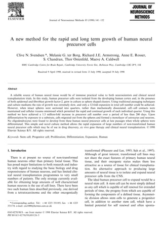
  • 5. C.N. S6endsen et al. / Journal of Neuroscience Methods 85 (1998) 141–152 145 Fig. 1. Cells isolated from the developing cortex and grown in EGF and FGF-2 formed mainly spheres at early passages (A1) but some discs could also be seen at later passages when grown in FGF-2, possibly as a result of transient attachment to the culture dish (A2). (B), (C), (D) and (E) show four quarters from a single sphere (A1) 1 h after sectioning. (F) Cumulative growth curves for Clon-5382 and BRC-44 based on expansion data in Table 2 and subsequent results not shown in Table 2. Note the steady and consistent exponential increase in total sphere number over time. Scale bar = 0.2 mm. Between 14 and 21 days of growth the spheres could be showed only modest expansion over this 14 day period gently dissociated to a mixed suspension of single cells as shown in Table 1. By 40 days of growth using the and sphere remnants before re-plating into growth sectioning method, some disc shaped clusters could be medium. Using this technique we have previously re- seen in addition to spheres (Fig. 1A1 and A2). The ported a 3-4-fold increase in cell number over the first discs mainly developed due to temporary attachment of few weeks in vitro, after which the absolute number of the newly sectioned spheres to the surface of the flask, cells harvested at each passage declined (Svendsen et and were often concave on one surface. This led to al., 1997b). In this study, seven separate experiments some disc/sphere aggregates which appeared hollow using either brain stem, spinal cord or cortical human when sectioned (approximately 30% at 100 days of fetal tissue showed similar results, with variable growth growth, data not shown). When individual spheres at for the first 4–6 weeks, after which their was very little 100 days growth were dissociated and the number of further growth with the maximum expansion of total cells counted, those with a size of between 0.35 and 0.45 cells only reaching 12-fold (Table 1). A variety of mm diameter contained an average of 61166+ 3498 modifications to the culture medium, growth factor viable cells (n= 25 spheres from CTX-44) with less than combinations and passaging strategies have been at- 5% non viable cells. Very similar results with regard to tempted. None of these had any effect on the slow cell number and sphere size were found at 50 and 150 growth rate and eventual senescence of these precur- days of growth (data not shown). There was a positive sors, although some cultures could be kept in a mitotic, correlation between sphere size and number of cells but non expanding, state (due to concomitant cell (Fig. 2A), although some variation existed due to the death) for up to 6 months (data not shown). technical difficulties of dissociating small single spheres A novel, non traumatic, passaging approach was (cells attaching to the side of the pipette, incomplete employed on several cultures. Data for two of these dissociation). We next assessed whether the size of the isolated from the cortex, which differed in both fetal sphere sections influenced their ability to grow back to age and method of cell isolation, are described in detail the size of the mother sphere. Regardless of the mother here. Instead of mechanical dissociation, individual sphere size, all quarters grew back to the same size in spheres were sectioned into quarters under a dissecting relation to the mother showing that the expansion is microscope using a scalpel blade (Fig. 1). The resulting not dependent on either large or small sphere sizes (Fig. 2B). [3H]Thymidine added to the spheres over the last four quarters were placed into fresh growth medium containing FGF-2 and over the next 24 h rounded to 24 h of growth at all stages in culture showed that there form new spheres which grew close to the size of the was a significant amount of uptake ( 5000 counts per mother sphere by 14 days. Using this method there was min/per sphere at each passage) indicating that active a steady and exponential increase in total sphere num- cell division was occurring. The total amount of expan- ber which did not decrease with time (Fig. 1; Table 2). sion achieved over the growth period (including a 5- Parallel cultures which were also switched to FGF-2 fold increase prior to the start of the sectioning alone but were dissociated rather than chopped, method) was greater than 1.5 million-fold for Clon-
  • 6. C.N. S6endsen et al. / Journal of Neuroscience Methods 85 (1998) 141–152 146 Fig. 2. Graphs showing (A) correlation between volume of sphere and number of cells per sphere. (B) Lack of any relationship between size of sectioned quarter and its subsequent growth relative to its own mother sphere. migrating out. Between 2 and 7 days, radiating pro- 5382 and both cultures showed a population doubling cesses often developed which sketched from the edge of time of approximately 4 days. Metaphase spreads the sphere onto the substrate (Fig. 3C). Along these showed that at 150 days of growth the human cells radiating strands, immature TuJ1 + neurons could be remained karyotypically normal with regard to chro- seen which often trailed processes in parallel lines and mosome number and appearance. Removal of FGF-2 may have been migrating out from the core of the from the growth medium at any stage resulted in sphere (Fig. 3D). Using confocal imaging the exact senescence and eventual death of cultures over a period anatomy of the sphere following 14 days of differentia- of 14 days. However, switching FGF-2 responsive tion could be seen and radial processes emanating from spheres to a medium containing EGF resulted in the the core were found to be GFAP positive (Fig. 4). growth of an EGF responsive spheres with very similar Migrating cells eventually formed a monolayer characteristics following plating and which also ex- around the plated sphere and labelled for either TuJ1 panded exponentially using this sectioning method. or GFAP but never with both markers at 14 days of However, the EGF responsive spheres were less prone differentiation (Fig. 5A, B and C). Detailed cell counts to attaching to the culture dish and subsequently within the monolayer around the sphere revealed that formed less discs. We are also systematically assessing the majority of the cells were either TuJ1 or GFAP the effects of sectioning on the exact growth rates of positive at both early and late passages and that no neural precursor cells isolated from other brain regions galactocerbroside (GAL-C) positive oligodendrocytes mentioned in Table 1, although bulk cultures spinal could be seen under these plating conditions (Fig. 5D). cord and brain stem do expand rapidly using this Although not analysed in detail, every whole sphere method (data not shown). plated (over 300) gave rise to both neurons and astro- cytes, strongly suggesting the presence of a common 3.2. Differentiation of human neural precursors precursor. At the very periphery of the migrating cells, lone GFAP+ astrocytes could often be found, onto Continual growth of the human neural spheres sug- which a number neurons had selectively migrated, the gested self renewal was occurring, but did not deter- processes from which were entirely confined to the mine the phenotypic potential of these cells. To assess astrocyte surface (Fig. 6A–C). This suggests that these this, whole spheres or differentiating spheres (at 100 precursor cell derived astrocytes provide an attractive days growth) were processed for indirect immunocyto- surface for the migrating neurons. Some cells also chemistry. The majority of cells (95%) within the expressed markers only found in mature neurons such growing spheres were found to be positive for nestin, as microtuble associated protein 2ab which is located (Fig. 3B) a marker for undifferentiated neuroepithelial mainly in dendrites (MAP-2ab; Fig. 6D) (Riederer and stem cells (Lendahl et al., 1990), but did not stain for Matus, 1985). Interestingly, following trituration to a TuJ1 (a specific early neuronal marker) (Menezes and single cell suspension and plating, very few TuJ1 + Luskin, 1997) or glial acidic fibriallary protein (GFAP; neurons were found while the majority of cells stained an astrocyte marker). When whole spheres were ex- for GFAP (data not shown), indicating that either the posed to a substrate, they rapidly attached and within temporal sequence of events following plating of whole hours cells with a glial morphology could be seen spheres, or the lack of physical trauma caused by trituration, may be required for neuronal survival and
  • 7. C.N. S6endsen et al. / Journal of Neuroscience Methods 85 (1998) 141–152 147 Fig. 3. Staining whole growing spheres at 100 days growth for Hoescht revealed that viable cell nuclei could be seen throughout in most cases (A). Scale bar: 100 vm. Immunocytochemical staining showed that the majority of cells within the growing spheres were nestin + and undifferentiated (B). Scale bar: 25 vm. Some spheres were plated onto poly-L-lysine coated coverslips in DMEM/B27 and 1% serum for 24 h to induce differentiation. Phase (C) and TuJ1 (D) staining of the same field from a sphere 4 days following plating. Note the radial fibre outgrowths along which TuJl + cells with extensive fibres could be seen. Arrow heads represent glial fibres with no TuJl + fibres, arrows represent TuJl + fibres along processes, double arrowhead shows single neuronal cell body with extended TuJl + processes. Scale bar: 25 vm. differentiation of cells arising from long term human 4. Discussion neural precursor cell cultures. The present study has demonstrated a new method 3.3. Automation of the sectioning technique for the long-term exponential expansion of non immor- talised or transformed human neural precursor cells, Through the detailed assessment of manually section- which maintained the capacity to generate a high per- ing individual spheres we have shown that sustained centage of neurons (see Fig. 7 for a schematic of the exponential growth of human neural precursor cells can technique). be achieved (Table 2). However, it is obviously not There are two main methods in the literature com- practical to section large numbers of spheres using this monly used to generate populations of neural precursor manual method. We therefore developed an automated cells. The first uses FGF-2 and a substrate to expand procedure using the McIlwain tissue chopper originally colonies of cells which grow attached to the culture designed to slice fresh brain tissue. Using this device up flask while the second uses EGF to expand aggregates to 1000 spheres can be automatically sectioned within a of cells (neurospheres) although it is now clear that few minutes. Each section rapidly rounds up after these growth factors are often interchangeable in their seeding into fresh medium and forms a new growing effects (for recent review, see Svendsen (1997)). We sphere as described previously using the manual tech- have not attempted to grow human cells attached to a nique, although obviously the spheres differ in size substrate in the presence of FGF-2 in this study, but depending on exactly where they were sectioned. As have focused instead on the neurosphere culture sphere size is not a critical determinant of cell expan- method. The advantage of the aggregate culture sion (see Fig. 2) we feel that this method should provide method is that large numbers of cells can be expanded an automated technique for growing large numbers of in a small volume of medium. There are no published human neural precursor cells. reports on the long term growth of human neural
  • 8. C.N. S6endsen et al. / Journal of Neuroscience Methods 85 (1998) 141–152 148 Fig. 4. Confocal image (Biorad Inc, UK) of a sphere plated for 14 days showing both TuJ1 positive neurons (red) and GFAP positive astrocytes (green) which had migrated from the core of a sphere (left) and formed a monolayer culture. Note the many radial fibres which stain for GFAP (arrowheads). Scale bar: 100 vm. Insert is a high power view of a plated region around the sphere ( × 4 magnification compared to rest of plate) showing how neuronal processes pass both under and over the astrocytes. precursor cells, although preliminary data suggests that published observations). Furthermore, we have found EGF can drive a human precursor for extended periods the expansion of human neurospheres to be very slow, of time (Vescovi et al., 1997). This cell may be similar and although division continued (based on incorpora- to the EGF responsive cell isolated from the developing tion of mitotic labels) real expansion stopped after 35 mouse striatum which can grown for long periods of days of growth (Svendsen et al., 1997a). Using the time in vitro as spheroid neurospheres and may repre- sectioning method presented here, human neurospheres sent a population of stem cells (Reynolds et al., 1992; continue to expand and give rise to high numbers of Reynolds and Weiss, 1996) although even as early as neurons even at late passages. We are currently identi- passage two (using conventional passaging methods) fying the phenotype of neurons generated from these these mouse neurospheres spontaneously gave rise to long term human cultures. virtually no neurons but rather glial cells (Arsenijevic What is the exact nature of the dividing cells within and Weiss, 1998). We have previously found that in the sectioned spheres?. Their growth rate was remark- contrast to mouse neurospheres, rat neurospheres enter ably stable and relatively slow, with a cell cycle time of a programmed senescence between 28 and 35 days of approximately 4 days throughout the culture period. growth using routine passaging methods (Svendsen et They were karyotypically normal on gross inspection al., 1997b) and also give rise to high numbers of which suggests they had not transformed but are main- astrocytes at later passages (Rosser and Svendsen, un- taining a normal cellular phenotype with a slow cell
  • 9. C.N. S6endsen et al. / Journal of Neuroscience Methods 85 (1998) 141–152 149 Fig. 5. Photomicrograph showing phase (A), TuJ1 (B) and GFAP (C) staining of an identical field of cells around the periphery of a sphere plated for 14 days. Many TuJI + and GFAP + cells could be found but no cells labelled with both markers. Arrows show neurons and arrowheads show astrocytes. Graph shows the numbers of labelled cells around the sphere as a percentage of total cells (n =12 spheres at each time) at either 50 or 150 days of growth (D). There was no significant difference between the numbers of neurons and astrocytes generated at early or late passages. cycle time consistent with a slowly dividing stem cell passages, but only neurons and astrocytes at late pas- population. Clonal analysis has classically been re- sages using the plating conditions in this study. To quired to prove pluri-potency, but is difficult to per- investigate this further we are currently assessing the form with this culture system since we have shown that effects of other growth factors and substrates on the groups of cells must maintain contact in order to grow differentiation of these long term human precursor for long periods. However, the observation that every cells, which have previously been shown to influence the sphere we have plated produced both neurons and fate rat hippocampal precursors and immortalised hu- astrocytes, but never only one phenotype, argues man neural precursors (Joh et al., 1996; Sah et al., strongly in favour of a common self renewing stem cell. 1997). It was of interest that the cultures generated It also remains possible that two uni-potent stem cells from widely differing fetal ages (8 and 21 weeks) gave with similar division rates and the capacity to produce rise to similar numbers of neurons and astrocytes at either neurons or astrocytes are dividing alongside each late passages. This would further suggest that a com- other. Oligodendrocytes were never seen to arise from mon cell is being driven in these cultures following an the late passage sectioned spheres, but have been seen initial period of instability where progenitors with a to develop from early passage spheres (Murray and more limited mitotic life span are filtered out. In sum- Dubois-Dalcq, 1997; Svendsen et al., unpublished ob- mary, the cell which is dividing in these cultures is servations). This suggests that either, (i) an oligoden- maybe best described as a precursor cell until we know drocyte precursor may be capable of dividing for a more about its exact phenotypic potential under a certain period of time and then be lost from the cul- variety of circumstances. tures during the extended period of growth which then There is currently some confusion regarding the dif- only consists of cells capable of giving rise to neurons ferential effects of EGF and FGF-2 on neural precursor and astrocytes or, (ii) a common precursor is able to cells. Based on clonal analysis, EGF was only found to make oligodendrocytes, neurons and astrocytes at early stimulate a glial progenitor from the mouse cortex late
  • 10. C.N. S6endsen et al. / Journal of Neuroscience Methods 85 (1998) 141–152 150 Fig. 6. Photomicrographs showing cells which had migrated out from the sphere and established colonies at the limit of the migration wave. Isolated epitheloid glial cells could be seen with many smaller cells on top which conformed exactly to the shape of the underlying cell suggesting a strong attraction of neurons to the developing astrocytes (A). The smaller cells were TuJI + neurons (B) and the epitheloid cells were GFAP + astrocytes (C) (C is the same field as A and B). MAP-2ab was found to label the dentritic processes of more mature neurons arising from the spheres (D). Scale bar: 15 vm. in development and had no effect on earlier cortical increase the mitotic effects of FGF-2 on embryonic precursors, in contrast to FGF-2 which was able to precursor cells (Caldwell and Svendsen, 1998) and it induce the division of a multipotent cell at both early was used throughout the current study. Interestingly, and late developmental ages using the same model we found that changing the growing human precursors (Kilpatrik and Bartlett, 1995). Furthermore, EGF ap- from FGF-2 to EGF had no obvious effect on the pears to stimulate glial division in adult subventricular ability to generate neurons following plating, although zones and may in fact repress neuronal development in this is the subject of a more detailed comparison cur- vivo (Kuhn et al., 1997). We have recently shown that rently in preparation. Clearly there is much more exper- following priming with FGF-2, the same cell responds imental work required to resolve these issues. to both EGF and FGF-2 in primary E14 mouse striatal The reason why the sectioning method is so effective tissue (Ciccolini and Svendsen, 1998). Perhaps in early in maintaining stable and rapid growth may be in part and late adulthood there are more restricted precursors due to the fact that there is no disruption of cell–cell which respond separately to these factors, whereas dur- contact within the intact regions of the spheres in ing development a common precursor exists. However, contrast to standard process of mechanical dissociation. the mixture of in vivo and in vitro data across different Membrane associated factors are known to be impor- species and culture conditions makes it impossible to tant for the division of neural precursor cells (Temple draw conclusions at present. It is of interest that the and Davis, 1994) and a ‘niche’ hypothesis has been adult mouse subventricular zone has recently been proposed which suggests stem cells will only retain their shown to contain FGF-2 responsive cells (Gritti et al., pluripotency within an appropriate environment 1996) which appear almost identical to the EGF re- (Schofield, 1978), both of which may be sustained using sponsive cells described originally by Reynolds and this sectioning method. Equally important may be the Weiss (1992) who claimed that FGF-2 was unable to reduction in cellular trauma that results from sectioning stimulate division of the same cells. This discrepancy rather than dissociating intact spheres. It is clear that may be due to the fact that heparin was added to the partial dissociation may also lead to intact remnants medium with FGF-2 in the later study. We have re- remaining which have cell–cell contact. Indeed, our cently shown that this proteoglycan can significantly normal passaging methods often result in non-complete
  • 11. C.N. S6endsen et al. / Journal of Neuroscience Methods 85 (1998) 141–152 151 Fig. 7. Schematic summarising the overall method. Conventional passaging techniques resulted in slow growth or senescence. By sectioning growing spheres there was less trauma to the cells and the quarters grew close to the size of the mother over a period of 14 days. This pattern continued for extensive periods of time in culture and allowed the exponential growth of human neural precursor cells. dissociation. The major disadvantage of this is that provide a valuable source of normal human neural tissue for both testing novel neuroactive compounds in spheres are generally stripped of cells leaving only the vitro and clinical neural transplantation programmes cores. Many of the stripped cells die but the cores may which are currently dependent on the collection of fresh continue to grow. However, this does not result in such human fetal tissues (Bjorklund, 1993; Svendsen, 1997). a rapid growth rate as simply sectioning the spheres and is far more inconsistent as it is difficult to control exactly how much dissociation is performed in one Acknowledgements culture to the next. It is tempting to speculate that the stages of sphere We thank S.B. Dunnett for his continual support, attachment, formation of radial processes and apparent Biorad Inc. for preparation of the confocal images and neuronal migration may recapitulate the normal pro- Ziggy Zhang and Irena Sarel of Blowhittiker Inc. for cess of primate development where neuronal precursors providing human neurospheres. The authors would also divide within ventricular zones before migrating to the like to thank Dr Scott Whittemore for critically ap- pial surface along radial glia (Rakic, 1985). However, praising an early version of this manuscript. This re- this needs to be substantiated with further studies search was funded by a Wellcome Fellowship to CNS which are the focus of ongoing work. Genetic manipu- and by the MRC. lations to these dividing cells should be possible, as achieved previously with both human and rat neural precursors (Sabate et al., 1995; Svendsen et al., 1996), References to allow the expression of specific proteins following grafting, thereby facilitating ex vivo CNS gene therapy Arsenijevic Y, Weiss S. Insulin-like growth factor-I is a differentiation (Friedmann, 1994). The spheres can be frozen and factor for post-mitotic CNS stem cell-derived neuronal precursors: stored which facilitates shipping and banking of such distinct actions from those of brain-derived neurotrophic factor. J tissue. Finally, as these sectioned neurospheres produce Neurosci 1998;18:2118 – 28. consistently large numbers of neurons, they may Bjorklund A. Better cells for brain repair. Nature 1993;362:414–5.
  • 12. C.N. S6endsen et al. / Journal of Neuroscience Methods 85 (1998) 141–152 152 Pleasure SJ, Lee VM-Y. NTera 2 cells: a human cell line which Buc-Caron MH. Neuroepithelial progenitor cells explanted from displays characteristics expected of a human committed neuronal human fetal brain proliferate and differentiate in vitro. Neurobiol progenitor cell. J Neurosci Res 1993;35:585 – 602. Dis 1995;2:37 – 47. Quinn SM, Walters WM, McKay RDG, Onifer SM, Whittemore SR. Caldwell MA, Svendsen CN. Potentiation of FGF-2 induced prolifer- Neuroepithelial precursor cells from fetal human spinal cord. Soc ation of mesencephalic neural precursors by heparin. Exp Neurol Neurosci Abstr 1997;23:320. 1998;in press. Rakic P. Limits of neurogenesis in primates. Science 1985;227:154–6. Chalmers-Redman RME, Priestley T, Kemp JA, Fine A. In vitro Reynolds BA, Weiss S. Generation of neurons and astrocytes from propagation and inducible differentiation of multipotential pro- isolated cells of the adult mammalian central nervous system. genitor cells from human fetal brain. Neuroscience 1997;76:1121 – Science 1992;255:1707– 10. 8. Reynolds BA, Weiss S. Clonal and population analyses demonstrate Ciccolini F, Svendsen CN. FGF-2 promotes acquisition of EGF that an EGF-responsive mammalian embryonic CNS precursor is responsiveness in mouse striatal precursor cells: Identification of a stem cell. Dev Biol 1996;175:1 – 13. neural precursor cells responding to both FGF-2 and EGF. J Reynolds BA, Tetzlaff W, Weiss S. A multipotent EGF-responsive Neurosci 1998;in press. striatal embryonic progenitor cell produces neurons and astro- Friedmann T. Gene therapy for neurological disorders. Trends Gene cytes. J Neurosci 1992;12:4565 – 74. Sci 1994;10:210 – 4. Riederer B, Matus A. Differential expression of distinct microtubule- Gage FH, Ray J, Fisher LJ. Isolation, characterisation, and use of associated proteins during brain development. Proc Natl Acad Sci stem cells from the CNS. In: Cowan WM, Shooter EM, Stevens USA 1985;82:6006 – 9. CF, Thompson RF, editors. Annual Review of Neuroscience, Sabate O, Horellou P, Vigne E, Colin P, Perricaudet M, Buc-Caron Palo Alto: Annual Reviews, 1995;18:159–92. M, Mallet J. Transplantation to the rat brain of human neural Gritti A, Parati EA, Cova L, Frolichsthal P, Galli P, Wanke E, progenitors that were genetically modified using adenoviruses. Faravelli L, Morassutti D, Roisen F, Nickel DD, Vescovi AL. Nat Genet 1995;9:256 – 60. Multipotential stem cells from the adult mouse brain proliferate Sah DWY, Ray J, Gage FH. Bi-potent progenitor cell lines from the and self-renew in response to basic fibroblast growth factor. J human CNS. Nat Biotechnol 1997;15:574 – 80. Neurosci 1996;16:1091 – 100. Schofield R. The relationship between the spleen colony forming cell and the haemopoietic stem cell. Blood Cells 1978;4:7 – 25. Hall PA, Watt FM. Stem cells: the generation and maintenance of Svendsen CN. Neural stem cells for brain repair. Alzheimer’s Res cellular diversity. Development 1989;106:619–33. 1997;3:131 – 5. Joh K, Hazel T, Muller T, Dugich-Djordjevic M, McKay RDG. Svendsen CN, Fawcett J, Bentlage C, Dunnett SB. Increased survival Single factors direct the differentiation of stem cells from the fetal of rat EGF-generated CNS progenitor cells using B27 supple- and adult central nervous system. Genes Dev 1996;10:3129–40. mented medium. Exp Brain Res 1995;102:407 – 14. Kilpatrik TJ, Bartlett PF. Cloned multipotential precursors from the Svendsen CN, Clarke DJ, Rosser AE, Dunnett SB. Survival and mouse cerebrum require FGF-2, whereas glial restricted precur- differentiation of rat and human epidermal growth factor-respon- sors are stimulated with either FGF-2 or EGF. J Neurosci sive precursor cells following grafting into the lesioned adult 1995;15:3653 – 61. central nervous system. Exp Neurol 1996;137:376 – 88. Kuhn HG, Winkler J, Kempermann G, Thal LJ, Gage FH. Epider- Svendsen CN, Caldwell MA, Shen J, ter Borg MG, Rosser AE, Tyers mal growth factor and fibroblast growth factor-2 have different P, Karmiol S, Dunnett SB. Long term survival of human central effects on neural progenitors in the adult rat brain. J Neurosci nervous system progenitor cells transplanted into a rat model of 1997;17:5820 – 9. Parkinson’s disease. Exp Neurol 1997a;148:135– 46. Lendahl U, Zimmerman LB, McKay RDG. CNS stem cells express a Svendsen CN, Skepper JN, Rosser AE, ter Borg M, Tyers P, Ryken new class of intermediate filament protein. Cell 1990;60:585–95. T. Restricted growth potential of rat striatal precursors as com- McKay RDG. Stem cells in the central nervous system. Science pared to mouse. Dev Brain Res 1997b;99:253 – 8. 1997;276:66 – 71. Temple S, Davis AD. Isolated rat cortical progenitor cells are main- Menezes JRL, Luskin MB. Expression of neuron-specific tubulin tained in division in vitro by membrane-associated factors. Devel- defines a novel population in the profliferative layers of the opment 1994;120:999 – 1008. developing telencephalon. J Neurosci 1997;14:5399–416. Vescovi AL, Daadi M, Asham R, Reynolds BA. Continuous genera- Murray K, Dubois-Dalcq M. Emergence of oligodendrocytes from tion of human catecholoaminergic neurons by embryonic CNS human neural spheres. J Neurosci Res 1997;50:146–56. stem cells. Soc Neurosci Abstr 1997;23:319. Palmer TD, Takahashi J, Gage FH. The adult rat hippocampus Weiss S, Reynolds BA, Vescovi AL, Morshead CM, Craig CG, van contains primordial neural stem cells. Mol Cell Neurosci der Kooy D. Is there a neural stem cell in the mammalian 1997;8:389 – 404. forebrain. Trends Neurosci 1996;19:387 – 93. . .