Minarcik robbins 2013_ch23-breast
•Transferir como PPT, PDF•
7 gostaram•1,758 visualizações


Recomendados
Mais conteúdo relacionado
Mais procurados
Mais procurados (18)
Semelhante a Minarcik robbins 2013_ch23-breast
Semelhante a Minarcik robbins 2013_ch23-breast (20)
Mais de Elsa von Licy
Mais de Elsa von Licy (20)
Último
Último (20)
Minarcik robbins 2013_ch23-breast
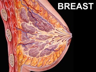
- 1. BREAST
- 4. HISTOLOGY • LOBE: (10 in whole breast) • LOBULE: (many per lobe) • ACINUS/I, aka ALVEOLUS/I: (many per lobule) • DUCT(S): INTRA- or INTERLOB(UL)AR, leading to the lactiferous ducts in the nipple
- 5. L O B E
- 6. LOBULE
- 9. THREE NORMAL PHASES • ACTIVE: about 50-50 Gland/Stroma ratio • LACTATING: Mostly Glands (like thyroid!!!), >>>50/50 • ATROPHIC: mostly stroma, <<<50/50
- 13. QUIZ ???
- 14. The most important thing to understand breast pathology is to get a solid IMAGE of the “NORMAL” breast lobule----ACINI, STROMA, BOUNDARIES
- 15. BREAST PATHOLOGY • DEVELOPMENTAL: • DEGENERATION: • INFLAMMATION: •NEOPLASM:
- 16. DEVELOPMENTAL • MILKLINE REMNANTS • ACCESSORY (axillary) BREAST TISSUE • NIPPLE INVERSION (fibrosis) • MACROMASTIA
- 20. 1) CONGENITAL 2) ACQUIRED: CARCINOMA 3) ACQUIRED: PIERCING
- 24. INFLAMMATION • ACUTE, staph most common • PERIDUCTAL • DUCT-ECTASIA • FAT NECROSIS, usually trauma • LYMPHOCYTIC, i.e., diabetic • GRANULOMATOUS, sarcoid, TB, etc., but mostly idiopathic
- 25. ACUTE MASTITIS
- 32. DUCTESIA
- 33. Ductesia CYSTS
- 35. FAT NECROSIS
- 36. FAT NECROSIS
- 39. NEOPLASIA • Benign epithelial • Benign stromal • Premalignant • Malignant epithelial (ductal, lobular) (adenocarcinomas) (insitu, infiltrating) • Malignant stromal
- 40. CLINICAL PRESENTATIONS •MASS , palpable or mammographic • NIPPLE DISCHARGE • PAIN
- 41. NEOPLASIA • BENIGN EPITHELIAL , aka, “FIBROCYSTIC” disease –NON-proliferative epithelium: i.e., cysts, fibrosis, adenosis –PROLIFERATIVE epithelium: hyperplasia, sclerosing adenosis, papilloma, fibroadenoma –ATYPICAL epithelium
- 42. CYST
- 46. FIBROSIS + CYSTS = FIBROCYSTIC DISEASE
- 47. NEOPLASIA • BENIGN EPITHELIAL , aka, “FIBROCYSTIC” disease –NON-proliferative epithelium: i.e., cysts, fibrosis, adenosis –PROLIFERATIVE epithelium: hyperplasia, sclerosing adenosis, papilloma, fibroadenoma –ATYPICAL epithelium
- 50. “COMPLEX” SCLEROSING ADENOSIS (RADIAL SCAR)
- 52. FIBROADENOMA: 1) EXTREMELY WELL DEFINED 2) YOUNGER WOMEN 3) ALWAYS BENIGN 4) CAN FIBROSE OR CALCIFY WITH AGE
- 53. PAPILLOMA
- 54. PAPILLOMA
- 55. PAPILLOMA
- 56. NEOPLASIA • BENIGN EPITHELIAL , aka, “FIBROCYSTIC” disease –NON-proliferative epithelium: i.e., cysts, fibrosis, adenosis –PROLIFERATIVE epithelium: hyperplasia, sclerosing adenosis, papilloma, fibroadenoma –ATYPICAL epithelium
- 57. FEATURES OF “ATYPIA” • • • • • • • • LOSS OF STROMA BETWEEN ACINI “SWISS CHEESE” HYPERPLASIA* CRIBRIFORMING** CELLULAR PLEOMORPHISM CELLULAR HYPERCHROMASIA INCREASED/ABNORMAL MITOSES* “ROMAN” BRIDGES*** NECROSIS*** (“COMEDO-carcinoma”)
- 59. DCIS
- 60. DCIS
- 61. DCIS
- 66. NORMAL lobule
- 69. LCIS • Usually hangs around MANY MANY years before it infiltrates, in contrast to DCIS • The BEST management may be judicious neglect, i.e., observation • If it does infiltrate, however, it is at least as bad as DCIS infiltrating, or probably WORSE, showing “indian” files
- 70. • • • • • • • • • • • • • • • • BREAST CANCER RISK FACTORS Age Menarche Age, early menarche is a risk First Live Birth First-Degree Relatives with Breast Cancer Breast Biopsies Race (caucasian the highest) Estrogen Exposure, prolonged, early menarche, late menopause Radiation Exposure Carcinoma of the contralateral breast or endometrium Geographic Influence Diet (high fat diet is riskiest) Obesity Exercise Lack of breast feeding is a risk, Lack of prior pregnancy is a risk. Environmental Toxins Tobacco • ABORTIONS?
- 71. BREAST CANCER PROGNOSTIC FACTORS • STAGING, especially POS or NEG lymph nodes, TNM, etc. • AGE • GENERAL HEALTH and IMMUNITY • Histologic degree of differentiation, i.e., GRADING • ERA/(PRA) • Her2, aka Her2-Neu
- 72. STAGING, TNM, based on biologic behavior • IN-SITU • EARLY disruption of the basal lamina, i.e., basement membrane • STROMAL infiltration • LYMPHATIC vessels • SENTINAL lymph node metastasis • MORE lymph node metastases • Adjacent structures, skin, ie, “inflammatory” • DISTANT, METASTASES, LIVER, BONE, LUNGS, BRAIN, EVERYWHERE
- 73. Total Cancers Per Cent In Situ Carcinoma 15–30 Ductal carcinoma in situ, DCIS 80 Lobular carcinoma in situ, LCIS 20 Invasive Carcinoma 70–85 No special type carcinoma ("ductal") 79 Lobular carcinoma 10 Tubular/cribriform carcinoma (Better prognosis than average) 6 Mucinous (colloid) carcinoma (Better prognosis than average) 2 Medullary carcinoma (Better prognosis than average) 2 Papillary carcinoma 1 Metaplastic carcinoma, (Squamous)
- 74. HISTOLOGIC TIDBITS • INFILTRATING DUCTAL • INFILTRATING LOBULAR (INDIAN FILE) • TUBULAR (LOOKS LIKE SCLEROSIS, BUT NO BASEMENT MEMBRANE) • MUCINOUS (COLLOID) • MEDULLARY (LOTS of LYMPHOCYTES)
- 76. INFILTRATING LOBULAR CA., “INDIAN” FILE PATTERN
- 78. INFILTRATING DUCTAL CA., “TUBULAR” PATTERN or TYPE
- 79. INFILTRATING DUCTAL CA., MUCINOUS (COLLOID) PATTERN or TYPE
- 81. NEOPLASIA, STROMAL Cysto-”SARCOMA” PHYLLODES (aka, PHYLLODES TUMOR), Looks like a giant fibroadenoma, really NOT a sarcoma SARCOMAS, true, are RARE!!!!
- 82. FIBROADENOMA
- 83. MALE BREAST • GYNECOMASTIA (related to hyperestrogenism) • CARCINOMA (1% of ♀ )
Notas do Editor
- Know the 2 major arteries (lateral and internal thoracic) and three lymph node groups which supply the breast.
- Know the 2 major arteries (lateral and internal thoracic) and three lymph node groups (axillary, internal thoracic (mammary) and supraclavicular) which supply the breast.
- Confusion between lobe, lobule, acini, alveolus, and duct is rampant in clinical medicine, but should never be confusing for you.
- Ther are an average of about 10 LOBES per breast. The suspensory ligament separates lobes.
- A lobule is part of a lobe composed of many acini. Lobules are separated from each other by bands of connective tissue.
- Acini are also known as alveoli.
- Acini are composed of glandular cells and myoepithelial cells.
- Active
- Pregnancy/Lactation
- Atrophic, i.e., post menopausal
- Breast tissue that is ~90% glandular and/or looks like “thyoid”, i.e., filled with milk, are lactating breasts
- Nipple lines extend from the axilla to the pubic regions, these are also called milklines.
- Go home and see if you hav any “moles” on your milk lines, and if you do, they may be accessory nipples.
- Breast tissue ALWAYS extends to the axilla, and when it does form an actual protuberance, it can be called an accessory breast. Breasts are modified apocrine sweat glands embryologically.
- Nipple retraction can be congenital or acquired, when acquired, it represents suspicion for underlying fibrosis due to neoplasm or inflammation.
- Macromastia.
- Atrophy is a NORMAL feature of postmenopausal breasts (estrogen withdrawal).
- Stroma>>>>>glands in atrophy, but lobules and acini are still present architecturally. Most carcinomas occur in atrophic (post menopausal,estrogen withdrawal) breasts
- All 4 of the classical signs of inflammation, heat redness, swelling, pain. What the the fifth?
- Intraductal and periductal inflammatory cells, mostly neutrophils in acute mastitis.
- Pap smear of nipple exudate in acute mastitis. What are most of these cells?
- Inflammatory carcinoma with its classic peau d’orange appearance.
- Note the tiny little “pits” in the orange peel.
- The tumor cells are INSIDE the skin dermal lymphatic spaces.
- Most of the inflammation here is PERI- ductal rather than INTRA- ductal. Acute or chronic? Why? Ans: Lymphocytes.
- Ductesia means dilated ducts.
- Dilated ducts are the same as cysts.
- “apocrine” refers both to a METHOD of SECRETION (as opposed to merocrine and holocrine), as well as a TYPE of CELL
- Classic cheesy appearance of fat in fat necrosis. Fat necrosis is usually due to mechanical trauma, surgical or otherwise.
- Giant cells and hemosiderin are usually easily found in fat necrosis. You should have no trouble finding either here.
- What is the principal inflammatory cell here? Ans: Lymphocyte. Because of this, would you like to call it “chronic” mastitis? Be my guest.
- The appearance is 100% exemplary of the diagnosis.
- All possibilities: Benign and malignant, glandular (i.e., epithelial) and stromal, and “borderline”.
- “Fibrocystic disease” is the waste basket term for benign breast disease characterized by fibrosis, cysts, inflammation, and a host of other benign changes. Certain features such as hyperplasia and papillomatosis, put it in a somewhat higher risk category for future carcinoma.
- Breast cyst, filled with fluid, in the pathology lab.
- Breast cyst, filled with fluid, in the ultrasound lab.
- This image speaks for itself. Do you think there is some apocrine metaplasia here too? Ans: YES
- Adenosis is defined as an increased number of acini per lobule.
- Hence the name, “fibrocystic” disease.
- “Benign” hyperplasia is characterized by, NO necrosis, the presence of MYOEPITHELIAL cells, and NO ATYPIA. Find a classical myoepithelial cell on the left.
- Sclerosing adenosis is often confused with malignancy. Why? Ans: the “sclerosis” can be mistaken for desmoplasia.
- VERy very very scary, but 100% benign, lesion.
- Note the myoepithelial cell. The presence of myoepithelial cells, means, BENIGN!!!
- Like meningiomas, fibroadenomas have the consistency of superballs, and you always feel like you want to bounce them!
- Our old friend the papillopma, i.e., a fingerlike proliferation of epithelium, growing over a fibrovascular core.
- Number 1 commandment in pathology: NEVER diagnosis a malignant papilloma on a frozen section!!!!! NEVER.
- The asterisked items, are more suspicious than the non-asterisked items. Intraductal NECROSIS is the most suspicious feature of all.
- Note the INTRADUCTAL NECROSIS.
- Note the atypia, “swiss cheese” hyperplasia, and early necrosis.
- Note the extreme artypia.
- Microcalcifications, seen on mammograms, are often the result of necrotic intraductal crud which has calcified. Lets make this quite simple: NECROSIS in a hyperplastic duct is usually DCIS
- This type of calcification represents about a 20% chance of malignancy and should be biopsied. This device helps pathologists to sample the areas of greatest concern more heavily.
- The Romans built many nice bridges, but not in China.
- A whole lobule filled with monotono0us cells of the same type can be called LCIS, or lobular carcinoma in situ. Note the COMPLETE LACK of atypia and necrosis, but it’s still CA-in-situ because this is a LOBULE!
- A whole lobule filled with monotonous cells of the same type can be called LCIS, or lobular carcinoma in situ.
- Statistical associations, risk factors, “causes?”, initiators, promotors
- HER2 is a proto-oncogene located at the long arm of human chromosome 17(17q11.2-q12). Approximately 25-30 percent of breast cancers have an amplification of the HER2/neu gene or overexpression of its protein product. Overexpression of this receptor in breast cancer is associated with increased disease recurrence and worse prognosis. Because of its prognostic role as well as its ability to predict response to trastuzumab, breast tumors are routinely checked for overexpression of HER2/neu. Overexpression also occurs in other cancer such as ovarian cancer and stomach cancer.
- 90% of infiltrating breast carcinomas are simply called “Infiltrating Ductal Carcinoma” on the pathology report.
- Indian file, British or American origin?
- The “tubular” pattern is somewhat better in behavior.
- The mucinous variant is also somewhat better in behavior.
- The medullary variant (i.e., lots of immune calls or lymphocytes) is also somewhat better in behavior. If you want to think that the reason for this is because there are a lot of immune cells “fighting” the tumor cells, you might be right, but it also tends to occur in younger women, who have the advantage of a younger age.
- Note that no matter how big a male’s breasts may get, they should never form lobules, but just end as blunt ducts.