
Recomendados
Mais conteúdo relacionado
Mais procurados
Mais procurados (20)
Mais de eadvisor
Mais de eadvisor (12)
Último
Último (20)
Visual field testing in pediatrics
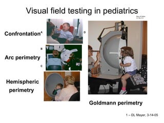
- 1. Visual field testing in pediatrics Confrontation Arc perimetry Hemispheric perimetry Goldmann perimetry 1 – DL Mayer, 3-14-05
- 2. The Count “eats” the white ball when the infant orients to it (looks at it) Confrontation with the Count and the white ball 2 – DL Mayer, 3-14-05
- 3. Arc perimetry Orienting to a flashing light on 4 oblique meridia, at 20 and 30 deg Child with CP – Detects lights in superior left 3 – DL Mayer, 3-14-05
- 4. Orienting to a flashing light at a fixed extent on one of 24 half meridia Hemispheric perimetry 4 – DL Mayer, 3-14-05
- 5. Goldmann perimetry Light moved from periphery to center of hemisphere Child signals detection of light with tap on buzzer Visual fields of a normal 4 year old Left eye Right eye 5 – DL Mayer, 3-14-05
- 6. 6 – DL Mayer, 3-14-05
- 7. Right and left visual field projection on visual pathway 7 – DL Mayer, 3-14-05
- 8. Child with markedly enlarged left ventricle 2a congenital hydrocephalus Right hemifield defect Confrontation 8– DL Mayer, 3-14-05
- 10. Child born at 35 wks gestation Bilateral PVL (white matter injury) Inferior field defect in each eye – dense, complete 10– DL Mayer, 3-14-05
- 12. Child later shown to have complete inferior field defect w/ Goldmann Missed w/ hemispheric perimetry (b/o head down & poor fixation) 12– DL Mayer, 3-14-05
- 15. Preterm infant – bilateral cerebral white matter damage MRI – 21 months 26 wks GA, 1 lb 15 oz, twin Spastic diplegia, seizures Mild ROP, X(T), VA sc OU 10/20 Complete, dense inferior field defect VFs-7 Goldmann perimetry, Age 5;8