

Recomendados
Mais conteúdo relacionado
Semelhante a Over view of nervous system
Semelhante a Over view of nervous system (20)
Mais de PS Deb
Mais de PS Deb (20)
Último
Último (20)
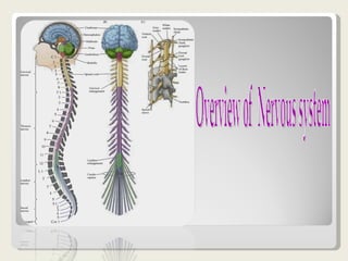
Over view of nervous system
- 1. Overview of Nervous system
- 2. Anatomical planes Anterior Posterior Lateral Medial
- 3. Nervous System Schema Brain Spinal Cord Nerves Gray Matter White matter Central Nuclei Brain Stem
- 5. Cerebrum
- 6. Thalamus
- 7. Hypothalamus
- 11. Brain Stem
- 12. Cranial Nerve
- 14. Spinal Cord
- 16. Brain Evolution
- 19. Neurons
- 20. Nerve Cell
- 21. Dendrite
- 22. Axon
- 23. Supporting cells
- 24. Propagation of nerve potential
- 25. Synapses
- 26. Types of Synapse
- 27. Functional Organization of NS
- 28. Neural network
- 29. Sensory Network
- 30. Motor Network
- 31. Motor System
- 33. Pain temperature and touch
- 34. Position sense
- 35. Stretch Reflex
- 36. Smell
- 37. Taste
- 38. Hearing
- 39. Vision
- 41. Sleeping
- 43. Memory
- 44. Intelligence
- 45. Thank You
Notas do Editor
- Overview Perhaps the major reason that neuroscience remains such an exciting field is the wealth of unanswered questions about the fundamental structure and functions of the human brain. To understand this remarkable organ (and the rest of the nervous system), the myriad cell types that constitute the nervous system must be identified, their interconnections traced, and the physiological role of the resulting circuits defined. Adding to these several challenges is the fact that a specialized anatomical vocabulary has arisen to describe the structure of the nervous system, as well as a specialized set of physiological terms to describe its functions. In light of these conceptual and semantic difficulties, comprehending the brain and the rest of the nervous system is greatly facilitated by a general picture of the organization of the nervous system, and by a review of the basic terms and anatomical conventions used in discussing its structure and function.
- A flexure in the long axis of the nervous system arose as humans evolved upright posture, leading to an approximately 120° angle between the long axis of the brainstem and that of the forebrain (A). The consequences of this flexure for anatomical terminology are indicated in (B). The terms anterior, posterior , superior , and inferior refer to the long axis of the body, which is straight. Therefore, these terms indicate the same direction for both the forebrain and the brainstem. In contrast, the terms dorsal , ventral , rostral , and caudal refer to the long axis of the central nervous system. The dorsal direction is toward the back for the brainstem and spinal cord, but toward the top of the head for the forebrain. The opposite direction is ventral. The rostral direction is toward the top of the head for the brainstem and spinal cord, but toward the face for the forebrain. The opposite direction is caudal. (C) The major planes of section used in cutting or imaging the brain. To understand the spatial organization of these systems, some additional vocabulary employed to describe them needs to be defined. The terms used to specify location in the central nervous system are the same as those used for the gross anatomical description of the rest of the body ( Figure 1.9 ). Thus, anterior and posterior indicate front and back; rostral and caudal, toward the head and tail; dorsal and ventral, top and bottom; and medial and lateral, the midline or to the side. Nevertheless, the comparison between these coordinates in the body versus the brain can be confusing. For the entire body these anatomical terms refer to the long axis, which is straight. The long axis of the central nervous system, however, has a bend in it. In human and other bipeds, a compensatory tilting of the rostral/caudal axis for the brain is necessary to properly compare body axes to brain axes. Once this adjustment has been made, the other axes for the brain can be easily assigned. The proper assignment of these anatomical axes then dictates the standard planes for histological sections or tomographic images used to study the internal anatomy of the brain (see Figure 1.9C) . Horizontal sections are taken parallel to the rostral/caudal axis of the brain. Sections taken in the plane dividing the two hemispheres are sagittal , and can be further categorized as median and paramedian according to whether the section is near the midline (median or midsagittal) or more lateral (paramedian). Sections in the plane of the face are called frontal or coronal. Different terms are usually used to refer to sections of the spinal cord. The plane of section orthogonal to the long axis of the cord is called transverse , whereas sections parallel to the long axis of the cord are called longitudinal . In a transverse section through the human spinal cord, the dorsal and ventral axes and the anterior and posterior axes indicate the same directions. Tedious though this terminology may be, it is essential for understanding the basic subdivisions of the nervous system.
- White Matter vs. Gray Matter Both the spinal cord and the brain consist of white matter = bundles of axons each coated with a sheath of myelin gray matter = masses of the cell bodies and dendrites — each covered with synapses . In the spinal cord, the white matter is at the surface, the gray matter inside. In the brain of mammals , this pattern is reversed. However, the brains of "lower" vertebrates like fishes and amphibians have their white matter on the outside of their brain as well as their spinal cord
- The subdivisions and components of the central nervous system. (A) A lateral view indicating the seven major components of the central nervous system. (Note that the position of the brackets on the left side of the figure refers to the vertebrae, not the spinal segments.) (B) The central nervous system in ventral view, indicating the emergence of the segmental nerves and the cervical and lumbar enlargements. (C) Diagram of several spinal cord segments, showing the relationship of the spinal cord to the bony canal in which it lies. The central nervous system (defined as the brain and spinal cord) is usually considered to have seven basic parts: the spinal cord, the medulla, the pons, the cerebellum, the midbrain, the diencephalon, and the cerebral hemispheres . The medulla, pons, and midbrain are collectively called the brainstem; the diencephalon and cerebral hemispheres are collectively called the forebrain. Within the brainstem are found cranial nerve nuclei that either receive input from cranial sensory ganglia via their respective cranial sensory nerves or give rise to axons that constitute cranial motor nerves . In addition, the brainstem is the conduit for several major tracts in the central nervous system. These tracts either relay sensory information from the spinal cord and brainstem to the midbrain and forebrain, or relay motor commands from the midbrain and forebrain back to motor neurons in the brainstem and spinal cord.
- Divisions of the Brain Generally, many body functions involve cells in several areas of the brain. However, certain areas of the brain tend to be more important in some functions while other areas dominate the control of other functions. Some major parts of the brain are listed below. Hindbrain : medulla oblongata, cerebellum, pons Midbrain Forebrain : thalamus, hypothalamus, cerebrum The human forebrain (prosencephalon) is made up of a pair of large cerebral hemispheres , called the telencephalon . Because of crossing over of the spinal tracts, the left hemisphere of the forebrain deals with the right side of the body and vice versa. a group of structures located deep within the cerebrum, that make up the diencephalon . Cerebrum The cerebrum became greatly enlarged as evolution progressed from fish to mammals. In reptiles, birds, and mammals, it receives sensory information and coordinates motor responses. Motor responses to the skeletal muscles originate in the cerebrum but are refined and coordinated by the cerebellum. In humans, the cerebrum is the largest part of the brain. Characteristics such as thinking, intelligence, and emotion are controlled here. Olfactory Bulbs - The anterior parts of the cerebral hemispheres are called the olfactory bulbs. It receives input from the olfactory nerves (smell). The olfactory bulbs of primitive vertebrates comprise a large proportion of the cerebrum. Cerebral Cortex - Over evolutionary time, gray matter developed over the cerebrum. This is the cerebral cortex and it is an information-processing center. It increased in size more rapidly than the skull so that it has become folded (convoluted) in order to fit in the skull. The human cerebral cortex is thin (1.5-4 mm thick) and is highly folded to increase its surface area. Intelligence, emotion, creativity, learning, and memory are localized in the cerebral cortex. Lobes of the cerebral cortex The cerebral cortex is divided into four lobes, each receives information from particular senses and processes the information into higher levels of consciousness. Lobe Function Frontal motor functions; permits conscious control of skeletal muscles; contains the primary motor cortex conscious thought Parietal sensory areas from the skin; contains the primary sensory cortex Occipital The primary visual cortex is located within the occipital lobe. Temporal hearing and smell Primary Sensory and Primary Motor Cortex - The primary sensory cortex is a narrow band of cortex tissue that extends from one side of the cortex near the ear over the top of the brain to the other side. Information from sensory receptors in the skin arrive at this area. The motor cortex is a band of cortex tissue directly anterior (in front) of the primary sensory cortex. Signals that control the skeletal muscles originate in this area. Corpus Callosum The corpus callosum contains neurons that cross from one side of the brain to the other, allowing each half to communicate with each other. The corpus callosum of people with severe epilepsy is sometimes cut to reduce the frequency and intensity of seizures. Researchers presented some of these people with words such as cowboy. When viewing this word, the first three letters (cow) are viewed in the left visual field of each eye and is projected onto the right half of the brain. These people could write the word cow with their left hand because their right brain controls the left side of the body and it is aware of the word cow but not boy. Their other hand could only write the word ''boy". Moreover, they could only say "boy" because language is controlled by the left hemisphere. Although they could see themselves write cow, they could only say boy.
- Thalamus the thalamus serves as a relay area to the cerebrum from other parts of the spinal cord and brain. For example, it receives sensory input (except smell) and sends to appropriate areas of the cerebral cortex. The cerebral cortex also sends information to the thalamus which then transmits this information to other areas of the brain and spinal cord. The Thalamus contains part of the reticular formation (see below).
- Hypothalamus Functions: Body Temperature, Emotions, Hunger, Thirst, Circadian Rhythms The hypothalamus is composed of several different areas and is located at the base of the brain. It is only the size of a pea (about 1/300 of the total brain weight), but it is responsible for some very important behaviors. One important function of the hypothalamus is the control of body temperature. The hypothalamus acts like a "thermostat" by sensing changes in body temperature and then sending out signals to adjust the temperature. For example, if you are too hot, the hypothalamus detects this and then sends out a signal to expand the capillaries in your skin. This causes blood to be cooled faster. The hypothalamus also controls the pituitary.
- Basal Ganglia Functions: Movement The basal ganglia are a group of structures, including the globus pallidus, caudate nucleus, subthalamic nucleus, putamen and substantia nigra, that are important in coordinating movement.
- Limbic System The limbic system contains neural pathways that connect portions of the cortex, thalamus, hypothalamus, and basal nuclei (several areas deep within the cerebrum). It causes pleasant or unpleasant feelings about experiences (rage, pain, pleasure, sorrow). This guides the individual into behavior that is likely to increase survival. The hippocampus is also important for memory
- Cerebellum The cerebellum coordinates and refines complex muscle movements. Movement information that is initiated in higher brain centers (the cerebral cortex) is compared to the actual position of the limbs. The cerebellum then adjusts and refines the movement. It is large in birds because flight requires considerable coordination. Functions: Movement, Balance, Posture The word "cerebellum" comes from the Latin word for "little brain." The cerebellum is located behind the brain stem. In some ways, the cerebellum is a bit like the cerebral cortex: the cerebellum is divided into hemispheres and has a cortex that surrounds these hemispheres.
- Brain stem Functions: Breathing, Heart Rate, Blood Pressure The brain stem is a general term for the area of the brain between the thalamus and spinal cord. Structures within the brain stem include the medulla, pons, tectum, reticular formation and tegmentum. Some of these areas are responsible for the most basic functions of life such as breathing, heart rate and blood pressure. Medulla oblongata The medulla looks like a swollen tip to the spinal cord. Nerve impulses arising here rhythmically stimulate the intercostal muscles and diaphragm — making breathing possible regulate heartbeat regulate the diameter of arterioles thus adjusting blood flow. The neurons controlling breathing have mu (µ) receptors , the receptors to which opiates , like heroin, bind. This accounts for the suppressive effect of opiates on breathing. Destruction of the medulla causes instant death. Pons The pons seems to serve as a relay station carrying signals from various parts of the cerebral cortex to the cerebellum. Nerve impulses coming from the eyes , ears , and touch receptors are sent on the cerebellum. The pons also participates in the reflexes that regulate breathing. The reticular formation is a region running through the middle of the hindbrain (and on into the midbrain). It receives sensory input (e.g., sound) from higher in the brain and passes these back up to the thalamus. The reticular formation is involved in sleep, arousal (and vomiting). The Midbrain The midbrain (mesencephalon) occupies only a small region in humans (it is relatively much larger in "lower" vertebrates). We shall look at only three features: the reticular formation : collects input from higher brain centers and passes it on to motor neurons. the substantia nigra : helps "smooth" out body movements; damage to the substantia nigra causes Parkinson's disease. the ventral tegmental area ( VTA ): packed with dopamine -releasing neurons that are activated by nicotinic acetylcholine receptors and whose projections synapse deep within the forebrain. The VTA seems to be involved in pleasure: nicotine, amphetamines and cocaine bind to and activate its dopamine-releasing neurons and this may account — at least in part — for their addictive qualities.
- Cranial Nerves and Spinal Nerves Humans have 12 pairs of cranial nerves and 31 pairs of spinal nerves . Cranial nerves are sensory, motor, or mixed, and all but the vagus are involved with the head and neck region; the vagus nerve manages the internal organs. Spinal nerves are all mixed nerves. Their regular arrangement reflects the segmentation of the human body. Spinal nerves are connected to the spinal cord by two branches called roots. The dorsal root contains sensory neurons . The dorsal root ganglion contains the cell bodies of sensory neurons. Sensory neurons therefore have long dendrites. The ventral root contains motor neurons. Motor neurons have short dendrites and long axons.
- The Ventricular System The cerebral ventricles are a series of interconnected, fluid-filled spaces that lie in the core of the forebrain and brainstem . The presence of ventricular spaces in the various subdivisions of the brain reflects the fact that the ventricles are the adult derivatives of the open space or lumen of the embryonic neural tube . Although they have no unique function, the ventricular spaces present in sections through the brain provide another useful guide to location. The largest of these spaces are the lateral ventricles (one within each of the cerebral hemispheres). These particular ventricles are best seen in frontal sections, where their ventral surface is usually defined by the basal ganglia, their dorsal surface by the corpus callosum, and their medial surface by the septum pellucidum , a membranous tissue sheet that forms part of the midline sagittal surface of the cerebral hemispheres. The third ventricle forms a narrow midline space between the right and left thalamus, and communicates with the lateral ventricles through a small opening at the anterior end of the third ventricle (called the interventricular foramen). The third ventricle is continuous caudally with the cerebral aqueduct , which runs though the midbrain. At its caudal end, the aqueduct opens into the fourth ventricle , a larger space in the dorsal pons and medulla. The fourth ventricle narrows caudally to form the central canal of the spinal cord. The ventricles are filled with cerebrospinal fluid , and the lateral, third, and fourth ventricles are the site of the choroid plexus , which produces this fluid. The cerebrospinal fluid percolates through the ventricular system and flows into the subarachnoid space through perforations in the thin covering of the fourth ventricle; it is eventually absorbed by specialized structures called arachnoid villi or granulations , and returned to the venous circulation. Figure 1.17. The ventricular system of the human brain. (A) Location of the ventricles as seen in a transparent left lateral view. (B) Table showing the ventricular spaces associated with each of the major subdivisions of the brain. (See Chapter 22 for an account of brain development that more fully explains the origin of the ventricular spaces.) Figure 1.18. The meninges. Upper left panel is a midsagittal view showing the three layers of the meninges in relation to the skull and brain. Right panels are blowups to show detail. The Meninges The cranial cavity is conventionally divided into three regions called the anterior, middle, and posterior cranial fossae. Surrounding and supporting the brain within this cavity are three protective tissue layers, which also extend down the brainstem and the spinal cord. Together these layers are called the meninges ( Figure 1.18 ). The outermost layer of the meninges is called the dura mater because it is thick and tough. The middle layer is called the arachnoid mater because of spiderlike processes called arachnoid trabiculae that extend from it toward the third layer, the pia mater , a thin, delicate layer of cells that closely invests the surface of the brain. Since the pia closely adheres to the brain as its surface curves and folds, whereas the arachnoid does not, there are placescalled cisterns where the subarachnoid space is especially large. The major arteries supplying the brain course through the subarachnoid space where they give rise to branches that penetrate the substance of the hemispheres. The subarachnoid space is therefore a frequent site of bleeding following trauma. A collection of blood between the meningeal layers is referred to as a subdural or subarachnoid hemorrhage, as distinct from bleeding within the brain itself.
- The Spinal Cord The vertebrae surround and protect the spinal cord. Cerebrospinal fluid within the central canal functions to cushion the spinal cord. Many sensory - motor reflex connections are in the spinal cord. Interneurons often lie between sensory and motor neurons . White matter White matter contains tracts that connect the brain and the spinal cord. The white color is due to the myelin sheaths. Gray matter Gray matter looks gray because it is unmyelinated. It contains the short interneurons that connect many sensory and motor neurons. Sensory neurons enter the gray matter and the axons of motor neurons leave it. The cell bodies of these motor neurons are located in the gray matter. 31 pairs of spinal nerves arise along the spinal cord. These are "mixed" nerves because each contain both sensory and motor axons. However, within the spinal column, all the sensory axons pass into the dorsal root ganglion where their cell bodies are located and then on into the spinal cord itself. all the motor axons pass into the ventral roots before uniting with the sensory axons to form the mixed nerves. The spinal cord carries out two main functions: It connects a large part of the peripheral nervous system to the brain. Information (nerve impulses) reaching the spinal cord through sensory neurons are transmitted up into the brain. Signals arising in the motor areas of the brain travel back down the cord and leave in the motor neurons. The spinal cord also acts as a minor coordinating center responsible for some simple reflexes like the withdrawal reflex . The interneurons carrying impulses to and from specific receptors and effectors are grouped together in spinal tracts .
- Nerves Nerves are bundles of neurons; either long dendrites and/or long axons. There are no cell bodies in nerves. The cell bodies are in the ganglia (PNS) or nuclei (in gray matter of the CNS). Most nerves contain both kinds of neurons (sensory and motor). The sensory neurons conduct information to the CNS, the motor neurons conduct away from the CNS. All of the neurons in some nerves conduct in the same direction. These nerves contain either sensory or motor neurons.
- Animal evolution has generated a wide range of species including single-celled animals and multicellular animals including invertebrates and vertebrates (left column, indicating time since common ancestor with humans). All of these animals show behavioural responses to their environment with vertebrates showing the most complex behaviours. Only the multicellular animals having anatomically specialised nerve cells forming their brains. The synapses that form the junctions between nerve cells are made of many proteins organised together into 'molecular signal processors' (middle column, Synapse protein complexity). In vertebrates and invertebrates, these proteins control psychological functions including learning and memory. Surprisingly, these synapse molecules exist in single-celled animals as a simple set of proteins (where they control response to environment), and this set was built upon to form a larger set used in the brains of invertebrates. This invertebrate set was expanded further in the brains of vertebrate species. The correlation between numbers of nerve cells in the brain of animals and the number of synaptic proteins shows that both contribute to the differences in species (right column).
- Cell Body In many ways, the cell body is similar to other types of cells. It has a nucleus with at least one nucleolus and contains many of the typical cytoplasmic organelles. It lacks centrioles, however. Because centrioles function in cell division, the fact that neurons lack these organelles is consistent with the amitotic nature of the cell. Dendrites Dendrites and axons are cytoplasmic extensions, or processes, that project from the cell body. They are sometimes referred to as fibers. Dendrites are usually, but not always, short and branching, which increases their surface area to receive signals from other neurons. The number of dendrites on a neuron varies. They are called afferent processes because they transmit impulses to the neuron cell body. There is only one axon that projects from each cell body. It is usually elongated and because it carries impulses away from the cell body, it is called an efferent process. Axon An axon may have infrequent branches called axon collaterals. Axons and axon collaterals terminate in many short branches or telodendria. The distal ends of the telodendria are slightly enlarged to form synaptic bulbs. Many axons are surrounded by a segmented, white, fatty substance called myelin or the myelin sheath. Myelinated fibers make up the white matter in the CNS, while cell bodies and unmyelinated fibers make the gray matter. The unmyelinated regions between the myelin segments are called the nodes of Ranvier.
- Cell Body In many ways, the cell body is similar to other types of cells. It has a nucleus with at least one nucleolus and contains many of the typical cytoplasmic organelles. It lacks centrioles, however. Because centrioles function in cell division, the fact that neurons lack these organelles is consistent with the amitotic nature of the cell. .
- Dendrites Dendrites and axons are cytoplasmic extensions, or processes, that project from the cell body. They are sometimes referred to as fibers. Dendrites are usually, but not always, short and branching, which increases their surface area to receive signals from other neurons. The number of dendrites on a neuron varies. They are called afferent processes because they transmit impulses to the neuron cell body. There is only one axon that projects from each cell body. It is usually elongated and because it carries impulses away from the cell body, it is called an efferent process.
- Axon An axon may have infrequent branches called axon collaterals. Axons and axon collaterals terminate in many short branches or telodendria. The distal ends of the telodendria are slightly enlarged to form synaptic bulbs. Many axons are surrounded by a segmented, white, fatty substance called myelin or the myelin sheath. Myelinated fibers make up the white matter in the CNS, while cell bodies and unmyelinated fibers make the gray matter. The unmyelinated regions between the myelin segments are called the nodes of Ranvier
- Saltatory action potential conduction along a myelinated axon. (A) Diagram of a myelinated axon. (B) Local current in response to action potential initiation at a particular site flows locally, as described in . However, the presence of myelin prevents the local current from leaking across the internodal membrane; it therefore flows farther along the axon than it would in the absence of myelin. Moreover, voltage-gated Na+ channels are present only at the nodes of Ranvier. This arrangement means that the generation of active, voltage-gated currents need only occur at these unmyelinated regions. The result is a greatly enhanced velocity of action potential conduction. Panel to the left of the figure legend shows the changing membrane potential as a function of time at the points indicated.
- Components Presynaptic terminal Synaptic cleft Postsynaptic membrane Neurotransmitters released by action potentials in presynaptic terminal Synaptic vesicles Diffusion Postsynaptic membrane Neurotransmitter removal When an impulse arrives at the end bulb , the end bulb membrane becomes more permeable to calcium . Calcium diffuses into the end bulb & activates enzymes that cause the synaptic vesicles to move toward the synaptic cleft. Some vesicles fuse with the membrane and release their neurotransmitter (a good example of exocytosis). The neurotransmitter molecules diffuse across the cleft and fit into receptor sites in the postsynaptic membrane. When these sites are filled, sodium channels open & permit an inward diffusion of sodium ions. This, of course, causes the membrane potential to become less negative (or, in other words, to approach the threshold potential). If enough neurotransmitter is released, and enough sodium channels are opened, then the membrane potential will reach threshold. If so, an action potential occurs and spreads along the membrane of the post-synaptic neuron (in other words, the impulse will be transmitted). Of course, if insufficient neurotransmitter is released, the impulse will not be transmitted. Impulse transmission - The nerve impulse (action potential) travels down the presynaptic axon towards the synapse, where it activates voltage-gated calcium channels leading to calcium influx, which triggers the simultaneous release of neurotransmitter molecules from many synaptic vesicles by fusing the membranes of the vesicles to that of the nerve terminal. The neurotransmitter molecules diffuse across the synaptic cleft, bind briefly to receptors on the postsynaptic neuron to activate them, causing physiological responses that may be excitatory or inhibitory depending on the receptor. The neurotransmitter molecules are then either quickly pumped back into the presynaptic nerve terminal via transporters, are destroyed by enzymes near the receptors (e.g. breakdown of acetylcholine by cholinesterase), or diffuse into the surrounding area.
- Types of neurotransmitters: 1- Excitatory - neurotransmitters that make membrane potential less negative (via increased permeability of the membrane to sodium) &, therefore, tend to 'excite' or stimulate the postsynaptic membrane 2 - Inhibitory - neurotransmitters that make membrane potential more negative (via increased permeability of the membrane to potassium) &, therefore, tend to 'inhibit' (or make less likely) the transmission of an impulse. One example of an inhibitory neurotransmitter is gamma aminobutyric acid (GABA; shown below). Medically, GABA has been used to treat both epilepsy and hypertension. Another example of an inhibitory neurotransmitter is beta-endorphin, which results in decreased pain perception by the CNS.
- Autonomic Nervous System This part of the nervous system sends signals to the heart, smooth muscle, glands, and all internal organs. It is generally without conscious control. The autonomic nervous system uses two or more motor neurons : The cell body of one of the motor neurons is in the CNS. The cell body of the other one is in a ganglion. Sympathetic The sympathetic nervous system prepares the body to deal with emergency situations. This is often called the "fight or flight" response. Stimulation from sympathetic nerves dilates the pupils, accelerates the heartbeat, increases the breathing rate, and inhibits the digestive tract. The neurotransmitter is norepinephrine (similar to epinephrine [adrenaline], the heart stimulant). Sympathetic nerves arise from the middle (thoracic-lumbar) portion of the spinal cord. Parasympathetic When there is little stress, the parasympathetic system tends to slow down the overall activity of the body. It causes the pupils to contract, it promotes digestion, and it slows the rate of heartbeat. The neurotransmitter is acetylcholine. The actual rate of stimulus to each organ is determined by the sum of opposing signals from the sympathetic and parasympathetic systems. Parasympathetic nerves arise from the brain and sacral (near the legs) portion of the cord.
- Somatic Nervous System The somatic nervous system provides conscious, voluntary control. It includes all of the nerves that serve the skeletal muscles and the exterior sense organs. Reflex arcs Reflexes are simple, stereotyped and repeatable motor actions (example: movements) brought about by a specific sensory stimulus. The reflex is involuntary but may involve the use of voluntary (skeletal) muscle and nerves. Reflexes are quick and produce behaviors that are typically beneficial. For example, when you fall, reflex arcs immediately act to extend your arm so that your arm prevents your head and body from hitting the ground. Some reflexes involve the brain, others do not. A whole series of responses may occur since some sensory neurons stimulate several interneurons which, in turn send impulses to other parts of the CNS. If you were to fall forward, interneurons would use information from the ears to determine the direction of the fall and extend the arms in a forward direction. If you were to fall toward the left side, interneurons would select neurons that activate muscles to extend your arm to the left side. Example: The stretch reflex The stretch reflex is involved in helping the body maintain its position without having to consciously think about it. When a muscle is stretched, stretch-sensitive receptors are stimulated. An action potential is conducted to the spinal cord. The axon terminals synapse with motor neurons leading right back to the muscles. This causes the muscle to contract to its original position.
- Reticular Formation The reticular formation is a net of nerve cells extending from the thalamus through the brain stem (midbrain, pons and medulla oblongata) to the spinal cord. It acts as a filter to incoming stimuli and discriminates important from unimportant. Hundreds of millions of sensory receptors flood the brain; the brain does not have the capacity to deal with even a small fraction of this information, so much of it must be ignored. Examples: You may be unaware of conversation in a crowded situation but the system alerts you when you hear your name. You can sleep in the presence of some kinds of sounds but others will wake you. The reticular activating system (RAS) is the part of the reticular formation that maintains wakefulness. Sleep centers are located in the reticular formation. Neurons in one sleep center secrete serotonin , a chemical that inhibits the RAS and thus causes drowsiness and sleep. Another sleep center secretes factors that counteract serotonin and bring about wakefulness. Damage to these centers can lead to unconsciousness or coma
- Memory The limbic system is involved in memory formation. The hippocampus , a structure that is deep in the cerebrum and a part of limbic system, is necessary to form new memories. People with a damaged hippocampus cannot remember things since the time the damage occurred but can remember from before. Short-term memory is probably stored as electrical differences because they can be removed by the application of an electrical shock. Long-term memory is probably stored as new or different synapses. Research on snails shows that learning is associated with an increased number of synapses. Forgetting is associated with a decreased number. Disuse can cause a synapse to wither and sever the connection between two neurons. Intensively stimulated synapses form stronger connections, grow, or sprout buds to form more connections. Memory appears to be stored in sensory areas of the cerebrum.