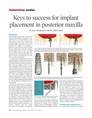
Implant placement in posterior maxilla by Dr. Ajay Singh
•
7 gostaram•1,722 visualizações

Implant placement in posterior maxilla. Dental implant therapy into the posterior maxilla has always been and continues to be a challenge due to various limitations in this region such as poor bone density, sinus pneumatization, lateral and vertical bone resorption, high occlusal forces and area of limited access. Further, if the implant is placed into poor density posterior maxilla, the bone which forms around the osseointegrated implants does not show very high bone to implant surface contact (BIC) percentage, thus in several cases the implant even after successful osseointegration may fail once it is restored in function.

Recomendados
Mais conteúdo relacionado
Mais procurados
Mais procurados (20)
Semelhante a Implant placement in posterior maxilla by Dr. Ajay Singh
Semelhante a Implant placement in posterior maxilla by Dr. Ajay Singh (20)
Último
Último (20)
Implant placement in posterior maxilla by Dr. Ajay Singh
- 1. Dental Practice // March-April 2013 // Vol 11 No 500 Keys to success for implant placement in posterior maxilla implantology section INTRODUCTION Dental implant therapy into the posterior maxilla has always been and continues to be a challenge due to various limitations in this region such as poor bone density, sinus pneumatization, lateral and vertical bone resorption, high occlusal forces and area of limited access. Further, if the implant is placed into poor density posterior maxilla, the bone which forms around the osseointe- grated implants does not show very high bone to implant surface contact (BIC) per- centage, thus in several cases the implant even after successful osseointegration may fail once it is restored in function. In past, several modifications to the con- ventional implant therapy have been done to make the implant successful into the posteri- or maxilla. Achieving the adequate initial sta- bility and bone implant contact are two major challenges into the posterior maxilla. Various protocols such as lateral bone aug- mentation using osteotomes, use of wide and long implant with deeper threads and high pitch value, use of implant with specific sur- face (HA coated implant), submerging implant platform apical to the ridge crest, bicortical stabilization of implant, and pro- gressive loading of implant have been applied to make the implant successful into the pos- terior maxilla. Besides the compromised bone density, sinus pneumatization is the great challenge in the posterior maxilla which results in limited bone height availability under the sinus. In such cases, maxillary sinus lifting and grafting has been providing promising results. The sinus-lift procedure was first performed by Dr. Hilt Tatum Jr. in 1974 during his period of preparation to begin sinus grafting. The first sinus graft was performed by Tatum in February, 1975 in Lee County Hospital in Opelika, Alabama. This was followed by the placement and successful restoration of two Endosteal implants. After this, suitable instruments were developed to manage the lining elevation from the different anatomical surfaces encountered in sinuses. Tatum first presented the concept at The Alabama Implant Congress in Birmingham, Alabama in 1976 and presented the evolution of tech- nique during multiple podium presentations each year until 1986 when he published an article describing the procedure. Dr. Philip Boyne was introduced to the procedure when he was invited, by Tatum, to be "The Discusser" of a presentation on sinus grafting given by Tatum at the annual meeting of The American Academy of Implant Dentistry in 1977. Boyne and James authored the first publication on the technique in 1980 when they published case reports of autogenous grafts placed into the sinus and allowed to heal for 6 months, which was followed by the placement of blade implants. LIMITATIONS WITH POSTERIOR MAXILLA 1. Poor bone quality (type IV/ D4) - challenge FIG 3 & 6: Dental implant with sharp and self tapping threads at the apical third and self condensing body can be placed after minimal drilling and achieves high initial stability even into poor density bone. FIG 7 & 8: Set of osteotomes which are used for lateral bone condensation and sub-crestal sinus elevation. FIG 1 & 2: After the osteotomy preparation for the implant 2mm short of sinus floor, the rest of the sinus floor is either grinded up using diamond tips/burs or fractured up using osteotome and the implant is placed by stabilis- ing its apex into the sinus floor and collar into the crest (Bicortical stabilization) DR. AJAY VIKRAM SINGH AND DR. SUNITA SINGH
- 2. Dental Practice // March-April 2013 // Vol 11 No 5 00 to achieve adequate initial stability of the implant 2. Limited bone height due to sinus pneuma- tisation and vertical bone resorption of ridge crest 3. Reduced bone width because of lateral resorption of posterior maxilla towards the hard palate, which also results in final pros- thesis with facial cantilevering 4. Area of less visibility and access 5. Proximity with sinus floor, posterior supe- rior artery etc. KEYS TO SUCCESSFUL IMPLANT THERAPY IN POSTERIOR MAXILLA 1. Longest and widest possible implant should be placed 2. Bicortical implant stabilization – implant platform is stabilized into high density cre- stal bone and its apex into high density sinus floor to achieve adequate initial implant stability. (Figures 1 & 2) 3. Using more number of implants for multi- ple unit prosthesis 4. The implant with sharp self tapping deeper threads should be preferred to achieve high primary stability in poor density trabecular bone (Figures 3 - 6). 5. Implants with faster osseointegrating sur- faces like Hydroxyapatite coated surface implants; SLA surface implants should be preferred. 6. Implant can be submerged 1.0 mm apical to the ridge crest to prevent its premature loading and micromovements during its healing phase. 7. Lateral bone condensation using special set of osteotomes to enhance the density of trabecular bone around the implant (Figures 7 & 8). 8. Longer submerged healing period for the implant. 9. Progressive bone loading to enhance the density of trabecular bone around the implant. 10. Sinus grafting to regenerate new bone into the sinus so that longer implant can be placed. (Figures 9 & 10). To approach the sinus for the Schneiderian membrane elevation and sinus floor grafting, Tatum advocated two approaches lateral as well as crestal. The lat- eral approach is usually preferred when sub antral bone is only 3-4 mm and a sinus membrane elevation is required to be per- formed to more than 4-5 mm. It should also be preferred when sinus grafting is per- formed for multiple number of implants. Sub crestal approach is less invasive and should be preferred in cases where 2-4 mm of sinus elevation is required to place an ade- quately long implant and to stabilize its apex into the high density sinus floor. The sub crestal approach of sinus elevation was first performed by Hilt Tatum in 1974 and pub- lished by Summer in 1994. ADVANTAGES OF THE CRESTAL APPROACH (SUMMER’S OSTEOTOME TECHNIQUE) FIG 18: Crestal bone is exposed using a soft tissue punch. FIG 19 & 20: Implant osteotomy is prepared in the usual fashion about 2.0 mm short of sinus floor. FIG 13 - 17: Various CT sections are showing only 8.0 mm sub-antral bone height and poor bone density. Thus the sinus lifting with sub-crestal approach and placement of 6 x 11.5 tapered implant is planned with CT. FIG 11 & 12: Missing maxillary molar (clinical view and radiograph)FIG 9 & 10: After the membrane has been lifted up to the desired height, sinus floor is grafted using autogenous bone or bone substitutes and implant is inserted.
- 3. Dental Practice // March-April 2013 // Vol 11 No 500 implantology section 1. Less invasive procedure. 2. Improves maxillary bone density, which allows greater initial stability of implants. 3. Less amount of grafting material is required to fill the lifted sinus membrane. 4. No barrier membrane is required 5. Limited flap elevation is required which maintain the blood supply to the lateral wall of the sinus. CASE REPORT A 48 year old female patient, medically fit for the implant therapy, reported for the replace- ment of missing tooth no.26. Radiographs and dental CT revealed the availability of only 8.0 mm bone under the maxillary sinus. A DentaScan also showed poor bone density at the implant site. To place an adequately long implant with adequate initial stability, a sinus elevation procedure with sub-crestal approach was planned. The ridge form was good and the marginal tissue was thick, stable and keratinised so the implant placement using tissue punch was planned. The osteoto- my for the implant was prepared in the usual fashion but 2.0mm short of the sinus floor. Further, an adequate size osteotome was used to fracture up the sinus floor and lift up the sinus membrane. The PRF plug which was prepared from the venous blood of the patient was introduced into the osteotomy to guard the sinus membrane against rupture. The bone substitute (HA + β-tcp) was intro- duced into the osteotome and pushed up using the osteotome which further elevated the sinus membrane to the desired height. The implant with deeper threads (6x11.5mm) was then placed and stabilized into the sinus floor. Because the implant is stabilised bicortically it achieved a primary stability of more than 35Ncm and the implant was left to heal with open protocol. The implant is restored in function after 4 month of open healing. The follow-up radi- ograph after one year showed stable crestal bone level and new bone regeneration into the grafted sinus. RESULT The author practiced conventional implant therapy in the posterior maxilla for years where he used hundreds of varying design root form implants in the posterior maxilla. The poor bone density, availability of limit- ed bone height (due to sinus pneumatiza- tion) and high force factors forced him to place the short length implant, submerging FIG 21 & 22: After the osteotomy has been prepared 2.0 mm short of sinus floor, an adequate size osteotome is used to fracture up the sinus floor and to lift up the Schneiderian membrane. FIG 27: The use of mechanical driver (torque ratchet) is showing that the implant has achieved primary sta- bility more than 35 Ncm because it has been stabilised bicortically FIG 28 & 29: Implant at the final position. Gingival former is inserted for transgingival healing of the implant FIG 23 & 24: The PRF (platelet-rich fibrin) is prepared from the patient’s venous blood which was withdrawn before surgery and centrifuged in a table top centrifuge machine. This fibrin is first inserted into the prepared osteotomy and pushed using osteotome to place it under the sinus membrane. It provides a protection to the membrane against rupture during its further elevation and grafting. It also enhances the bone regeneration potential of the graft material. FIG 25 & 26: The bone substitute (HA + β-Tcp) is now used to graft the elevated sinus floor and implant is inserted
- 4. it apical to ridge crest, uncovering the implant after more than 6 month submerged healing time, following the progressive load- ing protocols. This protocol required two surgical procedures, long healing time and longer time span to restore the implant. Further, the author faced a considerable number of implant failures with the conven- tional protocol because he was able to place only the short length implant in most cases. Moreover, to avoid the premature loading of the implant during its healing phase he was required to submerge implant sub crestal which often resulted in the loss of primary stability. Since the last couple of years, to avoid such problems and to maximize the success rate of implant in posterior maxilla, the Author switched over to long implants and stabilized them bicortically. Bicortical stabilization of the implant result in several advantages such as placement of the implant 4-5 mm longer than usual, high primary sta- bility of the implant, most of the implant are placed with non submerged protocol, shorter healing period for the implant (3-4 months), less implants are required to support multiu- nit prosthesis and no progressive loading is required in most cases. CONCLUSION Author placed more than 200 implants in the posterior maxilla in a period of 3 years where he stabilized the implant bicortically with or without sinus grafting. Most of the implants achieved primary stability more than 35 Ncm and hence placed with non submerged heal- ing protocol. Most implants restored in func- tion after the healing period of 3-4 months without following any progressive loading protocol. When the author compared this newer protocol with the conventional one, he found more promising results and a significantly higher success rate with the newer technique. Stabilizing the implant apex into the high density sinus floor resulted in a higher success rate. The author would like to mention here that this technique should only be performed by clinicians who have expertise in perform- ing sinus lifting with crestal approach to avoid inadvertent rupture of the Schneiderian membrane and post operative complication. Others with less expertise in this technique should follow protocols such as lateral bone condensation, placing the implant platform apical to the ridge crest, using widest possible implant with self tap- ping threads and self condensing body to maximize the initial stability and to enhance the bone implant surface contact, progressive bone loading to strengthen the trabecular bone around the osseointegrated implant etc. to achieve a higher success rate of implant placement in the posterior maxilla. For a complete list of references, email: info@dental-practice.biz Dr. Ajay Vikram Singh graduated in 2003 from Saraswati Dental College, Lucknow, and received PG. certificate training in Implantology from India. He received advanced level implant training at various centers and continuing education programmes in USA and also in Australia. He has authored a dental implant book “Clinical Implantology” published worldwide by “Elsevier”. He is the founder of International Implant Training Centre (IITC), Agra where he trains several den- tists from India and abroad in basic and advanced level implantology. He runs a private practice at Dr. Ajay Dental Clinic & Research Centre, Agra. He can be reached at drajaydentalclinic@gmail.com. Dr. Sunita Singh received continuing education in esthetic and implant dentistry, and fixed orthodontics at various centers in India and USA. She has received training in Cosmetic Dentistry from Washington University (USA). She is a member of various presti- gious implant associations and has co-authored the text book in implantology title “Clinical Implantology”. She has been practicing since 2003 at Dr. Ajay Dental Clinic and Research Center, Agra. About the AUTHORS Dental Practice // March-April 2013 // Vol 11 No 5 00 FIG 31 & 32: Clinical view 4 month after implant insertion. Gingival former is replaces with abutment. FIG 35: Radiograph one year after restoration is showing new bone formation into the grafted sinus and main- tained crestal bone level around the implant collar. FIG 33 & 34: Implant is restored in function using cement retained zirconium crown (CZAR- Monolithic, Katara Dental) FIG 30: Post operative radiograph in showing bicortically stabilized implant and grafted sinus.