SlideShare uma empresa Scribd logo
1 de 60
LEVELS OF THE LARYNX AND THEIR BOUNDARIES
The supraglottic division:
From the superior-most tip of the epiglottis -to a
transverse plane through the laryngeal ventricle.
 The glottis:
From this transverse plane to 1 cm inferiorly and
includes the true vocal cords.
 The subglottic region
From the inferior-most plane of the true cords -to the
inferior portion of the cricoid cartilage.
Supraglottis
• Extends from tip of epiglottis above to
laryngeal ventricle below.
• Contains vestibule, epiglottis, pre-epiglottic
fat, AE folds, FVC, paraglottic space, arytenoid
cartilages
• Pre-epiglottic space: Fat-filled space between
hyoid bone anteriorly & epiglottis posteriorly
• AE folds: Projects from cephalad tip of
arytenoid cartilages to inferolateral margin of
epiglottis
• Represents superolateral margin of
supraglottis, dividing it from pyriform sinus
(hypopharynx)
• False vocal cords: Mucosal surfaces of laryngeal
vestibule of supraglottis.
• Beneath FVC are paired paraglottic spaces
• Paraglottic spaces: Paired fatty regions beneath
false & true vocal cords
• Superiorly they merge into pre-epiglottic space
• Terminates inferiorly at under surface of TVC
Glottis
• TVC & anterior & posterior commissures
• Comprised of thyroarytenoid muscle (medial
fibers are "vocalis muscle")
• Anterior commissure: Midline, anterior
meeting point of TVC
Subglottis
• Subglottis extends from under surface of TVC
to inferior surface of cricoid cartilage
• Mucosal surface of subglottic area is closely
applied to cricoid cartilage
• Conus elasticus: Fibroelastic membrane
extends from medial margin of TVC above to
cricoid below
CASE 1
Dr. Mohit Goel
JR III
NAME : Mr. Harnapallu Baburao Abbanna Req No : 330
Patient Code : 140102135 CT No:330
AGE/SEX : 74 Yr(s) / Male Date: 09/05/2014
MSCT NECK WITH CONTRAST
HISTORY :
K/C/O CA hypopharynx.
Post radiotherapy status,
soft tissue density mass involving hypopharynx, larynx & upper oesophagus which is occluding
the laryngeal airway (supra-glottic space) as well as upper end of oesophagus.
soft tissue density mass is showing heterogenous post-contrast enhancement.
Valecula appears nomral.
Thyroid cartilage appears normal.
There is involvement of both thyro-arytenoid folds causing obliteration of both pyriform
sinuses (L>R)
Pre-epiglottic space appears normal.
There is involvement of para-glottic space on both sides.
Inferiroly the mass is extending upto the level of vocal cords.
NAME : Mr. Khan Ibrahim Amir Req No : 339
Patient Code : 140403479 / IPD CT No:339
AGE/SEX : 72 Yr(s) / Male Date: 10/05/2014
MSCT NECK /LARYNX
Clinical Profile: H/o change in voice.
Small mildly heterogeneously enhancing polypoidal mass lesion is noted involving right vocal
cord and right para-glottic space, protruding into the laryngeal lumen
Plain Post-contrast
There is small non enhancing hypodense area noted within the lesion s/o necrosis.
Anteriorly the lesion is extending upto the anterior commissure with minimal extension to
contralateral side. Thyroid cartilage appears normal.
There is minimal extension of the enhancing soft tissue into the subglottic region noted at
the anterior commissure. No obvious mass or thickening noted in posterior commissure.
Mucosal thickening in ethmoid sinus and right maxillary sinus.
Small lymph nodes noted in right level II
Calcified atherosclerotic changes are noted.
Supraglottic SCC
Approximately 30% of all laryngeal cancers arise in the supraglottis.
They often present in advanced stages, because symptoms
(hoarseness, due to vocal cord involvement) do not occur until late.
Due to the rich lymphatic network of the supraglottis, nodal
disease (level II and III) is a frequent finding in these patients.
Supraglottic SCC may arise in the
• anterior compartment (epiglottis) or
• the postero-lateral compartment (aryepiglottic fold and false
cords).
a. Epiglottic SCC
Spread:
PES (Pre-epiglottic space)
Vallecula
base of tongue
PGS (Paraglottic space)
PES glottis or subglottis
b. Aryepiglottic fold (AE fold) SCC
c. False cord SCC
Glottic SCC
Glottic SCCs represent about 65% of all laryngeal cancers.
Hoarseness of voice due to vocal cord involvement is the primary presenting
symptom in these patients.
Metastatic nodal disease is rare in glottic carcinomas due to the sparse lymphatic
drainage of the glottis.
Glottic SCCs commonly arise from the anterior half of the vocal cord and spread
into the anterior commissure.
Anterior commissural disease is seen on CT or MRI as soft tissue thickening of
more than 1-2 mm.
Spread to :
• contralateral cord
• thyroid cartilage
• posterior commissure
• Arytenoids
• cricoarytenoid joint and the cricoid cartilage
• superiorly to access the PES and the PGS
• inferiorly to reach the subglottis
Subglottic SCC
These cancers are rare, accounting for only 5% of all laryngeal cancers, clinically
silent and present late in the course.
Subglottic cancer is diagnosed if any tissue thickening is noted between the airway
and the cricoid ring.
Transglottic SCC
Laryngeal SCC encroaching on both, the glottis and supraglottis, with or without
subglottic component and when the site of origin is unclear, is termed as
transglottic tumor.
CORONAL
THANK YOU

Mais conteúdo relacionado

Mais procurados

ANATOMICAL VARIANTS OF CT PNS
ANATOMICAL VARIANTS OF CT PNSANATOMICAL VARIANTS OF CT PNS
ANATOMICAL VARIANTS OF CT PNSsusritha17
 
CT ANATOMY OF PARA NASAL SINUSES
CT ANATOMY OF PARA NASAL SINUSESCT ANATOMY OF PARA NASAL SINUSES
CT ANATOMY OF PARA NASAL SINUSESPrasanna Kumaravel
 
Diagnostic Imaging of Deep Neck Spaces
Diagnostic Imaging of Deep Neck SpacesDiagnostic Imaging of Deep Neck Spaces
Diagnostic Imaging of Deep Neck SpacesMohamed M.A. Zaitoun
 
Temporal bone radiology
Temporal bone radiologyTemporal bone radiology
Temporal bone radiologySatish Naga
 
IMAGING OF TEMPORAL BONE
IMAGING OF TEMPORAL BONEIMAGING OF TEMPORAL BONE
IMAGING OF TEMPORAL BONESameer Peer
 
Presentation1.pptx, radiological imaging of the nasopharyngeal diseases.
Presentation1.pptx, radiological imaging of the nasopharyngeal diseases.Presentation1.pptx, radiological imaging of the nasopharyngeal diseases.
Presentation1.pptx, radiological imaging of the nasopharyngeal diseases.Abdellah Nazeer
 
Presentation1.pptx, radiological imaging of adult neck masses.
Presentation1.pptx, radiological imaging of adult neck masses.Presentation1.pptx, radiological imaging of adult neck masses.
Presentation1.pptx, radiological imaging of adult neck masses.Abdellah Nazeer
 
Diagnostic Imaging of Pineal Region Masses
Diagnostic Imaging of Pineal Region MassesDiagnostic Imaging of Pineal Region Masses
Diagnostic Imaging of Pineal Region MassesMohamed M.A. Zaitoun
 
Normal Anatomy of Buccal mucosa and cancer
 Normal Anatomy of  Buccal mucosa and cancer Normal Anatomy of  Buccal mucosa and cancer
Normal Anatomy of Buccal mucosa and cancerKanhu Charan
 
Imaging in Skull base
Imaging in Skull baseImaging in Skull base
Imaging in Skull baseRakesh Ca
 
Radiological approach to neck spaces
Radiological approach to neck spacesRadiological approach to neck spaces
Radiological approach to neck spacesMilan Silwal
 
Diagnostic Imaging of Temporal bone
Diagnostic Imaging of Temporal boneDiagnostic Imaging of Temporal bone
Diagnostic Imaging of Temporal boneMohamed M.A. Zaitoun
 
Anatomy of neck spaces
Anatomy of neck spacesAnatomy of neck spaces
Anatomy of neck spacesWalid Rezk
 
Lesions of parapharyngeal region
Lesions of parapharyngeal regionLesions of parapharyngeal region
Lesions of parapharyngeal regionairwave12
 
Diagnostic Imaging of the Pituitary Gland
Diagnostic Imaging of the Pituitary GlandDiagnostic Imaging of the Pituitary Gland
Diagnostic Imaging of the Pituitary GlandMohamed M.A. Zaitoun
 
Presentation1.pptx, radiological imaging of pediatric neck masses.
Presentation1.pptx, radiological imaging of pediatric neck masses.Presentation1.pptx, radiological imaging of pediatric neck masses.
Presentation1.pptx, radiological imaging of pediatric neck masses.Abdellah Nazeer
 
Sulcal and gyral neuroanatomy
Sulcal and gyral neuroanatomySulcal and gyral neuroanatomy
Sulcal and gyral neuroanatomyNavni Garg
 
Anatomy of neck spaces and levels of cervical
Anatomy of neck spaces and levels of cervicalAnatomy of neck spaces and levels of cervical
Anatomy of neck spaces and levels of cervicalairwave12
 
Radiologic anatomy of the cranial nerves
Radiologic anatomy of the cranial nervesRadiologic anatomy of the cranial nerves
Radiologic anatomy of the cranial nerveshazem youssef
 

Mais procurados (20)

ANATOMICAL VARIANTS OF CT PNS
ANATOMICAL VARIANTS OF CT PNSANATOMICAL VARIANTS OF CT PNS
ANATOMICAL VARIANTS OF CT PNS
 
CT ANATOMY OF PARA NASAL SINUSES
CT ANATOMY OF PARA NASAL SINUSESCT ANATOMY OF PARA NASAL SINUSES
CT ANATOMY OF PARA NASAL SINUSES
 
Diagnostic Imaging of Deep Neck Spaces
Diagnostic Imaging of Deep Neck SpacesDiagnostic Imaging of Deep Neck Spaces
Diagnostic Imaging of Deep Neck Spaces
 
Temporal bone radiology
Temporal bone radiologyTemporal bone radiology
Temporal bone radiology
 
IMAGING OF TEMPORAL BONE
IMAGING OF TEMPORAL BONEIMAGING OF TEMPORAL BONE
IMAGING OF TEMPORAL BONE
 
Presentation1.pptx, radiological imaging of the nasopharyngeal diseases.
Presentation1.pptx, radiological imaging of the nasopharyngeal diseases.Presentation1.pptx, radiological imaging of the nasopharyngeal diseases.
Presentation1.pptx, radiological imaging of the nasopharyngeal diseases.
 
Presentation1.pptx, radiological imaging of adult neck masses.
Presentation1.pptx, radiological imaging of adult neck masses.Presentation1.pptx, radiological imaging of adult neck masses.
Presentation1.pptx, radiological imaging of adult neck masses.
 
Diagnostic Imaging of Pineal Region Masses
Diagnostic Imaging of Pineal Region MassesDiagnostic Imaging of Pineal Region Masses
Diagnostic Imaging of Pineal Region Masses
 
Normal Anatomy of Buccal mucosa and cancer
 Normal Anatomy of  Buccal mucosa and cancer Normal Anatomy of  Buccal mucosa and cancer
Normal Anatomy of Buccal mucosa and cancer
 
Imaging in Skull base
Imaging in Skull baseImaging in Skull base
Imaging in Skull base
 
Radiological approach to neck spaces
Radiological approach to neck spacesRadiological approach to neck spaces
Radiological approach to neck spaces
 
Diagnostic Imaging of Temporal bone
Diagnostic Imaging of Temporal boneDiagnostic Imaging of Temporal bone
Diagnostic Imaging of Temporal bone
 
Anatomy of neck spaces
Anatomy of neck spacesAnatomy of neck spaces
Anatomy of neck spaces
 
Lesions of parapharyngeal region
Lesions of parapharyngeal regionLesions of parapharyngeal region
Lesions of parapharyngeal region
 
Diagnostic Imaging of the Pituitary Gland
Diagnostic Imaging of the Pituitary GlandDiagnostic Imaging of the Pituitary Gland
Diagnostic Imaging of the Pituitary Gland
 
Presentation1.pptx, radiological imaging of pediatric neck masses.
Presentation1.pptx, radiological imaging of pediatric neck masses.Presentation1.pptx, radiological imaging of pediatric neck masses.
Presentation1.pptx, radiological imaging of pediatric neck masses.
 
Sulcal and gyral neuroanatomy
Sulcal and gyral neuroanatomySulcal and gyral neuroanatomy
Sulcal and gyral neuroanatomy
 
Anatomy of neck spaces and levels of cervical
Anatomy of neck spaces and levels of cervicalAnatomy of neck spaces and levels of cervical
Anatomy of neck spaces and levels of cervical
 
Radiologic anatomy of the cranial nerves
Radiologic anatomy of the cranial nervesRadiologic anatomy of the cranial nerves
Radiologic anatomy of the cranial nerves
 
Skull base imaging
Skull base imagingSkull base imaging
Skull base imaging
 

Destaque

CA larynx Presentation - diag. & treatment
CA larynx Presentation - diag. & treatmentCA larynx Presentation - diag. & treatment
CA larynx Presentation - diag. & treatmentShubham Yadav
 
Carcinoma larynx- A wider perspective
Carcinoma larynx- A wider perspectiveCarcinoma larynx- A wider perspective
Carcinoma larynx- A wider perspectivePriyanko Chakraborty
 
Carcinoma larynx recent trends in management
Carcinoma larynx recent trends in managementCarcinoma larynx recent trends in management
Carcinoma larynx recent trends in managementZia Qazi
 
Tumours of hypopharynx
Tumours of hypopharynxTumours of hypopharynx
Tumours of hypopharynxVinay Bhat
 
Rehabilitation after laryngectomy
Rehabilitation after laryngectomyRehabilitation after laryngectomy
Rehabilitation after laryngectomyMd Roohia
 
Radiation therapy for laryngeal function preservation by Brian O'Sullivan
Radiation therapy for laryngeal function preservation by Brian O'SullivanRadiation therapy for laryngeal function preservation by Brian O'Sullivan
Radiation therapy for laryngeal function preservation by Brian O'SullivanEurasian Federation of Oncology
 
Laryngeal transplantation
Laryngeal transplantationLaryngeal transplantation
Laryngeal transplantationDražen Shejbal
 
Management of ca. larynx
Management of  ca. larynxManagement of  ca. larynx
Management of ca. larynxshafiddin
 
Neck dissection-020116-slides
Neck dissection-020116-slidesNeck dissection-020116-slides
Neck dissection-020116-slidesmarcello ribas
 
Neck Dissections
Neck DissectionsNeck Dissections
Neck Dissectionsguest26910d
 
Conservative surgery for head and neck cancer
Conservative surgery for head and neck cancerConservative surgery for head and neck cancer
Conservative surgery for head and neck cancerDr Zeeshan Ahmad
 
Cancer of the hypopharynx
Cancer  of  the hypopharynxCancer  of  the hypopharynx
Cancer of the hypopharynxMohamed Barakat
 

Destaque (20)

CA larynx Presentation - diag. & treatment
CA larynx Presentation - diag. & treatmentCA larynx Presentation - diag. & treatment
CA larynx Presentation - diag. & treatment
 
Cancer of larynx
Cancer of larynxCancer of larynx
Cancer of larynx
 
Ca larynx
Ca larynxCa larynx
Ca larynx
 
Carcinoma larynx- A wider perspective
Carcinoma larynx- A wider perspectiveCarcinoma larynx- A wider perspective
Carcinoma larynx- A wider perspective
 
4 the larynx(anatomy)
4 the larynx(anatomy)4 the larynx(anatomy)
4 the larynx(anatomy)
 
Carcinoma larynx recent trends in management
Carcinoma larynx recent trends in managementCarcinoma larynx recent trends in management
Carcinoma larynx recent trends in management
 
Tumours of hypopharynx
Tumours of hypopharynxTumours of hypopharynx
Tumours of hypopharynx
 
Laryngeal Cancer
Laryngeal CancerLaryngeal Cancer
Laryngeal Cancer
 
H&N post ttt
H&N post tttH&N post ttt
H&N post ttt
 
Rehabilitation after laryngectomy
Rehabilitation after laryngectomyRehabilitation after laryngectomy
Rehabilitation after laryngectomy
 
Radiation therapy for laryngeal function preservation by Brian O'Sullivan
Radiation therapy for laryngeal function preservation by Brian O'SullivanRadiation therapy for laryngeal function preservation by Brian O'Sullivan
Radiation therapy for laryngeal function preservation by Brian O'Sullivan
 
Laryngeal transplantation
Laryngeal transplantationLaryngeal transplantation
Laryngeal transplantation
 
Management of ca. larynx
Management of  ca. larynxManagement of  ca. larynx
Management of ca. larynx
 
laryngeal carcinoma
laryngeal carcinomalaryngeal carcinoma
laryngeal carcinoma
 
Neck dissection-020116-slides
Neck dissection-020116-slidesNeck dissection-020116-slides
Neck dissection-020116-slides
 
Neck Dissections
Neck DissectionsNeck Dissections
Neck Dissections
 
Conservative surgery for head and neck cancer
Conservative surgery for head and neck cancerConservative surgery for head and neck cancer
Conservative surgery for head and neck cancer
 
Cancer of the hypopharynx
Cancer  of  the hypopharynxCancer  of  the hypopharynx
Cancer of the hypopharynx
 
Voice rehabilitation following laryngectomy
Voice rehabilitation following laryngectomyVoice rehabilitation following laryngectomy
Voice rehabilitation following laryngectomy
 
Hypopharyngeal cancer
Hypopharyngeal cancer Hypopharyngeal cancer
Hypopharyngeal cancer
 

Semelhante a Larynx anatomy and laryngeal ca

Seminar on cancer of larynx
Seminar on cancer of larynxSeminar on cancer of larynx
Seminar on cancer of larynxYousuf Choudhury
 
larynx anatomy, radiology and diagnostic work up.pptx
larynx anatomy, radiology and diagnostic work up.pptxlarynx anatomy, radiology and diagnostic work up.pptx
larynx anatomy, radiology and diagnostic work up.pptxRenuSingh171087
 
malignancies of the larynx
malignancies of the larynxmalignancies of the larynx
malignancies of the larynxSarthak Moharir
 
Imaging in laryngeal malignancy
Imaging in laryngeal malignancyImaging in laryngeal malignancy
Imaging in laryngeal malignancyRutvij Modh
 
Carcinoma anatomy and epidemiology
Carcinoma anatomy and epidemiologyCarcinoma anatomy and epidemiology
Carcinoma anatomy and epidemiologySatyajeet Rath
 
Anatomy of Larynx by Kanato.T. Assumi
Anatomy of Larynx by Kanato.T. AssumiAnatomy of Larynx by Kanato.T. Assumi
Anatomy of Larynx by Kanato.T. AssumiKanato Assumi
 
Cancer of the larynx presentation
Cancer  of  the larynx presentationCancer  of  the larynx presentation
Cancer of the larynx presentationMohamed Barakat
 
Nasopharyngeal cancer
Nasopharyngeal cancerNasopharyngeal cancer
Nasopharyngeal cancerDeepika Malik
 
Anatomy of larynx
Anatomy of larynxAnatomy of larynx
Anatomy of larynxJinu Iype
 
Ca larynx - etio & types
Ca larynx - etio & typesCa larynx - etio & types
Ca larynx - etio & typesShubham Yadav
 
hypopharyngealcancer2-151012175726-lva1-app6892.pptx
hypopharyngealcancer2-151012175726-lva1-app6892.pptxhypopharyngealcancer2-151012175726-lva1-app6892.pptx
hypopharyngealcancer2-151012175726-lva1-app6892.pptxegodoc222
 
larynx Grossiing, Total laryngectomy,
 larynx Grossiing, Total laryngectomy,  larynx Grossiing, Total laryngectomy,
larynx Grossiing, Total laryngectomy, Kiran Gore
 
Larynx Imaging 2nd part laryngeal congenital inflammatory traumatic CT MRI Dr...
Larynx Imaging 2nd part laryngeal congenital inflammatory traumatic CT MRI Dr...Larynx Imaging 2nd part laryngeal congenital inflammatory traumatic CT MRI Dr...
Larynx Imaging 2nd part laryngeal congenital inflammatory traumatic CT MRI Dr...AHMED ESAWY
 
Clinical Anatomy Of Laryngeal Cancer.ppt
Clinical Anatomy Of Laryngeal Cancer.pptClinical Anatomy Of Laryngeal Cancer.ppt
Clinical Anatomy Of Laryngeal Cancer.pptAsifaAndleeb
 

Semelhante a Larynx anatomy and laryngeal ca (20)

Seminar on cancer of larynx
Seminar on cancer of larynxSeminar on cancer of larynx
Seminar on cancer of larynx
 
larynx anatomy, radiology and diagnostic work up.pptx
larynx anatomy, radiology and diagnostic work up.pptxlarynx anatomy, radiology and diagnostic work up.pptx
larynx anatomy, radiology and diagnostic work up.pptx
 
malignancies of the larynx
malignancies of the larynxmalignancies of the larynx
malignancies of the larynx
 
Larynx.ppt
Larynx.pptLarynx.ppt
Larynx.ppt
 
Imaging in laryngeal malignancy
Imaging in laryngeal malignancyImaging in laryngeal malignancy
Imaging in laryngeal malignancy
 
Carcinoma anatomy and epidemiology
Carcinoma anatomy and epidemiologyCarcinoma anatomy and epidemiology
Carcinoma anatomy and epidemiology
 
Anatomy of Larynx
Anatomy of Larynx Anatomy of Larynx
Anatomy of Larynx
 
Anatomy of Larynx by Kanato.T. Assumi
Anatomy of Larynx by Kanato.T. AssumiAnatomy of Larynx by Kanato.T. Assumi
Anatomy of Larynx by Kanato.T. Assumi
 
Cancer of the larynx
Cancer  of  the larynxCancer  of  the larynx
Cancer of the larynx
 
Ca larynx
Ca larynxCa larynx
Ca larynx
 
Cancer of the larynx presentation
Cancer  of  the larynx presentationCancer  of  the larynx presentation
Cancer of the larynx presentation
 
Nasopharyngeal cancer
Nasopharyngeal cancerNasopharyngeal cancer
Nasopharyngeal cancer
 
Anatomy of larynx
Anatomy of larynxAnatomy of larynx
Anatomy of larynx
 
Canaer of larynx
Canaer of larynxCanaer of larynx
Canaer of larynx
 
Ca larynx - etio & types
Ca larynx - etio & typesCa larynx - etio & types
Ca larynx - etio & types
 
hypopharyngealcancer2-151012175726-lva1-app6892.pptx
hypopharyngealcancer2-151012175726-lva1-app6892.pptxhypopharyngealcancer2-151012175726-lva1-app6892.pptx
hypopharyngealcancer2-151012175726-lva1-app6892.pptx
 
larynx Grossiing, Total laryngectomy,
 larynx Grossiing, Total laryngectomy,  larynx Grossiing, Total laryngectomy,
larynx Grossiing, Total laryngectomy,
 
Larynx Imaging 2nd part laryngeal congenital inflammatory traumatic CT MRI Dr...
Larynx Imaging 2nd part laryngeal congenital inflammatory traumatic CT MRI Dr...Larynx Imaging 2nd part laryngeal congenital inflammatory traumatic CT MRI Dr...
Larynx Imaging 2nd part laryngeal congenital inflammatory traumatic CT MRI Dr...
 
Carcinoma Nasopharynx
Carcinoma NasopharynxCarcinoma Nasopharynx
Carcinoma Nasopharynx
 
Clinical Anatomy Of Laryngeal Cancer.ppt
Clinical Anatomy Of Laryngeal Cancer.pptClinical Anatomy Of Laryngeal Cancer.ppt
Clinical Anatomy Of Laryngeal Cancer.ppt
 

Mais de Anish Choudhary

Mais de Anish Choudhary (20)

Pulmonary embolism radiology
Pulmonary embolism radiologyPulmonary embolism radiology
Pulmonary embolism radiology
 
Anatomy of esophgus
Anatomy of esophgusAnatomy of esophgus
Anatomy of esophgus
 
Malignant liver masses
Malignant liver massesMalignant liver masses
Malignant liver masses
 
KEYS OF RADIOLOGY SPOTTERS GIT
KEYS OF RADIOLOGY SPOTTERS GITKEYS OF RADIOLOGY SPOTTERS GIT
KEYS OF RADIOLOGY SPOTTERS GIT
 
RADIOLOGY SPOTTERS GIT
RADIOLOGY SPOTTERS GITRADIOLOGY SPOTTERS GIT
RADIOLOGY SPOTTERS GIT
 
radiology Spotters mixed bag
radiology Spotters mixed bagradiology Spotters mixed bag
radiology Spotters mixed bag
 
Radiology Spotters
Radiology Spotters Radiology Spotters
Radiology Spotters
 
Usg gb & biliary t
Usg gb & biliary tUsg gb & biliary t
Usg gb & biliary t
 
Ultrasoud hernia
Ultrasoud herniaUltrasoud hernia
Ultrasoud hernia
 
Tvs image gallery
Tvs image galleryTvs image gallery
Tvs image gallery
 
Treat. contrast reaction
Treat. contrast reactionTreat. contrast reaction
Treat. contrast reaction
 
Transitional vertebrae
Transitional vertebraeTransitional vertebrae
Transitional vertebrae
 
Spots with keys
Spots with keysSpots with keys
Spots with keys
 
Spots with keys (2)
Spots with keys (2)Spots with keys (2)
Spots with keys (2)
 
Shoulder ultrasound
Shoulder ultrasoundShoulder ultrasound
Shoulder ultrasound
 
Sectional anatomy of abdomen
Sectional anatomy of abdomenSectional anatomy of abdomen
Sectional anatomy of abdomen
 
Retroperitoneal masses
Retroperitoneal massesRetroperitoneal masses
Retroperitoneal masses
 
Renal doppler
Renal dopplerRenal doppler
Renal doppler
 
Precocious puberty
Precocious pubertyPrecocious puberty
Precocious puberty
 
Pre fess pns ct
Pre fess pns ctPre fess pns ct
Pre fess pns ct
 

Último

Presentació "Real-Life VR Integration for Mild Cognitive Impairment Rehabilit...
Presentació "Real-Life VR Integration for Mild Cognitive Impairment Rehabilit...Presentació "Real-Life VR Integration for Mild Cognitive Impairment Rehabilit...
Presentació "Real-Life VR Integration for Mild Cognitive Impairment Rehabilit...Badalona Serveis Assistencials
 
History and Development of Pharmacovigilence.pdf
History and Development of Pharmacovigilence.pdfHistory and Development of Pharmacovigilence.pdf
History and Development of Pharmacovigilence.pdfSasikiranMarri
 
Informed Consent Empowering Healthcare Decision-Making.pptx
Informed Consent Empowering Healthcare Decision-Making.pptxInformed Consent Empowering Healthcare Decision-Making.pptx
Informed Consent Empowering Healthcare Decision-Making.pptxSasikiranMarri
 
Introduction to Sports Injuries by- Dr. Anjali Rai
Introduction to Sports Injuries by- Dr. Anjali RaiIntroduction to Sports Injuries by- Dr. Anjali Rai
Introduction to Sports Injuries by- Dr. Anjali RaiGoogle
 
call girls in Dwarka Sector 21 Metro DELHI 🔝 >༒9540349809 🔝 genuine Escort Se...
call girls in Dwarka Sector 21 Metro DELHI 🔝 >༒9540349809 🔝 genuine Escort Se...call girls in Dwarka Sector 21 Metro DELHI 🔝 >༒9540349809 🔝 genuine Escort Se...
call girls in Dwarka Sector 21 Metro DELHI 🔝 >༒9540349809 🔝 genuine Escort Se...saminamagar
 
call girls in green park DELHI 🔝 >༒9540349809 🔝 genuine Escort Service 🔝✔️✔️
call girls in green park  DELHI 🔝 >༒9540349809 🔝 genuine Escort Service 🔝✔️✔️call girls in green park  DELHI 🔝 >༒9540349809 🔝 genuine Escort Service 🔝✔️✔️
call girls in green park DELHI 🔝 >༒9540349809 🔝 genuine Escort Service 🔝✔️✔️saminamagar
 
Statistical modeling in pharmaceutical research and development.
Statistical modeling in pharmaceutical research and development.Statistical modeling in pharmaceutical research and development.
Statistical modeling in pharmaceutical research and development.ANJALI
 
LUNG TUMORS AND ITS CLASSIFICATIONS.pdf
LUNG TUMORS AND ITS  CLASSIFICATIONS.pdfLUNG TUMORS AND ITS  CLASSIFICATIONS.pdf
LUNG TUMORS AND ITS CLASSIFICATIONS.pdfDolisha Warbi
 
97111 47426 Call Girls In Delhi MUNIRKAA
97111 47426 Call Girls In Delhi MUNIRKAA97111 47426 Call Girls In Delhi MUNIRKAA
97111 47426 Call Girls In Delhi MUNIRKAAjennyeacort
 
See the 2,456 pharmacies on the National E-Pharmacy Platform
See the 2,456 pharmacies on the National E-Pharmacy PlatformSee the 2,456 pharmacies on the National E-Pharmacy Platform
See the 2,456 pharmacies on the National E-Pharmacy PlatformKweku Zurek
 
epilepsy and status epilepticus for undergraduate.pptx
epilepsy and status epilepticus  for undergraduate.pptxepilepsy and status epilepticus  for undergraduate.pptx
epilepsy and status epilepticus for undergraduate.pptxMohamed Rizk Khodair
 
Presentation on General Anesthetics pdf.
Presentation on General Anesthetics pdf.Presentation on General Anesthetics pdf.
Presentation on General Anesthetics pdf.Prerana Jadhav
 
PULMONARY EMBOLISM AND ITS MANAGEMENTS.pdf
PULMONARY EMBOLISM AND ITS MANAGEMENTS.pdfPULMONARY EMBOLISM AND ITS MANAGEMENTS.pdf
PULMONARY EMBOLISM AND ITS MANAGEMENTS.pdfDolisha Warbi
 
VarSeq 2.6.0: Advancing Pharmacogenomics and Genomic Analysis
VarSeq 2.6.0: Advancing Pharmacogenomics and Genomic AnalysisVarSeq 2.6.0: Advancing Pharmacogenomics and Genomic Analysis
VarSeq 2.6.0: Advancing Pharmacogenomics and Genomic AnalysisGolden Helix
 
SYNDESMOTIC INJURY- ANATOMICAL REPAIR.pptx
SYNDESMOTIC INJURY- ANATOMICAL REPAIR.pptxSYNDESMOTIC INJURY- ANATOMICAL REPAIR.pptx
SYNDESMOTIC INJURY- ANATOMICAL REPAIR.pptxdrashraf369
 
call girls in munirka DELHI 🔝 >༒9540349809 🔝 genuine Escort Service 🔝✔️✔️
call girls in munirka  DELHI 🔝 >༒9540349809 🔝 genuine Escort Service 🔝✔️✔️call girls in munirka  DELHI 🔝 >༒9540349809 🔝 genuine Escort Service 🔝✔️✔️
call girls in munirka DELHI 🔝 >༒9540349809 🔝 genuine Escort Service 🔝✔️✔️saminamagar
 
The next social challenge to public health: the information environment.pptx
The next social challenge to public health:  the information environment.pptxThe next social challenge to public health:  the information environment.pptx
The next social challenge to public health: the information environment.pptxTina Purnat
 
Hematology and Immunology - Leukocytes Functions
Hematology and Immunology - Leukocytes FunctionsHematology and Immunology - Leukocytes Functions
Hematology and Immunology - Leukocytes FunctionsMedicoseAcademics
 
SWD (Short wave diathermy)- Physiotherapy.ppt
SWD (Short wave diathermy)- Physiotherapy.pptSWD (Short wave diathermy)- Physiotherapy.ppt
SWD (Short wave diathermy)- Physiotherapy.pptMumux Mirani
 

Último (20)

Presentació "Real-Life VR Integration for Mild Cognitive Impairment Rehabilit...
Presentació "Real-Life VR Integration for Mild Cognitive Impairment Rehabilit...Presentació "Real-Life VR Integration for Mild Cognitive Impairment Rehabilit...
Presentació "Real-Life VR Integration for Mild Cognitive Impairment Rehabilit...
 
History and Development of Pharmacovigilence.pdf
History and Development of Pharmacovigilence.pdfHistory and Development of Pharmacovigilence.pdf
History and Development of Pharmacovigilence.pdf
 
Informed Consent Empowering Healthcare Decision-Making.pptx
Informed Consent Empowering Healthcare Decision-Making.pptxInformed Consent Empowering Healthcare Decision-Making.pptx
Informed Consent Empowering Healthcare Decision-Making.pptx
 
Introduction to Sports Injuries by- Dr. Anjali Rai
Introduction to Sports Injuries by- Dr. Anjali RaiIntroduction to Sports Injuries by- Dr. Anjali Rai
Introduction to Sports Injuries by- Dr. Anjali Rai
 
call girls in Dwarka Sector 21 Metro DELHI 🔝 >༒9540349809 🔝 genuine Escort Se...
call girls in Dwarka Sector 21 Metro DELHI 🔝 >༒9540349809 🔝 genuine Escort Se...call girls in Dwarka Sector 21 Metro DELHI 🔝 >༒9540349809 🔝 genuine Escort Se...
call girls in Dwarka Sector 21 Metro DELHI 🔝 >༒9540349809 🔝 genuine Escort Se...
 
call girls in green park DELHI 🔝 >༒9540349809 🔝 genuine Escort Service 🔝✔️✔️
call girls in green park  DELHI 🔝 >༒9540349809 🔝 genuine Escort Service 🔝✔️✔️call girls in green park  DELHI 🔝 >༒9540349809 🔝 genuine Escort Service 🔝✔️✔️
call girls in green park DELHI 🔝 >༒9540349809 🔝 genuine Escort Service 🔝✔️✔️
 
Statistical modeling in pharmaceutical research and development.
Statistical modeling in pharmaceutical research and development.Statistical modeling in pharmaceutical research and development.
Statistical modeling in pharmaceutical research and development.
 
LUNG TUMORS AND ITS CLASSIFICATIONS.pdf
LUNG TUMORS AND ITS  CLASSIFICATIONS.pdfLUNG TUMORS AND ITS  CLASSIFICATIONS.pdf
LUNG TUMORS AND ITS CLASSIFICATIONS.pdf
 
97111 47426 Call Girls In Delhi MUNIRKAA
97111 47426 Call Girls In Delhi MUNIRKAA97111 47426 Call Girls In Delhi MUNIRKAA
97111 47426 Call Girls In Delhi MUNIRKAA
 
See the 2,456 pharmacies on the National E-Pharmacy Platform
See the 2,456 pharmacies on the National E-Pharmacy PlatformSee the 2,456 pharmacies on the National E-Pharmacy Platform
See the 2,456 pharmacies on the National E-Pharmacy Platform
 
epilepsy and status epilepticus for undergraduate.pptx
epilepsy and status epilepticus  for undergraduate.pptxepilepsy and status epilepticus  for undergraduate.pptx
epilepsy and status epilepticus for undergraduate.pptx
 
Presentation on General Anesthetics pdf.
Presentation on General Anesthetics pdf.Presentation on General Anesthetics pdf.
Presentation on General Anesthetics pdf.
 
PULMONARY EMBOLISM AND ITS MANAGEMENTS.pdf
PULMONARY EMBOLISM AND ITS MANAGEMENTS.pdfPULMONARY EMBOLISM AND ITS MANAGEMENTS.pdf
PULMONARY EMBOLISM AND ITS MANAGEMENTS.pdf
 
VarSeq 2.6.0: Advancing Pharmacogenomics and Genomic Analysis
VarSeq 2.6.0: Advancing Pharmacogenomics and Genomic AnalysisVarSeq 2.6.0: Advancing Pharmacogenomics and Genomic Analysis
VarSeq 2.6.0: Advancing Pharmacogenomics and Genomic Analysis
 
SYNDESMOTIC INJURY- ANATOMICAL REPAIR.pptx
SYNDESMOTIC INJURY- ANATOMICAL REPAIR.pptxSYNDESMOTIC INJURY- ANATOMICAL REPAIR.pptx
SYNDESMOTIC INJURY- ANATOMICAL REPAIR.pptx
 
call girls in munirka DELHI 🔝 >༒9540349809 🔝 genuine Escort Service 🔝✔️✔️
call girls in munirka  DELHI 🔝 >༒9540349809 🔝 genuine Escort Service 🔝✔️✔️call girls in munirka  DELHI 🔝 >༒9540349809 🔝 genuine Escort Service 🔝✔️✔️
call girls in munirka DELHI 🔝 >༒9540349809 🔝 genuine Escort Service 🔝✔️✔️
 
The next social challenge to public health: the information environment.pptx
The next social challenge to public health:  the information environment.pptxThe next social challenge to public health:  the information environment.pptx
The next social challenge to public health: the information environment.pptx
 
Hematology and Immunology - Leukocytes Functions
Hematology and Immunology - Leukocytes FunctionsHematology and Immunology - Leukocytes Functions
Hematology and Immunology - Leukocytes Functions
 
SWD (Short wave diathermy)- Physiotherapy.ppt
SWD (Short wave diathermy)- Physiotherapy.pptSWD (Short wave diathermy)- Physiotherapy.ppt
SWD (Short wave diathermy)- Physiotherapy.ppt
 
Epilepsy
EpilepsyEpilepsy
Epilepsy
 

Larynx anatomy and laryngeal ca

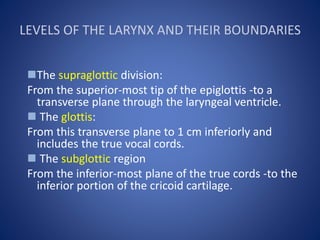
  • 1. LEVELS OF THE LARYNX AND THEIR BOUNDARIES The supraglottic division: From the superior-most tip of the epiglottis -to a transverse plane through the laryngeal ventricle.  The glottis: From this transverse plane to 1 cm inferiorly and includes the true vocal cords.  The subglottic region From the inferior-most plane of the true cords -to the inferior portion of the cricoid cartilage.
  • 2.
  • 3. Supraglottis • Extends from tip of epiglottis above to laryngeal ventricle below. • Contains vestibule, epiglottis, pre-epiglottic fat, AE folds, FVC, paraglottic space, arytenoid cartilages
  • 4. • Pre-epiglottic space: Fat-filled space between hyoid bone anteriorly & epiglottis posteriorly • AE folds: Projects from cephalad tip of arytenoid cartilages to inferolateral margin of epiglottis • Represents superolateral margin of supraglottis, dividing it from pyriform sinus (hypopharynx)
  • 5. • False vocal cords: Mucosal surfaces of laryngeal vestibule of supraglottis. • Beneath FVC are paired paraglottic spaces • Paraglottic spaces: Paired fatty regions beneath false & true vocal cords • Superiorly they merge into pre-epiglottic space • Terminates inferiorly at under surface of TVC
  • 6.
  • 7.
  • 8.
  • 9. Glottis • TVC & anterior & posterior commissures • Comprised of thyroarytenoid muscle (medial fibers are "vocalis muscle") • Anterior commissure: Midline, anterior meeting point of TVC
  • 10.
  • 11.
  • 12. Subglottis • Subglottis extends from under surface of TVC to inferior surface of cricoid cartilage • Mucosal surface of subglottic area is closely applied to cricoid cartilage • Conus elasticus: Fibroelastic membrane extends from medial margin of TVC above to cricoid below
  • 13.
  • 14.
  • 15.
  • 16.
  • 17.
  • 18.
  • 19.
  • 20.
  • 21.
  • 22.
  • 23.
  • 24.
  • 25.
  • 26.
  • 27.
  • 28.
  • 29.
  • 30.
  • 31.
  • 32. CASE 1 Dr. Mohit Goel JR III
  • 33. NAME : Mr. Harnapallu Baburao Abbanna Req No : 330 Patient Code : 140102135 CT No:330 AGE/SEX : 74 Yr(s) / Male Date: 09/05/2014 MSCT NECK WITH CONTRAST HISTORY : K/C/O CA hypopharynx. Post radiotherapy status,
  • 34. soft tissue density mass involving hypopharynx, larynx & upper oesophagus which is occluding the laryngeal airway (supra-glottic space) as well as upper end of oesophagus.
  • 35. soft tissue density mass is showing heterogenous post-contrast enhancement.
  • 36. Valecula appears nomral. Thyroid cartilage appears normal.
  • 37. There is involvement of both thyro-arytenoid folds causing obliteration of both pyriform sinuses (L>R)
  • 38. Pre-epiglottic space appears normal. There is involvement of para-glottic space on both sides.
  • 39. Inferiroly the mass is extending upto the level of vocal cords.
  • 40. NAME : Mr. Khan Ibrahim Amir Req No : 339 Patient Code : 140403479 / IPD CT No:339 AGE/SEX : 72 Yr(s) / Male Date: 10/05/2014 MSCT NECK /LARYNX Clinical Profile: H/o change in voice.
  • 41. Small mildly heterogeneously enhancing polypoidal mass lesion is noted involving right vocal cord and right para-glottic space, protruding into the laryngeal lumen Plain Post-contrast
  • 42. There is small non enhancing hypodense area noted within the lesion s/o necrosis.
  • 43. Anteriorly the lesion is extending upto the anterior commissure with minimal extension to contralateral side. Thyroid cartilage appears normal.
  • 44. There is minimal extension of the enhancing soft tissue into the subglottic region noted at the anterior commissure. No obvious mass or thickening noted in posterior commissure.
  • 45. Mucosal thickening in ethmoid sinus and right maxillary sinus.
  • 46. Small lymph nodes noted in right level II
  • 48. Supraglottic SCC Approximately 30% of all laryngeal cancers arise in the supraglottis. They often present in advanced stages, because symptoms (hoarseness, due to vocal cord involvement) do not occur until late. Due to the rich lymphatic network of the supraglottis, nodal disease (level II and III) is a frequent finding in these patients. Supraglottic SCC may arise in the • anterior compartment (epiglottis) or • the postero-lateral compartment (aryepiglottic fold and false cords).
  • 49. a. Epiglottic SCC Spread: PES (Pre-epiglottic space) Vallecula base of tongue PGS (Paraglottic space) PES glottis or subglottis
  • 50.
  • 51. b. Aryepiglottic fold (AE fold) SCC
  • 53. Glottic SCC Glottic SCCs represent about 65% of all laryngeal cancers. Hoarseness of voice due to vocal cord involvement is the primary presenting symptom in these patients. Metastatic nodal disease is rare in glottic carcinomas due to the sparse lymphatic drainage of the glottis. Glottic SCCs commonly arise from the anterior half of the vocal cord and spread into the anterior commissure. Anterior commissural disease is seen on CT or MRI as soft tissue thickening of more than 1-2 mm.
  • 54. Spread to : • contralateral cord • thyroid cartilage • posterior commissure • Arytenoids • cricoarytenoid joint and the cricoid cartilage • superiorly to access the PES and the PGS • inferiorly to reach the subglottis
  • 55.
  • 56. Subglottic SCC These cancers are rare, accounting for only 5% of all laryngeal cancers, clinically silent and present late in the course. Subglottic cancer is diagnosed if any tissue thickening is noted between the airway and the cricoid ring.
  • 57.
  • 58. Transglottic SCC Laryngeal SCC encroaching on both, the glottis and supraglottis, with or without subglottic component and when the site of origin is unclear, is termed as transglottic tumor.

Notas do Editor

  1. Free edge of epiglottis is attached to hyoid bone via hyoepiglottic ligament which is covered by glossoepiglottic fold, a ridge of mucous membrane.
  2. Graphic at mid-supraglottic level shows hyoepiglottic ligament dividing lower pre-epiglottic space. No fascia separates pre-epiglottic space from paraglottic space. These two endolaryngeal spaces are submucosal locations where tumors hide from clinical detection. Aryepiglottic fold represents junction between larynx & hypopharynx.
  3. Graphic at low supraglottic level shows false vocal cords (FVC) formed by mucosal surfaces of laryngeal vestibule. Paraglottic space is beneath FVC, a common location for submucosal tumor spread.
  4. (Top) Graphic at glottic, true vocal cord level shows thyroarytenoid. Medial fibers of thyroarytenoid muscle are known as vocalis muscle. Pyriform sinus apex is seen at glottic level. Thyroarytenoid gap is location where tumors may spread between larynx & hypopharynx.
  5. Graphic at level of undersurface of true vocal cord shows posterior lamina of cricoid cartilage. Post-cricoid hypopharynx represents anterior wall of lower hypopharynx & extends from cricoarytenoid joints to lower edge of cricoid cartilage at cricopharyngeus muscle. Posterior wall of hypopharynx represents inferior continuation of posterior oropharyngeal wall & extends to cervical esophagus.
  6. Graphic at subglottic level shows cricothyroid joint immediately adjacent to recurrent laryngeal nerve, located in tracheoesophageal groove.
  7. Coronal graphic posterior view shows false & true vocal cords separated by laryngeal ventricle. Quadrangular membrane is a fibrous membrane which extends from upper arytenoid & corniculate cartilages to lateral epiglottis. Conus elasticus is a fibroelastic membrane which extends from vocal ligament of true vocal cord to cricoid. There membranes represent a relative barrier to tumor spread but are not seen on conventional imaging.
  8. Sagittal graphic of midline larynx shows laryngeal ventricle, air-space which separates false vocal cords above with true vocal cords below. Aryepiglottic folds project from tip of arytenoid cartilage to inferolateral margin of epiglottis. Aryepiglottic folds represent junction between supraglottis & hypopharynx. Medial wall of aryepiglottic fold is endolaryngeal while posterolateral wall is anteromedial margin of pyriform sinus.
  9. (Top) First of nine axial CECT images presented from superior to inferior of larynx & hypopharynx with patient in quiet respiration. Hyoid bone represents the level of the roof of larynx & hypopharynx Glossoepiglottic & pharyngoepiglottic folds represent transition from oropharynx above to larynx & hypopharynx below.
  10. (Middle) Image of high supraglottic level of larynx shows C-shaped pre-epiglottic space, a common location for tumors to hide. If supraglottic tumor extends to pre-epiglottic space, it becomes a T3 tumor
  11. (Bottom) Image of high supraglottic level shows pre-epiglottic & paraglottic spaces are continuous, with no intervening fascia. This allows tumors to spread submucosally in these locations. Posterolateralwall of aryepiglottic fold is anteromedial margin of pyriform sinus.
  12. (Top) Image of mid-supraglottic level shows thyroepiglottic ligament dividing the pre-epiglottic space. Aryepiglottic folds are at margin of pyriform sinus & larynx & a tumor primary to aryepiglottic fold is considered a "marginal supraglottic" tumor.. II
  13. (Middle) Image of low supraglottic level shows false vocal cord level. Paraglottic space represents deep fatty space beneath false vocal cords. Tumors that cross laryngeal ventricle & involve false & true vocal cords are considered transglottic
  14. . (Bottom) Image at glottic level shows true vocal cords in abduction in quiet respiration. True vocal cord level is identified on CT when arytenoid and cricoid cartilages are seen and muscle fills inferior paraglottic space. Anterior and posterior commissures of true vocal cords should be less than 1 mm in normal patients. Post-cricoid hypopharynx is typically collapsed.
  15. In this image through the undersurface of true cord level the cricothyroid space is seen. Lack of arytenoid cartilage identifies undersurface of true cord level.
  16. (Middle) Image more inferior shows subglottic level with cricoid ring nearly complete. Cricoid is only complete cartilage ring in larynx & provides structural integrity. Dislocations of cricothyroid joint may result in vocal cord paralysis secondary to recurrent laryngeal nerve injury. There may be associated atrophy of posterior cricoarytenoid muscle on involved side of vocal cord paralysis
  17. (Bottom) At the level of the inferior cricoid cartilage the inferior margin of larynx & hypopharynx are transitioning to the trachea & cervical esophagus.
  18. First of three axial CECT images from superior to inferior in patient with breath holding shows adduction of false & true vocal cords as well as aryepiglottic folds.
  19. (Middle) Image at low supraglottic level shows level of false vocal cords in adduction. Note mucosa of aryepiglottic folds contacts posterior hypopharyngeal wall
  20. (Bottom) Image at glottic level shows adduction of true vocal cords. With breath holding, true vocal cords oppose in midline. A cord that remains paramedian is either paralyzed or mechanically fixed. Vocal cord paralysis typically results in a paramedian true vocal cords with associated abnormal location of arytenoid cartilage which is fixed in an anterior-medial position. With breath holding, paralyzed cord remains fixed while opposite normal cord crosses midline in attempt to close glottis. There may be an associated patulous pyriform sinus.
  21. In this image the laryngeal ventricle is visible as an air space between false vocal cords above & true vocal cords below. When a tumor crosses laryngeal ventricle to involve true & false cords it is transglottic, which has important treatment implications. Coronal imaging is particularly useful for evaluation of transglottic disease.
  22. This image reveals pre-epiglottic fat to be continuous with paraglottic fat. These are the most important spaces of endolarynx as they allow submucosal spread of tumors which is undetectable by clinical exam.
  23. Pre-epiglottic fat is seen at midline posterior & inferior to hyoid bone. Diseases of posterior hypopharyngeal wall are well seen on sagittal imaging. Sagittal imaging also helps define cranial to caudal extent of lesions.