The Human Jungle: Exploring the Microbiome
•
12 gostaram•20,972 visualizações

This is a keynote lecture I gave at a scientific conference on how to translate discoveries about the genome into medical applications. ( http://jointsummits2011.amia.org/keynote-presentations ) I urged them to think of the human body as a lake, and to think of themselves as ecologists. For more information, visit http://carlzimmer.com

Recomendados
Recomendados
Mais conteúdo relacionado
Semelhante a The Human Jungle: Exploring the Microbiome
Semelhante a The Human Jungle: Exploring the Microbiome (20)
Último
Último (20)
The Human Jungle: Exploring the Microbiome
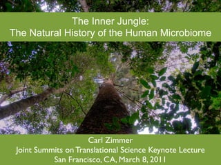
- 1. The Inner Jungle: The Natural History of the Human Microbiome Carl Zimmer Joint Summits on Translational Science Keynote Lecture San Francisco, CA, March 8, 2011
- 4. G. Evelyn Hutchinson (1903-1991)
- 8. Linsley Pond as an energy flow Raymond Lindeman www.cedarcreek.umn.edu/
- 9. Real food webs are even more complex than this...
- 10. The biodiversity of a little lake: 4 species of mammals 3 birds 20 fish ~200 zooplankton ~1,000 algae ~20,000 species total
- 11. Why so many species? http://www.glerl.noaa.gov/pubs/brochures/foodweb/LMfoodweb.pdf
- 12. Hutchinson’s Niche From The Art of Ecology, 2011
- 13. A complicated space From The Art of Ecology, 2011
- 14. Heating of Linsley Pond From The Art of Ecology, 2011
- 15. Dissolved substances From The Art of Ecology, 2011
- 16. From The Art of Ecology, 2011 Time opens new niches
- 17. From The Art of Ecology, 2011 “The History of a Lake”
- 18. Transforming invasions: the alewife
- 20. Brooks and Dodson 1965, Science 150:28-35
- 22. Erosion and eutrophication leave their mark Brugham, Ecology 1979 59:19-36
- 23. Brugham, Ecology 1979 59:19-36
- 25. Max Delbruck (1906-1981) http://en.wikipedia.org/wiki/ File:Max_Delbruck.jpg
- 28. Max Delbruck and Salvador Luria
- 30. Cold Spring Harbor Archives http://www.flickr.com/photos/cshlarchives/4276595071/
- 31. “Molecular genetics, our latest wonder, has taught us to spell out the connectivity of the tree of life in such palpable detail that we may say in plain words, ‘This riddle of life has been solved.’ The ideas of information storage, of the replication of the stored information and of its Max Delbruck, programmed readout have Nobel Lecture, 1969 become commonplace and have filtered down into the popular magazines and grade school textbooks.”
- 32. blogs.discover magazine.com/ loom
- 33. It will unlock new insights into our origins and history as a species; and it points to new ways of combating disease. The people of many countries have invested in the Human Genome Project's determination of the sequence, and it is hard to see how that investment could have received better returns. http://www.nature.com/nature/journal/v409/n6822/full/409745a0.html
- 34. A flood of data...
- 36. The Genomic Hype Cycle http://en.wikipedia.org/wiki/Hype_cycle
- 37. Yes, we’re like viruses...
- 38. ...but we are also like lakes
- 40. “Wee animacules”
- 42. Diaper ecology
- 44. Wang et al, Appl Microbiol Biotechnol (2010) 88:1333–1342
- 45. Stalking the Wild Microbiome Wang et al, Appl Microbiol Biotechnol (2010) 88:1333–1342
- 46. 100 trillion microbes in your body: more microbes than all the humans who have ever lived sigen.org
- 47. Imagine producing an elephant’s worth of microbes
- 48. Now multiply times five
- 49. Microbial abundance raises the question: how human are we? Human: 10 trillion human cells 20,000 human genes Microbiota: Microbiota: 100 trillion microbial cells 20 million microbial genes 99.9% of our genomes the same, but our microbes...?
- 50. The microbiome is like an extra organ Sekirov, I. et al. Physiol. Rev. 90: 859-904 2010; doi:10.1152/physrev.00045.2009 Copyright ©2010 American Physiological Society
- 51. The microbiome helps digest food & metabolize drugs Annu. Rev. Nutr. 2002. 22:283–307 doi: 10.1146/annurev.nutr.22.011602.092259
- 52. The microbiome manages the immune system Fraune and Bosch 2010 Bioessays 32: 571–580
- 53. The microbiome kills invading pathogens The skin bacteria Staphylococcus epidermis makes δ-toxin and kills S. aureus Cogen et al, PLoS ONE 5(1): e8557. doi:10.1371/journal.pone.0008557
- 54. The microbiome heals wounds Left ear: wound Right ear: wound without S. epidermis with S. epidermis Lai 2009 Nature Medicine 15, 1377 - 1382 (2009)
- 55. The microbiome: an organ that works as an ecosystem
- 56. Diversity from nose to nose Wos-Oxley et al, The ISME Journal (2010) 4, 839–851
- 57. Mouth to mouth: 818 bacterial species lives in three people’s mouths, 387 shared by all three. Zaura et al BMC Microbiol (2009) vol. 9 (1) pp. 259
- 58. What is the shape of the core microbiome? Hamady M , Knight R Genome Res. 2009;19:1141-1152
- 59. Or is it a core of genes, not of species? Hamady M , Knight R Genome Res. 2009;19:1141-1152
- 60. Diversity within the core Periphery (not in all people) Core (in all people) Costello et al, Science Vol. 326 no. 5960 pp. 1694-1697
- 61. r Hair The body has many niches Hair ehead Forehead Forehead ernal ear (L) External nose External nose ernal ear (R) External ear (R) External ear (R) el External ear (L) External ear (L) mpit (R) Palm (R) Palm (R) mpit (L) Index finger (R) Index finger (R) ernal nose Palm (L) Palm (L) tril (R) Index finger (L) Index finger (L) tril (L) Forearm (R) Forearm (R) ex finger (R) Forearm (L) Forearm (L) m (R) Nostril (R) Nostril (R) ex finger (L) Nostril (L) Nostril (L) earm (R) EAC (R) Armpit (R) earm (L) EAC (L) Armpit (L) m (L) Armpit (R) Sole of foot (R) k of knee (R) Armpit (L) Sole of foot (L) k of knee (L) Sole of foot (R) Back of knee (R) e of foot (R) Sole of foot (L) Back of knee (L) e of foot (L) Back of knee (R) Navel C (R) Back of knee (L) EAC (R) C (L) Navel EAC (L) ns penis Glans penis Glans penis ia minora Labia minora Labia minora l cavity Oral cavity Oral cavity sal tongue Dorsal tongue Dorsal tongue (Sp) Gut (Sp) Gut (Sp) (Sw) Gut (Sw) Gut (Sw) 0.02 0.05 weighted quantitative symmetric UniFrac (Kulczynski) Costello et al, Science Vol. 326 no. 5960 pp. 1694-1697 Fig. S11
- 62. Detailed biogeography of the human face
- 63. Detailed biogeography of the human face
- 64. Wang et al, Appl Microbiol Biotechnol (2010) 88:1333–1342
- 65. From The Art of Ecology, 2011 Time opens new niches
- 66. Time opens new niches A 0.9 0.8 UniFrac distance 0.7 0.6 variation within people (day-to-day) variation between people 0.5 (on any given day) 0.4 Oral Gut Skin Nostril Hair EAC cavity Habitat B 0.3 eighted UniFrac distance 0.2 variation within people 0.1 (day-to-day) Costello et al, Science Vol. 326 no. 5960 pp. 1694-1697 variation between people
- 67. Diversity through co-dependence: the case of the elusive mouth resident, Synergistetes Vartoukian et al Environmental Microbiology (2010) 12(4), 916–928
- 68. Top: Synergistetes alone in culture grows extremely slowly Bottom: Parvimonas micra speeds up growth of Synergistetes (streak) Vartoukian et al Environmental Microbiology (2010) 12(4), 916–928
- 69. It’s a jungle in there
- 70. Four mouth microbes that cannot grow alone Actinomyces Streptococcus naeshindii oralis Fusobacterium Porphyromonas nucleatum gigivalis Robinson et al, Microbiol Mol Biol Rev. 2010 Sep;74(3):453-76.
- 71. Four mouth microbes that cannot grow alone supports the growth of Actinomyces Streptococcus naeshindii oralis supports the growth of Fusobacterium Porphyromonas nucleatum gigivalis Robinson et al, Microbiol Mol Biol Rev. 2010 Sep;74(3):453-76.
- 72. Four mouth microbes that cannot grow alone supports the growth of Actinomyces Streptococcus naeshindii oralis supports the growth of supports the growth of Fusobacterium Porphyromonas nucleatum gigivalis Robinson et al, Microbiol Mol Biol Rev. 2010 Sep;74(3):453-76.
- 73. Four mouth microbes that cannot grow alone supports the growth of Actinomyces Streptococcus naeshindii oralis supports the growth of supports the growth of supports the growth of Fusobacterium Porphyromonas nucleatum gigivalis Robinson et al, Microbiol Mol Biol Rev. 2010 Sep;74(3):453-76.
- 74. Four mouth microbes that cannot grow alone supports the growth of Actinomyces Streptococcus naeshindii oralis supports the growth of supports the cannot support the growth of growth of supports the growth of Fusobacterium Porphyromonas nucleatum gigivalis Robinson et al, Microbiol Mol Biol Rev. 2010 Sep;74(3):453-76.
- 75. Four mouth microbes that cannot grow alone supports the growth of Actinomyces Streptococcus naeshindii oralis supports the growth of supports the cannot support on its cannot support the growth of own, but can speed growth of growth if A. naeshindii is also present supports the growth of Fusobacterium Porphyromonas nucleatum gigivalis Robinson et al, Microbiol Mol Biol Rev. 2010 Sep;74(3):453-76.
- 76. The Tangled Bank Kolenbrander et al 2010 Nature Reviews Microbiology 8:471
- 77. “The History of a Lake” From The Art of Ecology, 2011
- 78. The History of A Microbiome Reid et al Nat Rev Micro (2011) vol. 9 (1) pp. 27-38
- 79. From sterile to lush in a matter of weeks Fierer et al, Research in Microbiology 161 (2010) 635e642
- 80. Sampling of the microbiota 20 minutes after birth
- 81. Koenig et al, PNAS 2010
- 82. A baby’s microbiome grows more diverse Koenig et al, PNAS 2010
- 83. A lesson from ecology: diversity is healthy Salmonella invades more successfully if mice are treated with antibiotics, have low-diversity microbiomes (LCM), or are germ-free Photo- http://flic.kr/p/3vhoNg Stecher and Hardt Current Opinion in Microbiology 2010, 14:1–10
- 84. Reid et al Nat Rev Micro (2011) vol. 9 (1) pp. 27-38
- 85. Reid et al Nat Rev Micro (2011) vol. 9 (1) pp. 27-38
- 86. Reid et al Nat Rev Micro (2011) vol. 9 (1) pp. 27-38
- 87. Reid et al Nat Rev Micro (2011) vol. 9 (1) pp. 27-38
- 88. Reid et al Nat Rev Micro (2011) vol. 9 (1) pp. 27-38
- 89. Two invaders that alter ecosystems Alewife Helicobacter hepatica
- 90. As Helicobacter hepaticus becomes more common in mouse cecae (A), microbiome diversity falls (B) KUEHL ET AL, INFECTION AND IMMUNITY, Oct. 2005, p. 6952–6961
- 91. A Manmade Catastrophe (for the Microbiome)
- 92. Jernberg et al, Microbiology (2010), 156, 3216–3223
- 93. Antonopoulos et al, Infection and Immunity (2009) vol. 77 (6) pp. 2367-2375
- 94. Robinson et al, Microbiol Mol Biol Rev. 2010 Sep;74(3):453-76.
- 95. Microbes guide the flow of energy
- 96. Do My Bacteria Make Me Look Fat? Ley RE, Backhed F, Turnbaugh P, et al. 2005. Proc Natl Acad Sci USA 102: 11070–5.
- 97. • Germ-free mice given ob/ob or wild-type gut microbes • Chow consumption and exercise the same for both groups • Both sets of mice had similar starting weight and percentage of body fat Turnbaugh, et al. Nature 2006; 444:1027
- 98. Sandoval et al, Science 328, 179 (2010)
- 99. Kinross et al. Genome Medicine 2011, 3:14
- 100. Becoming Ecosystem Engineers Alexander Khoruts, University of Minnestoa
- 101. Khoruts’s dilemma: 61 year old patient --Antibiotics for lung infection led to intestinal C. difficile infection Chronic Diarrhea - 8 months Loose bowel movements every 15 minutes Wore diapers at all times Confined to a wheel chair Lost 27 Kg Antibiotic therapy for C. difficile uneffective Khoruts, et al. J. Clin. Gastroenterol. 44, 354–360 (2010)
- 102. Microbiome transplant Khoruts, et al. J. Clin. Gastroenterol. 44, 354–360 (2010)
- 103. Patient had first solid bowel movement 2 days after treatment On 6 month follow up visit, reported daily stools that were well formed Khoruts, et al. J. Clin. Gastroenterol. 44, 354–360 (2010)
- 106. Thanks to Jonathan Eisen, Karen Jansson, Rob Knight, Sarkis Mazmanian, David Post, and David Skelly For more information, visit carlzimmer.com
Notas do Editor
- \n
- \n
- \n
- \n
- \n
- \n
- \n
- \n
- \n
- \n
- \n
- \n
- \n
- \n
- \n
- \n
- \n
- \n
- \n
- \n
- \n
- \n
- \n
- \n
- \n
- \n
- \n
- \n
- \n
- \n
- \n
- \n
- \n
- \n
- \n
- \n
- \n
- \n
- \n
- \n
- \n
- \n
- \n
- \n
- \n
- \n
- \n
- \n
- \n
- \n
- \n
- \n
- \n
- \n
- \n
- \n
- \n
- Models of a core microbiome. The circles represent the microbial communities in different individuals and can be thought of as either representing different taxa (species, genera, etc.) or representing different genes. (A) “Substantial core” model. Most individuals share most components of the microbiota. (B) “Minimal core” model. All individuals share a few components, and any individual shares many components with a few other individuals, but very little is shared across all individuals. (C) “No core” model. Nothing is shared by all individuals, and most diversity is unique to a given individual. (D) “Gradient” model. Individuals next to each other on a gradient, for example, age or obesity, share many components, but individuals at opposite ends share little or nothing. (E) “Subpopulation” model. Different subpopulations, for example, those defined by geography or disease, have different cores, but nothing is shared across subpopulations. Scenarios C–E would represent situations in which the strategy of identifying core species for sequencing, then using these as a scaffold for “omics” studies, would be problematic.\n
- Models of a core microbiome. The circles represent the microbial communities in different individuals and can be thought of as either representing different taxa (species, genera, etc.) or representing different genes. (A) “Substantial core” model. Most individuals share most components of the microbiota. (B) “Minimal core” model. All individuals share a few components, and any individual shares many components with a few other individuals, but very little is shared across all individuals. (C) “No core” model. Nothing is shared by all individuals, and most diversity is unique to a given individual. (D) “Gradient” model. Individuals next to each other on a gradient, for example, age or obesity, share many components, but individuals at opposite ends share little or nothing. (E) “Subpopulation” model. Different subpopulations, for example, those defined by geography or disease, have different cores, but nothing is shared across subpopulations. Scenarios C–E would represent situations in which the strategy of identifying core species for sequencing, then using these as a scaffold for “omics” studies, would be problematic.\n
- \n
- \n
- \n
- \n
- \n
- \n
- \n
- \n
- \n
- \n
- \n
- \n
- \n
- \n
- \n
- \n
- \n
- \n
- \n
- \n
- \n
- \n
- \n
- \n
- \n
- \n
- \n
- \n
- \n
- \n
- \n
- \n
- \n
- \n
- \n
- \n
- \n
- \n
- \n
- \n
- \n
- \n
- \n
- \n
- \n
- \n