SlideShare uma empresa Scribd logo
1 de 51
doi:10.1136/thx.2006.073080
2007;62;1-46Thorax
W S Lim
pandemic
with an influenza-like illness during an influenza
Pandemic flu: clinical management of patients
http://thorax.bmj.com/cgi/content/full/62/suppl_1/1
Updated information and services can be found at:
These include:
Data supplement
http://thorax.bmj.com/cgi/content/full/62/suppl_1/1/DC1
"erratum"
References
http://thorax.bmj.com/cgi/content/full/62/suppl_1/1#BIBL
This article cites 144 articles, 53 of which can be accessed free at:
service
Email alerting
at the top right corner of the article
Receive free email alerts when new articles cite this article - sign up in the box
Notes
http://journals.bmj.com/cgi/reprintform
To order reprints of this article go to:
http://journals.bmj.com/subscriptions/
go to:ThoraxTo subscribe to
on 27 July 2009thorax.bmj.comDownloaded from
January 2007 Vol 62 Supplement I
Pandemic flu: clinical management of
patients with an influenza-like illness
during an influenza pandemic
Provisional guidelines from the British Infection
Society, British Thoracic Society and Health
Protection Agency in collaboration with the
Department of Health
AN INTERNATIONAL JOURNAL OF RESPIRATORY MEDICINE
thorax62_supp_I_cover.qxd 12/14/2006 6:05 PM Page 1
on 27 July 2009thorax.bmj.comDownloaded from
President, British Thoracic Society: Professor P M Calverley
Editor-in-Chief
J A Wedzicha (UK)
Editors
S L Johnston (UK) D M Mitchell (UK)
Associate Editors
P M A Calverley (UK)
M Dusmet (UK)
J S Elborn (N Ireland)
J M FitzGerald (Canada)
J A Fleetham (Canada)
N M Foley (UK)
I Hall (UK)
R Hubbard (UK)
D A Lomas (UK)
D M Mannino (USA)
F D Martinez (USA)
C Robertson (Australia)
B Schonhofer (Germany)
G A Silvestri (USA)
G I Town (New Zealand)
M K B Whyte (UK)
Statistical editors
S A Lewis (UK) T M McKeever (UK)
Images editors
J M FitzGerald (Canada)
J R Mayo (Canada)
J C Hogg (Canada)
Letters editor
T Wilkinson (UK)
Lung Alert editors
A Bhowmik (UK) J R Hurst (UK)
International advisory board
N Ambrosino (Italy)
J N Baraniuk (USA)
P J Barnes (UK)
C R W Beasley (New Zealand)
J R Britton (UK)
A S Buist (USA)
E R Chilvers (UK)
S-H Cho (Korea)
S-E Dahlen (Sweden)
G C Donaldson (UK)
M W Elliott (UK)
Y Fukuchi (Japan)
D M Geddes (UK)
P Goldstraw (UK)
R Goldstein (Canada)
C Griffiths (UK)
J C Hogg (Canada)
S T Holgate (UK)
P Hopewell (USA)
M Ichinose (Japan)
A Kendrick (UK)
T King (USA)
A J Knox (UK)
C K W Lai (China)
G J Laurent (UK)
P LeSouef (Australia)
W MacNee (UK)
C Mayaud (France)
J Moore-Gillon (UK)
A Morice (UK)
A Papi (Italy)
R Panettieri (USA)
N G Papadopoulos (Greece)
M R Partridge (UK)
I D Pavord (UK)
M G Pearson (UK)
T A E Platts Mills (USA)
L Restrick (UK)
D S Robinson (UK)
R M Rudd (UK)
S Sethi (USA)
T Sethi (UK)
A K Simonds (UK)
P Sliwinski (Poland)
R A Stockley (UK)
J K Stoller (USA)
M J Tobin (USA)
A Torres (Spain)
J Vestbo (Denmark)
E H Walters (Australia)
S T Weiss (USA)
A Wells (UK)
J W Wilson (Australia)
A A Woodcock (UK)
M Woodhead (UK)
R Zuwallack (USA)
Editor, BMJ
Disclaimer
Thorax is published by BMJ Publishing Group Ltd, a wholly owned
subsidiary of the British Medical Association, and the British Thoracic
Society.
The BMA grants editorial freedom to the Editor of Thorax. Thorax follows
guidelines on editorial independence produced by the World Association of
MedicalEditors andthe codeon goodpublicationpractice ofthe Committee
on Publication Ethics.
Thorax is intended for medical professionals and is provided without
warranty, express or implied. Statements in the journal are the
responsibility of their authors and advertisers and not authors’ institutions,
the BMJ Publishing Group or the BMA unless otherwise specified or
determined by law. Acceptance of advertising does not imply endorsement.
To the fullest extent permitted by law, the BMJ Publishing Group shall not
be liable for any loss, injury or damage resulting from the use of Thorax or
any information in it whether based on contract, tort, or otherwise. Readers
are advised to verify any information they choose to rely on.
Guidelines for authors and reviewers
Full instructions are available online at http://thorax.bmj.com/ifora
All papers must be submitted via Bench.Press at
http://submit-thorax.bmj.com
Subscription information
See page ifc
Copyright
E 2007 BMJ Publishing Group Ltd and British Thoracic Society. All rights
reserved; no part of this publication may be reproduced, stored in a retrieval
system, or transmitted inany form or by any means, electronic,mechanical,
photocopying, recording, or otherwise without the prior permission of
Thorax
Authors are required to grant Thorax an exclusive licence to publish; further
details available online at www.thoraxjnl.com/ifora
N Thorax is published by BMJ Publishing Group, copyedited by Macmillan
India, typeset by The Charlesworth Group and printed in UK on acid-free
paper by Latimer Trend & Co Ltd, Plymouth
N Periodicals postage paid, Rahway, NJ. Postmaster: send address changes
to: Thorax, c/o Mercury Airfreight International Ltd, 365 Blair Road,
Avenel, NJ 07001, USA
contents THORAX
Volume 62 Supplement 1 January 2007
. . . . . . . . . . . . . . . . . . . . . . . . . . . . . . . . . . . . . . . . . . . . . . . . . . . . . . . . . . . . . . . . . . . . . . . . . . . . . . . . . . .
Pandemic flu: clinical management of patients with an
influenza-like illness during an influenza pandemic
Provisional guidelines from the British Infection
Society, British Thoracic Society, and Health Protection
Agency in collaboration with the Department of Health
SYNOPSIS AND INTRODUCTION
i1 Synopsis of main recommendations
i1 Synopsis 1 Clinical management of adults referred to hospital
i3 Synopsis 2 Clinical management of children referred to hospital
i5 1 Introduction
i6 2 Epidemiology and health impact projections
i8 3 Clinical features in adults
i11 4 Clinical features in children
PART 1 CLINICAL MANAGEMENT IN PRIMARY CARE
i14 5 General management and investigations
i15 6 Criteria for hospital referral
i16 7 Antiviral use
i17 8 Antibiotic use
PART 2 CLINICAL MANAGEMENT OF ADULTS REFERRED TO HOSPITAL
i19 9 Severity assessment
i19 10 General investigations
i20 11 Microbiological investigations
i21 12 General management
i23 13 Antiviral use
i26 14 Antibiotic use
PART 3 CLINICAL MANAGEMENT OF CHILDREN REFERRED TO HOSPITAL
i30 15 Severity assessment
i30 16 General investigations
i31 17 Microbiological investigations
i32 18 General management in hospital
i33 19 Antiviral use
i34 20 Antibiotic use
i35 21 Acknowledgements and Committee members
i36 APPENDICES
i44 REFERENCES
These guidelines have also been published in the Journal of Infection
2006;53(Suppl 1):S1–S58.
AIMS AND SCOPE
Thorax, a leading journal in respiratory medicine, is the official journal of the British
Thoracic Society. It enjoys an enviable and longstanding reputation for publishing
clinical and experimental research articles covering many disciplines, including
pathology, immunology and surgery. In addition it provides coverage of recent
developments in basic biomedical sciences and publishes a regular review series to
keep thoracic physicians abreast of advances.
Impact Factor 6.15
EDITOR-IN-CHIEF
J A Wedzicha
EDITORS
S L Johnston
D M Mitchell
ASSOCIATE EDITORS
P M A Calverley D A Lomas
M Dusmet D M Mannino
J S Elborn F D Martinez
J M FitzGerald C Robertson
J A Fleetham B Schonhofer
N M Foley G A Silvestri
I Hall G I Town
R Hubbard M K B Whyte
STATISTICAL EDITORS
S A Lewis
T M McKeever
CONTACT DETAILS
Editorial Office
Thorax, BMJ Journals,
BMA House, Tavistock Square,
London WC1H 9JR, UK
Tel: +44 (0)20 7383 6147
Fax: +44 (0)207 383 6668
Email: thorax@bmjgroup.com
IMAGES EDITORS
J M FitzGerald
J R Mayo
J C Hogg
LETTERS EDITOR
T Wilkinson
LUNG ALERT EDITORS
A Bhowmik
J R Hurst
MANAGING EDITOR
A Horgan
DEVELOPMENT EDITOR
G Stewart
PRODUCTION EDITOR
M Dodd
JOURNAL ASSISTANT
J Cresswell
ADVISORY BOARD
See table of contents
Supplement Enquiries
Gavin Stewart, Development Editor
Tel: +44 (0)20 7383 6170
Fax: +44 (0)20 7383 6668
Email: gstewart@bmjgroup.com
Subscriptions (except USA)
Subscription Manager, BMJ Journals, BMJ Publishing Group Ltd,
PO Box 299, London WC1H 9TD, UK
Tel: +44 (0)20 7383 6270
Fax: +44 (0)20 7383 6402
Email: subscriptions@bmjgroup.com
http://thorax.bmjjournals.com/subscriptions
US Subscriptions
BMJ Publishing Group, PO Box 281, Annapolis Junction, MD 20701-0281, USA
Tel: +1 800 348 6473 (toll free)
Fax: +1 301 206 9789
Email: bmjpg@pmds.com
Advertising
Advertising Manager, BMJ Journals
Tel: +44 (0)20 7383 6181
Fax: +44 (0)20 7383 6556
Email: ecurrer@bmjgroup.com
http://bmjpg.com/advertising
Author Reprints
Sheila Williams
Tel: +44 (0)20 7383 6305
Fax: +44 (0)20 7383 6699
Email: swilliams@bmjgroup.com
Commercial Reprints (except USA & Canada)
Nadia Gurney-Randall
Tel: +44 (0)20 8445 5825
Fax: +44 (0)20 8445 5870
Email: ngurneyrandall@bmjgroup.com
Commercial Reprints (USA & Canada)
Marsha Fogler, PO Box 3227, Cherry Hill, NJ 08034, USA
Tel: +1 800 482 1450
Fax: +1 856 489 4449
Email: fogler@erols.com
Permissions
See www.bmj.com/permissions
British Thoracic Society
17 Doughty Street,
London, WC1N 2PL, UK
Tel: +44 (0)20 7831 8778
Fax: +44 (0)20 7831 8766
Email: bts@brit-thoracic.org.uk
www.brit-thoracic.org.uk
GUIDELINES FOR AUTHORS AND REVIEWERS
All papers must be submitted via Bench.Press at
http://submit-thorax.bmj.com/
SUBSCRIPTION INFORMATION
Thorax is published monthly (subscribers receive all supplements)
ISSN 0040-6376 (print); 1468-3296 (online)
INSTITUTIONAL RATES
Print – 2007 rates: £410; US$759; J607
Online – Site licences are priced on FTE basis and allow access by the whole institution. Print is available at deeply discounted rates for
online subscribers; details available online at www.bmjjournals.com/subscriptions or contact the Subscription Manager in the UK (see above)
PERSONAL RATES
Print (includes online access at no additional cost) – £174; US$322; J258
Online only – £95; US$176; J141
HOW TO SUBSCRIBE
N Subscribers may pay by cheque*, Switch, or credit card (Mastercard, Visa, American Express)
N Orders may be placed with any leading subscription agent or bookseller
N Call our subscription hotline on +44 (0)20 7383 6270; fax hotline +44 (0)20 7383 6402
N All enquiries and single copy sales should be addressed to the UK office
N Personal print or online only and institutional print subscriptions may be purchased online at www.bmjjournals.com/subscriptions
(payment by Visa/Mastercard only)
*UK cheques must be drawn on a UK bank account; US cheques must be drawn on a US bank account
Residents of some EC countries and Canada must pay VAT; for details, call us or visit www.bmjjournals.com/subscriptions/cost2007.dtl
SYNOPSIS AND INTRODUCTION
Pandemic flu: clinical management of patients with an
influenza-like illness during an influenza pandemic
Provisional guidelines from the British Infection Society, British Thoracic Society, and Health
Protection Agency in collaboration with the Department of Health
. . . . . . . . . . . . . . . . . . . . . . . . . . . . . . . . . . . . . . . . . . . . . . . . . . . . . . . . . . . . . . . . . . . . . . . . . . . . . . . . . . . . . . . . . . . . . . . . . . . . . . . . . . . . . . . . . . . . . . . . . . . . . . . . . . .
Thorax 2007;62(Suppl I):i1–i13. doi: 10.1136/thx.2006.073080
. . . . . . . . . . . . . . . . . . . . . . . .
Correspondence to:
Dr W S Lim, Department of
Respiratory Medicine,
Nottingham University
Hospitals, City Hospital
Campus, Nottingham NG5
1PB, UK; weishen.lim@nuh.
nhs.uk
. . . . . . . . . . . . . . . . . . . . . . . .
SYNOPSIS OF MAIN RECOMMENDATIONS
Scope and purpose
N This document is intended for use in the UK in
the event that the World Health Organization
declares that an influenza pandemic has
started,1
and the Department of Health in
England (UK-wide lead agency on pandemic
influenza, including the devolved administra-
tions) has declared UK Pandemic Alert Level 2
(cases of pandemic influenza identified within
the UK).
N These guidelines are not relevant for the
management of patients affected by seasonal/
interpandemic influenza, lower respiratory tract
infections, community acquired pneumonia or
exacerbations of chronic obstructive pulmonary
disease (COPD).
N Once an influenza pandemic is under way, users
are strongly urged to ensure that they refer to
the most up-to-date version of these guidelines
(from web-based access points).
SYNOPSIS 1 CLINICAL MANAGEMENT OF
ADULTS REFERRED TO HOSPITALS
S1.1 Severity assessment in hospital
N Patients with uncomplicated influenza infection
would be expected to make a full recovery and
do not require hospital care.
N In uncomplicated infection, the illness usually
resolves in seven days although cough, malaise
and lassitude may persist for weeks.
N Patients with worsening of pre-existing comor-
bid medical conditions should be managed
according to best practice for that condition
with reference to published disease-specific
guidelines, if available, for example the
National Institute for Health and Clinical
Excellence’s COPD guidelines.
S1.2 Influenza-related pneumonia
N In hospital, patients with influenza-related
pneumonia and who have a CURB-65 score of
3, 4 or 5 (see Box A) are at high risk of death
and should be managed as having severe
pneumonia.
N Patients with bilateral lung infiltrates on chest
radiography consistent with primary viral pneu-
monia should be managed as having severe
pneumonia regardless of CURB-65 score.
N Patients who have a CURB-65 score of 2 are
at increased risk of death. They should be
considered for short stay inpatient treatment or
hospital supervised outpatient treatment. This
decision is a matter of clinical judgment.
N Patients who have a CURB-65 score of 0 or 1 are
at low risk of death. They can be treated as
having non-severe pneumonia and may be
suitable for home treatment.
S1.3 High dependency or intensive care unit
transfer
N Patients with primary viral pneumonia or a
CURB-65 score of 4 or 5 should be considered
for high dependency unit (HDU)/intensive care
unit (ICU) transfer.
N General indications for HDU/ICU transfer
include:
(1) persisting hypoxia with PaO2 ,8 Kpa
despite maximal oxygen administration
(2) progressive hypercapnia
(3) severe acidosis (pH,7.26)
(4) septic shock
N Patients with influenza admitted to intensive
care units should be managed by specialists
with appropriate training in intensive care,
respiratory medicine and/or infectious diseases.
S1.4 General investigations
N The investigations shown in table A are
recommended in patients referred to hospital.
Abbreviations: ARDS, acute respiratory distress syndrome;
CAP, community acquired pneumonia; CNS, central
nervous system; COPD, chronic obstructive pulmonary
disease; CSF, cerebrospinal fluid; GP, general practitioner;
HDU, high dependency unit; ICU, intensive care unit; ILI,
influenza-like illness; NIV, non-invasive ventilation; PCT,
primary care trust
Box A CURB-65 score
Score 1 point for each feature present:
N Confusion (mental test score of (8, or new
disorientation in person, place or time)
N Urea .7 mmol/l
N Respiratory rate >30/min
N Blood pressure (SBP ,90 mmHg or DBP
(60 mmHg)
N Age >65 years
i1
www.thoraxjnl.com
N In those patients who are subsequently followed up in a
hospital outpatient clinic or by a general practitioner (GP), a
repeat chest x ray should be obtained at around six weeks if
respiratory symptoms or signs persist or where there is a
higher risk of underlying malignancy (especially smokers
and those over 50 years of age).
N Further investigations including a CT thoracic scan and
bronchoscopy should be considered if the chest x ray remains
abnormal at follow up.
S1.5 Microbiological investigations
S1.5.1 Early in a pandemic (UK alert levels 1, 2 and 3)
Virology—all patients
1. Nose and throat swabs in virus transport medium.
2. If presentation is more than seven days after onset of
illness, an ‘‘acute’’ serum (5–10 ml clotted blood) should be
collected and a ‘‘convalescent’’ sample (5–10 ml clotted
blood) obtained after an interval of not less than seven
days.
Bacteriology—patients with influenza-related
pneumonia
1. Blood culture (preferably before antibiotic treatment is
commenced)
2. Pneumococcal urine antigen (20 ml urine sample)
3. Legionella urine antigen (20 ml urine sample)
4. Sputum gram stain, culture and antimicrobial suscept-
ibility tests on samples obtained from patients who:
(i) are able to expectorate purulent samples, and
(ii) have not received prior antibiotic treatment.
5. Paired serological examination for influenza/other agents.
Acute serum should be collected and a ‘‘convalescent’’
sample obtained after an interval not less than seven days
(both 5–10 ml clotted blood).
S1.5.2 Once a pandemic is established (UK alert level 4)
Virology—not routinely recommended
Bacteriology—patients with influenza-related
pneumonia in accordance to the severity of illness
(a) Non-severe pneumonia (CURB-65 Score 0, 1 or 2)
– No routine testing.
– In patients who do not respond to empirical antibiotic
therapy, sputum samples should be sent for Gram stain
culture and antimicrobial susceptibility tests.
(b) Severe pneumonia (CURB-65 Score 3, 4 or 5, or bilateral
chest x ray changes)
– Blood culture, preferably before antibiotic treatment is
commenced
– Pneumococcal urine antigen (20 ml urine)
– Sputum gram stain, culture and antimicrobial
susceptibility tests on samples obtained from patients
who are able to expectorate purulent samples, and have
not received prior antibiotic treatment.
– Paired serological examination for influenza/other
agents. ‘‘Acute’’ serum should be collected and a
‘‘convalescent’’ sample obtained after an interval not less
than seven days (both 5–10 ml clotted blood).
– Tracheal or endotracheal aspirate samples, if avail-
able, should be sent for Gram stain, culture and
antimicrobial susceptibility testing.
S1.6 General management
S1.6.1 Initial management
N Hypoxic patients should receive appropriate oxygen therapy
with monitoring of oxygen saturations and inspired oxygen
concentration with the aim to maintain PaO2 >8 Kpa and
SaO2 >92%. High concentrations of oxygen can safely be
given in uncomplicated pneumonia.
N Oxygen therapy in patients with pre-existing COPD compli-
cated by ventilatory failure should be guided by repeated
arterial blood gas measurements. Non-invasive ventilation
(NIV) may be helpful.
N In patients without pre-existing COPD who develop respira-
tory failure, NIV may be of value as a bridge to invasive
ventilation in specific circumstances when level 3 beds are in
high demand. Respiratory and/or critical care units experi-
enced in the use of NIV are best placed to ensure the
appropriate infection control measures are adopted at all
times.
N Patients should be assessed for cardiac complications and
also volume depletion and their need for additional
intravenous fluids.
N Nutritional support should be given in severe or prolonged
illness.
S1.6.2 Monitoring in hospital
N Temperature, respiratory rate, pulse, blood pressure, mental
status, oxygen saturation and inspired oxygen concentration
should be monitored and recorded initially at least twice
daily and more frequently in those with severe illness or
requiring regular oxygen therapy. An Early Warning Score
system is a convenient way to perform this.
N In patients who are not progressing satisfactorily a full
clinical reassessment and a repeat chest radiograph are
recommended.
S1.6.3 Discharge and follow up
N Patients should be reviewed 24 hours prior to discharge.
Those with two or more of the following unstable clinical
factors should be considered for remaining in hospital:
(1) temperature .37.8˚C
(2) heart rate .100/min
(3) respiratory rate .24/min
(4) systolic blood pressure ,90 mmHg
(5) oxygen saturation ,90%
Table A General investigations
Test Who this applies to
Full blood count All patients
Urea and electrolytes All patients
Liver function tests All patients
Chest x ray All patients
Pulse oximetry All patients. If ,92% on air, then arterial
blood gases
Electrocardiogram Patients with cardiac and respiratory
complications or comorbid illnesses
C-reactive protein If influenza-related pneumonia is suspected
i2 Provisional guidelines from the BIS/BTS/HPA with the Department of Health
www.thoraxjnl.com
(6) inability to maintain oral intake
(7) abnormal mental status.
N Follow up clinical review should be considered for all
patients who suffered significant complications or who had
significant worsening of their underlying disease, either with
their GP or in a hospital clinic.
N At discharge or at follow up, patients should be offered
access to information about their illness, take home
medication and any follow up arrangements.
N It is the responsibility of the hospital team to arrange the
follow up plan with the patient and the GP.
S1.7 Use of antivirals
N Individuals should only be considered for treatment with
antivirals (neuraminidase inhibitors) if they have all of the
following:
(1) an acute influenza-like illness (ILI)
(2) fever (.38˚C) and
(3) been symptomatic for two days or less.
N Treatment schedule: adults, oseltamivir 75 mg every
12 hours for five days (dose to be reduced by 50% if
creatinine clearance is less than 30 ml/min—that is, 75 mg
od)
N Patients who are unable to mount an adequate febrile
response—for example, the immunocompromised or very
elderly—may still be eligible for antiviral treatment despite
lack of documented fever.
N Hospitalised patients who are severely ill, particularly if also
immunocompromised, may benefit from antiviral treatment
started more than 48 hours from disease onset, although
there is no evidence to demonstrate benefit, or lack of, in
such circumstances.
S1.8 Antibiotic management
S1.8.1 Influenza not complicated by influenza-related
pneumonia
N Previously well adults with acute bronchitis complicating
influenza, in the absence of pneumonia, do not routinely
require antibiotics.
N Antibiotics should be considered in those previously well
adults who develop worsening symptoms (recrudescent
fever or increasing dyspnoea).
N Patients at high risk of complications or secondary infection
(Appendix 2) should be considered for antibiotics in the
presence of lower respiratory features.
N Most patients can be adequately treated with oral antibiotics.
N The preferred choice includes co-amoxiclav or a tetracycline.
N A macrolide such as clarithromycin (or erythromycin) or
a fluoroquinolone active against Streptococcus pneumoniae
(S pneumoniae) and Staphylococcus aureus (S aureus) is an
alternative choice in certain circumstances.
S1.8.2 Non-severe influenza-related pneumonia
N Most patients can be adequately treated with oral antibiotics.
N Oral therapy with co-amoxiclav or a tetracycline is preferred.
N When oral therapy is contraindicated, recommended par-
enteral choices include intravenous co-amoxiclav, or a
second or third generation cephalosporin (cefuroxime or
cefotaxime).
N A macrolide (erythromycin or clarithromycin) or a fluor-
oquinolone active against S pneumoniae and S aureus is an
alternative regimen where required—for example, for those
intolerant of penicillins. Currently levofloxacin and moxi-
floxacin are the only recommended fluoroquinolones
licensed in the UK.
N Antibiotics should be administered within four hours of
admission.
S1.8.3 Severe influenza-related pneumonia
N Patients with severe pneumonia should be treated immedi-
ately after diagnosis with parenteral antibiotics.
N An intravenous combination of a broad spectrum beta-
lactamase stable antibiotic such as co-amoxiclav or a second
(for example, cefuroxime) or third (for example, cefotaxime)
generation cephalosporin together with a macrolide (for
example, clarithromycin or erythromycin) is preferred.
N An alternative regimen includes a fluoroquinolone with
enhanced activity against pneumococci together with a
broad spectrum b-lactamase stable antibiotic or a macrolide.
Currently levofloxacin is the only fluoroquinolone with an
intravenous formulation licensed in the UK.
S1.8.4 Route and duration of antibiotic
N Patients treated initially with parenteral antibiotics should
be transferred to an oral regimen as soon as clinical
improvement occurs and the temperature has been normal
for 24 hours, providing there is no contraindication to the
oral route.
N For most patients admitted to hospital with non-severe and
uncomplicated pneumonia, seven days of appropriate anti-
biotics is recommended.
N For those with severe, microbiologically undefined pneumo-
nia, 10 days’ treatment is proposed. This should be extended
to 14–21 days where S aureus or Gram negative enteric bacilli
pneumonia is suspected or confirmed.
S1.8.5 Failure of empirical antibiotics
N For those with non-severe pneumonia in hospital on
combination therapy, changing to a fluoroquinolone with
effective pneumococcal and staphylococcal cover is an
option.
N Adding further antibiotics effective against MRSA is an
option for those with severe pneumonia not responding to
combination antibiotic therapy.
SYNOPSIS 2 CLINICAL MANAGEMENT OF CHILDREN
REFERRED TO HOSPITAL
S2.1 Severity assessment in children (see Appendix 5)
S2.1.1 In the community
N Coughs and mild fevers. These children should be treated
at home by parents with antipyretics and fluids (note:
aspirin should not be used in children).
N High fever (.38.5˚C) and cough or influenza-like
symptoms. These children should seek advice from a
community health professional. If there are no features that
put them at high risk of complications they should be
treated with oseltamivir, and given advice on antipyretics
and fluids. Children aged ,1 year and those at risk of
complications (Appendix 2) should be seen by a genral
practitioner.
N High fever (.38.5˚C) and cough or influenza-like
symptoms, plus at risk group. These children should be
seen by a GP or in A&E. Children may be considered at
increased risk of complications if they have cough and fever
(or ILI) and temperature .38.5˚C, plus either chronic
comorbid disease or one of following features:
Pandemic flu: clinical management of patients with an influenza-like illness during an influenza pandemic i3
www.thoraxjnl.com
– breathing difficulties
– severe earache
– vomiting .24 hours
– drowsiness.
These patients should be offered an antibiotic as well as
oseltamivir (in those .1 year of age) and advice on antipyretics
and fluids. Children aged ,1 year with none of the above
features should be treated with antipyretics and fluids with a
low threshold for antibiotics if they become more unwell.
S2.1.2 Hospital admission
Indicators for hospital admission are:
(1) Signs of respiratory distress
– markedly raised respiratory rate
– grunting
– intercostal recession
– breathlessness with chest signs
(2) Cyanosis
(3) Severe dehydration
(4) Altered conscious level
(5) Complicated or prolonged seizure
(6) Signs of septicaemia—extreme pallor, hypotension, floppy
infant
Most children admitted to hospital are likely to need oxygen
therapy and/or intravenous support as well as antibiotics and
oseltamivir.
Indications for transfer to high dependency or intensive care
are:
(1) failure to maintain a SaO2 of .92% in FiO2 of .60%
(2) the child is shocked
(3) severe respiratory distress and a raised PaCO2 (.6.5 Kpa)
(4) rising respiratory rate and pulse rate with clinical evidence
of severe respiratory distress with or without a raised
PaCO2
(5) recurrent apnoea or slow irregular breathing
(6) evidence of encephalopathy
When there are no paediatric intensive care unit beds
available, children will have to be triaged on the basis of the
severity of their acute and coexisting disease, and the likelihood
of their achieving full recovery.
S2.2 General investigations for children in hospital
N A full blood count with differential, urea, creatinine and
electrolytes, liver enzymes and a blood culture should be
done in all severely ill children.
N A chest x ray should be performed in children who are
hypoxic, have severe illness or who are deteriorating despite
treatment.
N Pulse oximetry should be performed in every child being
assessed for admission to hospital with pneumonia.
S2.3 Microbiological investigations in hospital
S2.3.1 Early in a pandemic (UK alert levels 1, 2 and 3)
Virology—all children
(1) Nasopharyngeal aspirate or nose and throat swabs.
(2) If presentation is more than 7 days after onset of illness, an
‘‘acute’’ serum (2–5 ml clotted blood) should be collected
and a ‘‘convalescent’’ sample (2–5 ml clotted blood)
obtained after an interval of not less than 7 days.
Bacteriology—children with influenza-related
pneumonia
(1) Blood culture (before antibiotic treatment is commenced).
(2) Sputum samples obtained from older children.
(3) Paired serological examination for influenza/other agents.
S2.3.2 Once a pandemic is established (UK alert level 4)
Virology—not routinely recommended
Bacteriology—children with influenza-related pneumonia
(1) Blood culture (before antibiotic treatment is commenced).
(2) Sputum samples obtained from older children.
(3) Paired serological examination for influenza/other agents.
S2.4 General management of children admitted to
hospital
N Patients whose oxygen saturation is 92% or less while
breathing air should be treated with oxygen given by nasal
cannulae, head box, or face mask to maintain oxygen
saturation above 92%.
N When children are unable to maintain oral intake, supple-
mentary fluids should, when possible, be given by the
enteral route. Intravenous fluids in those with severe
pneumonia should be given at 80% basal levels.
N Children can be safely discharged from hospital when they
(1) are clearly improving
(2) are physiologically stable
(3) can tolerate oral feeds
(4) have a respiratory rate ,40/min (,50/min in infants)
(5) have an awake oxygen saturation of .92% in air.
S2.5 Antiviral therapy in children
N In the setting of a pandemic, children should only be
considered for treatment with antivirals if they have all of
the following:
(1) an acute ILI
(2) fever (.38.5˚C) and
(3) been symptomatic for two days or less.
N Oseltamivir is the antiviral agent of choice.
N In children who are severely ill in hospital oseltamivir may
be used if the child has been symptomatic for ,6 days (but
there is no evidence to demonstrate benefit or lack of it in
such circumstances).
S2.6 Antibiotic therapy in children
N Children (a) who are at risk of complications of influenza or
(b) with disease severe enough to merit hospital admission
during an influenza pandemic should be treated with an
antibiotic that will provide cover against S pneumoniae,
S aureus and Haemophilus influenzae (H influenzae).
N For children under 12 years co-amoxiclav is the drug of
choice. Clarithromycin or cefuroxime should be used in
children allergic to penicillin. For children over 12 years
doxycycline is an alternative.
N Oral antibiotics should be given if oral fluids are tolerated.
i4 Provisional guidelines from the BIS/BTS/HPA with the Department of Health
www.thoraxjnl.com
N Children who are severely ill with pneumonia complicating
influenza should have a second agent added to the regime
(for example, clarithromycin or cefuroxime) and the drugs
should be given intravenously to ensure high serum and
tissue antibiotic levels.
1 INTRODUCTION
1.1 Scope and purpose
This document contains guidance for health professionals
regarding the treatment of pandemic influenza, agreed by
experts from the British Infection Society, the British Thoracic
Society and the Health Protection Agency. It is published as
official UK guidance by the Department of Health in England
and covers treatment in hospitals and the community, of both
adults and children. It is intended for use in the UK in the event
that the World Health Organization declares that an influenza
pandemic has started,1
and the Department of Health in
England (UK-wide lead agency on pandemic influenza,
including the devolved administrations) has declared UK
Pandemic Alert Level 2 (cases of pandemic influenza identified
within the UK; see Appendix 1).2
This guidance should be read in conjunction with UK
Infection Control Guidance for Pandemic Influenza,3
the
Department of Health UK Pandemic Influenza Contingency
Plan,2
Operational Guidance for Health Service Planners,4
and
the Operational Framework for stockpiling, distributing and
using antiviral drugs in the event of pandemic influenza5
and
the Primary Care Operational Plan.
To facilitate preparedness planning, this document has been
written in advance of the emergence of the next influenza
pandemic, at a time when the identity of the causative virus
remains unknown.
These guidelines are based on the best evidence available
from previous pandemic and interpandemic influenza periods.
The guidance may evolve as clinicopathological information on
the eventual pandemic virus emerges. Once an influenza
pandemic is under way, users are strongly urged to refer to
the most up-to-date version of these guidelines (from web-
based access points).
1.2 Context
Seasonal influenza is a familiar infection in the UK, especially
during winter. Every year strains of influenza (type A or B)
circulate, giving rise to clinical consultations in primary care
(age-specific impact varies by season), episodes of hospital
treatment (mainly in older persons and young children, but
occasionally in working age adults), and deaths (mainly in the
elderly). Treatment in primary care and hospital may be
required due to the direct effects of influenza virus infection
or its possible complications, most commonly secondary
bacterial pneumonia. Increases in GP consultations for ILI
and winter bed pressures are frequently associated with periods
of known community influenza activity.6
Pandemic influenza occurs when a new influenza A virus
subtype emerges which is markedly different from recently
circulating subtypes and strains, and is able to:
N infect humans;
N spread efficiently from person to person;
N cause significant clinical illness in a high proportion of those
infected.
Because the virus is novel in humans, a high proportion of
the population will have little or no immunity, producing a
large pool of susceptible persons; accordingly the disease
spreads widely and rapidly.
Influenza pandemics occur sporadically and unpredictably.
In 1918, a devastating and unusual pandemic caused by
influenza A/H1N1 (‘‘Spanish flu’’) killed between 20 and 40
million people worldwide. Other pandemics that followed had a
less devastating impact but were nevertheless severe. Influenza
A/H2N2 (‘‘Asian flu’’) emerged in 1957 and H3N2 (‘‘Hong Kong
flu’’) in 1968; both caused roughly 1 million excess deaths
worldwide.7
The circumstances still exist for a new influenza virus with
pandemic potential to emerge and spread, and the longest
interval so far recorded between pandemics is 39 years
(1918–57). The unpredictability of the timing of the next
pandemic is underlined by the occurrence of several large
outbreaks of highly pathogenic avian influenza associated
with epizootic transmission to humans.8
By far the most
serious has been the massive and unprecedented outbreak of
highly pathogenic influenza (A/H5N1) affecting poultry in
East and South East Asia in late 2003, which is still
continuing. This outbreak has so far been associated with a
small number of human cases but a high proportion of
deaths. Recently, epidemiological and virological changes
have been reported from northern Viet Nam which may
indicate that the virus is beginning to adapt to humans.9
Although the emergence of an A/H5N1 strain with capacity to
spread efficiently between humans is neither inevitable nor
imminent, international concern has increased regarding the
possibility that avian influenza A/H5N1 may evolve to
produce the next pandemic.
Other events and developments that inform the creation of
this guidance are the development and licensing of a new class
of drug (neuraminidase inhibitors) active against influenza,
and UK government’s announcement of plans to procure 14.6
million treatment courses of oseltamivir (TamifluH)10
for use in
the UK in the event of a pandemic.
1.3 Who are these guidelines aimed at?
These guidelines are offered for the guidance of all UK hospital
doctors and primary care physicians. In the event of a
pandemic, it is envisaged that all healthcare practitioners,
regardless of individual specialisation, may be involved in the
management of patients with influenza. It is intended that
these guidelines also be of value to healthcare practitioners who
do not usually manage patients with influenza but may be
called upon to do so in a pandemic situation. Modification of
some recommendations at a local level may be necessary in
specific instances.
These guidelines are not relevant for the management of
patients affected by seasonal influenza, sporadic acute exacer-
bations of COPD, lower respiratory tract infections or commu-
nity acquired pneumonia (CAP).
1.4 Primary care
At the primary care level, a national Operational Plan including
the following three broad areas is deemed important:
(1) clinical management of patients with influenza
(2) management of patient demand, including patients who do
not have influenza
(3) health service delivery plans.
These guidelines cover the first of these areas and will serve
as the source document for the Primary Care Operational
Plan. The Primary Care Operational Plan will incorporate all
three areas within a single reference and is being developed by
the Department of Health in collaboration with the Royal
College of General Practitioners and the British Medical
Association.
Pandemic flu: clinical management of patients with an influenza-like illness during an influenza pandemic i5
www.thoraxjnl.com
1.5 Healthcare delivery modes
Even though it is impossible to predict with certainty the
impact of the next pandemic, based upon the available
epidemiological and modelling information, it is clear that it
will generate demands for health care which may saturate or
overwhelm normal NHS acute services for a period of time,
perhaps several weeks or months. Accordingly, it should be
anticipated that the NHS (in common with all health systems
around the world) will need to revert to emergency arrange-
ments. These are laid out in further detail in Operational
Guidance for Health Service Planners,4
the UK Operational
Framework for stockpiling, distributing and using antiviral
drugs in the event of pandemic influenza5
and in the Primary
Care Operational Plan. With regard to the delivery of medical
care for patients with influenza this is normally achieved
through:
N GP treatment of community patients ‘‘well’’ enough to be
managed in the community
N hospital care in acute medicine for persons considered too ill
to be managed at home.
In the event of a pandemic, the following additional care
settings may have to be considered as the threshold for hospital
admission rises:
N treatment of patients in the community (who would
normally receive care from a GP) by other healthcare
professionals (nurses, paramedics, pharmacists, etc) follow-
ing treatment guidance laid out in this publication and using
prescription-only medicines according to Patient Group
Directives
N treatment of patients in their own homes or in temporary
intermediate care facilities by a GP, following treatment
guidance laid out in this publication when, under normal
circumstances, such patients would have been admitted for
hospital care
N treatment of severely ill patients in hospital by medical and
nursing teams who do not normally manage patients with
influenza or CAP, in areas of the hospital not normally used
for providing medical care (for example, surgical teams and
bed space diverted from routine elective work towards
pandemic response).
1.6 Grading of recommendations
The recommendations offered in the current guidelines are
based on a matrix of evidence centred mainly around seasonal
influenza, expert opinion and group consensus. Grading of
these recommendations based on the strength of the evidence
base was deemed inappropriate.
2 EPIDEMIOLOGY AND HEALTH IMPACT
PROJECTIONS
Summary
1. The scale and severity of illness (and hence consequences)
caused by pandemic influenza generally exceed those of
even the most severe winter epidemics.
2. Mortality in the UK is likely to exceed 50,000 deaths,
possibly much higher.
3. Besides the elderly, excess mortality is also likely in
younger adults and children.
4. Modelling studies suggest that after a case occurs in Hong
Kong, because of international travel, it will take less than
one month for the virus to reach the UK.
5. Once cases begin to occur in the UK it will take only 2–3
weeks before activity is widespread and roughly a further
three weeks (six weeks after initial cases in UK) until
activity peaks.
6. It is possible that there will be more than one epidemic
wave (with an interval of several months) and, if a second
wave occurs, it may be more severe than the first.
7. Cumulative clinical and serological attack rates across all
waves together may be in the order of 25% and 50%
respectively.
8. Increases in demand for healthcare services are likely to be
very substantial in both primary care and hospital settings.
2.1 Introduction
When an influenza pandemic occurs, a substantial proportion
(possibly all) of the population is likely to be non-immune,
producing a large pool of susceptible persons. In past
pandemics, the scale and severity of illness (and hence
consequences) have been variable but broadly of a higher order
than even the most severe winter epidemics. It is reasonable to
expect this to be the case with the next pandemic as well.
2.2 Excess mortality
Excess mortality due to influenza occurs in most winter seasons
but is especially marked during epidemics. The average annual
excess mortality attributable to influenza in recent years is
around 12,000 deaths per annum in England and Wales,11
although there is considerable yearly variation and some years
are notably much higher than the average (estimated 26,000 in
1989/90 epidemic). Excess mortality in England and Wales
associated with the three pandemics of the twentieth century
has also varied widely; this was estimated at 198,000 civilians
in 1918/19, and 37,500 in 1957/58. In 1968/69 and 1969/70
(both seasons considered to be associated with the influenza A/
H3N2 pandemic), there were an estimated 31,000 and 47,000
deaths respectively.7
Therefore the extent of mortality asso-
ciated with the next pandemic cannot be reliably predicted
although it is reasonable to plan for a scenario worse than a
severe winter epidemic of normal influenza.
2.3 Age distribution of morbidity and mortality
Typically, there are changes in the age distribution of cases
compared with seasonal influenza. Mortality, which in typical
seasonal influenza is usually confined to age groups over 65
years, tends to be increased in younger age groups. The size of
any increase in morbidity and mortality and the extent to
which a shift in age distribution occurs depend on a variety of
factors including the nature of the pandemic virus and pre-
existing immunity but appears to be a consistent phenom-
enon.12
Therefore, clinicians can expect to see relatively larger
amounts of influenza-related illness in younger adults com-
pared with normal winter activity. At least one third of all
excess deaths may be expected in persons under 65 years of age.
2.4 Geographical and temporal spread
Virological and clinical surveillance of influenza have improved
markedly since the last pandemic in 1968. However, the extent
of international travel has also grown. Modelling studies using
transmission characteristics based on the 1968/69 pandemic
and international air-traffic data from 2002 indicate that the
approximate delay between a first case in Hong Kong and first
introduction to UK will be less than one month13
In terms of the
spread within the UK, it will probably take only 2–3 weeks from
the initial introduction(s) until activity is widespread and a
further three weeks (six weeks from initial UK cases) until
activity peaks.
The temporal and spatial spread of a pandemic strain is
important, particularly in terms of the demand placed on
healthcare services. Pandemic activity taking the form of a brief
i6 Provisional guidelines from the BIS/BTS/HPA with the Department of Health
www.thoraxjnl.com
but severe peak in cases will be more difficult for all services to
cope with, compared with an identical number of cases distributed
over a longer time course. For example, during the A/H3N2
pandemic a long first wave occurred in the winter of 1968/69 with
morbidity and mortality approximately at the same level as the
previous seasonal influenza; but in the following winter of 1969/
70 a short and more severe epidemic occurred with a threefold
higher peak in general practice consultation rates and a fourfold
higher peak in mortality attributed to influenza, bronchitis and
pneumonia. The high peak in consultation rates is well illustrated
in figure 2.1.
2.5 Pandemic waves
In 1918/19, the A/H1N1 pandemic occurred in three distinct
epidemic waves: early spring 1918, autumn 1918 and late
winter 1919. The second wave was by far the largest and case-
fatality rates were also higher than in the first wave. The A/
H3N2 pandemic caused an epidemic wave in the winter of
1968/69 but a more severe one in 1969/70. In contrast, the
second wave of the 1957/58 pandemic in the UK was very small
in comparison to the first.7
Thus it should be considered a
possibility that more than one wave of influenza will occur
within a few months of the emergence of a pandemic virus and
a subsequent wave could be worse than the first.
2.6 Health impact projections
It is impossible to predict reliably with precision the level of
excess mortality that will be experienced in the next pandemic.
However, table 2.1 illustrates the broad range of excess
mortality that it is reasonable to consider, based on various
realistic combinations of case fatality rate and clinical attack
rates derived from previous pandemics and epidemics.
A case fatality rate of 0.37% corresponds to the aggregate rate
observed in recent epidemic seasons (1989/90, 1991/92, 1993/
94, 1995/96, 1996/97, 1997/98 and 1998/99) and the 1957
pandemic, although the overall case-fatality rate observed in
the 1918–19 pandemic was in the region of 1–2%. A clinical
attack rate of around 25% corresponds to the approximate
clinical attack rate seen in all three previous pandemics of the
twentieth century. Thus, a figure of at least 50,000 excess
deaths is likely.
Using mathematical projections, it is possible to illustrate the
potential impact of the next pandemic, but these do not
amount to accurate predictions. Table 2.2 summarises the
number of events that might be expected by a GP with 1000
patients on his/her list and by a Primary Care Trust (PCT)
serving a population of 100,000 persons.
Using the same assumptions, table 2.3 illustrates the number
of events by week over an assumed 15 week (single wave)
pandemic period in a typical PCT population of 100 000. Most
0
100
200
300
400
500
600
700
800
900
1000
1100
1200
1300
1966/67
1967/68
1968/69
1969/70
1970/71
1971/72
1972/73
1973/74
1974/75
1975/76
1976/77
1977/78
1978/79
1979/80
1980/81
1981/82
1982/83
1983/84
1984/85
1985/86
1986/87
1987/88
1988/89
1989/90
1990/91
1991/92
1992/93
1993/94
1994/95
1995/96
1996/97
1997/98
1998/99
1999/00
2000/01
2001/02
2002/03
2003/04
2004/05
Year
Rateper100000population
1969/70
1972/73
1975/76
1989/90
1967/68
Figure 2.1 Royal College of General Practitioners’ index for influenza and influenza-like illness, 1966 and 2005 (year marked at start of season, that is,
week 40 (October)).
Table 2.1 (A) Range of possible excess deaths based on
various permutations of case-fatality rates and clinical attack
rates for England and Wales
Overall case
fatality rate
Clinical attack rate
10% 25% 50%
0.37% 19,300 48,400* 96,700
1.00% 51,700 129,200 258,400
1.5% 77,100 192,700 385,400
2.5% 129,200 323,000 645,900
*Corresponds to aggregate for recent epidemics (see text).
Table 2.1 (B) Range of possible excess deaths based on
various permutations of case-fatality rates and clinical attack
rates for the UK
Overall case
fatality rate
Clinical attack rate
10% 25% 50%
0.37% 21,500 53,700* 107,500
1.00% 56,700 141,800 283,700
1.5% 85,100 212,800 425,500
2.5% 141,800 354,600 709,300
*Corresponds to aggregate for recent epidemics (see text).
Pandemic flu: clinical management of patients with an influenza-like illness during an influenza pandemic i7
www.thoraxjnl.com
major acute trusts receive patients from a catchment area
spanning several PCTs and the figures below require pro-rata
adjustment before applying to individual hospitals.
3 CLINICAL FEATURES IN ADULTS
Summary
1. Influenza is clinically defined as the presence of fever and
new (or, in those with chronic lung disease, worsening)
cough of acute onset in the context of influenza circulating
in the community. This clinical definition may be modified
once a pandemic occurs.
2. The spectrum of clinical disease associated with a
pandemic strain cannot be forecast.
3. Pneumonia, either primary viral or secondary bacterial, is
the commonest complication of influenza in adults.
4. Neurological complications are rare in adults.
3.1 How reliable is a clinical diagnosis of influenza
infection during a pandemic?
The clinical manifestations of infection by influenza viruses are
diverse, ranging from asymptomatic infection to fulminant
respiratory distress leading to respiratory failure and death.
Furthermore, the presence of an ILI comprising of a combina-
tion of fever, cough, sore throat, myalgia and headache is not
specific for influenza infection. Other respiratory pathogens
that may present with an ILI include viruses such as respiratory
syncytial virus (RSV), adenovirus, rhinovirus and parainfluenza
virus, as well as bacterial pathogens such as Chlamydia
pneumoniae, Legionella sp, Mycoplasma pneumoniae and S pneumo-
niae.14–16
Studies that have examined the value of a clinical definition
of ILI in the diagnosis of influenza infection have not always
used the same clinical definition for an ILI and have included
different study populations, making comparison between
studies complicated. A systematic review of the literature in
this area identified the threefold combination of the presence of
fever, cough and acute onset to be the most predictive clinical
features. The accuracy of this clinical definition was higher in
persons aged 60 years and above compared to patient groups
without age restrictions (positive likelihood ratio (95% CI) 5.4
(3.8 to 7.7) v 2.0 (1.8 to 2.1)).17
The probability of influenza
infection also increases with increasing level of fever.18 19
Importantly, the predictive value of clinical definitions based
on an ILI increases when influenza virus is known to be
Table 2.2 Estimated burden of illness attributable to pandemic influenza over the entire
pandemic based on a 25% clinical attack rate and illustrative case hopitalisation and case-
fatality rates of 0.55% and 0.37% respectively
Population
People with clinical
symptoms/Health
Care Contacts GP consultations A&E presentations
Minimum excess
hospitalisations
Minimum
excess deaths
Population
of 1000
250 (100–500) 25 (10–50) 13 (5–25) 1 (0–3) 1 (0–2)
Population
of 100,000
25,000 (10,000–
50,000)
2500 (1000–5000) 1250 (500–2500) 140 (50–300) 90 (40–180)
Health Care Contacts represent the equivalent of GP consultations outside the pandemic period. It is envisaged that
individuals experiencing symptoms will be diverted away from GPs in a pandemic. General practitioner consultations
represent the remaining contacts required to deal with complications and with young children (see text for explanation).
Figures are rounded and represent work additional to normal background health service activity. (Figures in parentheses
illustrate the range from 10% (lower limit) to 50% (upper limit) attack rates.)
Table 2.3 Demand for Health Care Contacts by primary care unit: weekly totals for the number of new clinical cases, and thus
potential demand for Heath Care Contacts, per 100 000 population, and per Primary Care Trust (PCT), community pharmacy, GP
practice or GP list of various sizes (see footnote for definition of ‘‘small’’, ‘‘medium’’ and ‘‘large’’ as they are used in the table)
Period Clinical cases
Cases per
100,000
% of
total
cases
Cases per PCT Cases per pharmacy Cases per GP practice Cases per GP
Small Medium Large Small Medium Large Small Medium Large Small Medium Large
Week 1 21,367 36 0.1% 28 54 109 1 2 3 1 2 3 0 1 1
Week 2 30,400 51 0.2% 40 77 155 2 3 4 2 3 5 1 1 1
Week 3 121,886 205 0.8% 162 310 620 7 11 18 8 13 19 3 3 4
Week 4 464,219 780 3.1% 617 1181 2360 28 41 67 29 49 72 10 12 15
Week 5 1,569,434 2638 10.6% 2086 3992 7977 94 137 226 99 166 242 33 42 52
Week 6 3,206,013 5388 21.6% 4261 8155 16,295 192 280 462 203 339 494 67 85 106
Week 7 3,147,669 5290 21.2% 4183 8007 15,999 189 275 454 199 333 485 66 84 105
Week 8 2,122,779 3568 14.3% 2821 5400 10,790 127 185 306 134 224 327 44 56 70
Week 9 1,444,925 2428 9.7% 1920 3676 7344 87 126 208 91 153 223 30 38 48
Week 10 1,122,055 1886 7.5% 1491 2854 5703 67 98 162 71 119 173 23 30 37
Week 11 778,167 1308 5.2% 1034 1980 3955 47 68 112 49 82 120 16 21 26
Week 12 387,404 651 2.6% 515 985 1969 23 34 56 25 41 60 8 10 13
Week 13 232,944 392 1.6% 310 593 1184 14 20 34 15 25 36 5 6 8
Week 14 128,240 216 0.9% 170 326 652 8 11 18 8 14 20 3 3 4
Week 15 97,498 164 0.7% 130 248 496 6 9 14 6 10 15 2 3 3
All weeks 14,875,000 25,000 100% 19,770 37,839 75,606 891 1299 2145 942 1572 2292 311 396 494
‘‘Small’’, ‘‘medium’’ and ‘‘large’’ refer to the 2.5th, 50th and 97.5th percentiles for the population served by a PCT, community pharmacy, GP practice or GP list, as
follows.
Small: PCT, 80,000; Pharmacy, 3600; GP practice, 3800; GP list, 1200.
Medium: PCT, 150,000; Pharmacy, 5200; GP practice, 6300; GP list, 1600.
Large: PCT, 300,000; Pharmacy, 8600; GP practice 9200; GP list, 2000.
i8 Provisional guidelines from the BIS/BTS/HPA with the Department of Health
www.thoraxjnl.com
circulating in the community.15 17 20
In cohort studies, correla-
tion of ILI with laboratory-confirmed influenza infection ranges
from 25–45%, while in clinical trials rates of 70% have been
consistently reported.15 21–23
These findings relate to influenza infections during inter-
pandemic periods. During a global influenza pandemic, when a
pandemic strain is known to be circulating locally in an
immunologically susceptible population, the presence of an ILI
would be expected to be highly predictive for influenza
infection. (However, the extent to which a clinical diagnosis
of ILI becomes predictive during a pandemic will also be
determined by the behaviour of the public. If many, who would
not normally present to a health professional, are prompted to
present, then the predictive value of a clinical diagnosis of ILI
will be reduced.)
3.2 What are the clinical features of uncomplicated
influenza?
The following description will relate mainly to interpandemic
influenza A infections. Influenza B and C are not considered
pandemic threats. Different strains may be associated with
different clinical presentations and disease severity. For instance,
there is evidence to suggest that the H3N2 subtype causes more
severe disease than H1N1 subtype.24
The spectrum of clinical
disease associated with a new influenza A subtype (for example,
a pandemic strain) cannot be determined currently and may
differ from that described for interpandemic influenza.
The incubation period prior to the onset of symptoms is
commonly 2–4 days (range 1–7 days). In adults, the illness
typically presents as an abrupt onset of fever accompanied by a
range of other symptoms as listed in Box 3.2.25–29
Fever is the paramount symptom and may reach 41˚C although
more usually it ranges between 38–40˚C. The peak occurs within
24 hours of onset and lasts typically for three days (range 1–5
days).25–29
The cough is generally dry although in up to 40% of
cases it may be productive. A productive cough together with chest
tightness and substernal soreness is more common in patients
with underlying chronic lung disease. Myalgia affects mainly the
back and limbs. Gastrointestinal symptoms such as vomiting and
diarrhoea are uncommon (,10%) in adults. Abdominal pain is
rare.
Clinical findings include a toxic appearance in the initial
stages, hot and moist skin, a flushed face, injected eyes and
hyperaemic mucous membranes around the nose and pharynx.
Tender cervical lymphadenopathy is found in a minority
(,10%) of cases. Wheezing or lung crackles are recognised
findings (,10%) more commonly noted in patients with
coexisting chronic lung disease.
Although the overall clinical picture of uncomplicated
influenza in any specific age group is similar for different
influenza A subtypes, the frequency of certain symptoms may
vary. For instance, during the ‘‘Asian’’ pandemic of 1957
(H2N2), headache and sore throat were frequent initial
symptoms.30
In uncomplicated infection, the illness usually resolves in
seven days although cough, malaise and lassitude may persist
for weeks.
Box 3.1 Clinical case definition of influenza
(March 2006)
The presence of fever and new (or, in those with chronic lung
disease, worsening) cough of acute onset in the context of
influenza circulating in the community. (Important note: this
definition may be modified once a pandemic occurs.)
Box 3.2 Range of symptoms associated with
uncomplicated influenza infection
N Cough (,85%)
N Malaise (,80%)
N Chills (,70%)
N Headache (,65%)
N Anorexia (,60%)
N Coryzal symptoms (,60%)
N Myalgia (,53%) and
N Sore throat (,50%).
Table 3.1 Complications associated with influenza infection in adults
Complication Incidence Comments
Respiratory
Acute bronchitis Common More common in elderly and those with chronic medical
conditions
Primary viral pneumonia Uncommon Onset within 48 hours of start of fever
Secondary bacterial pneumonia Common Typically occurs four to five days after onset of illness
Cardiovascular
ECG abnormalities Common Non-specific T wave and rhythm changes, ST segment
deviation. Mostly not associated with cardiac symptoms
Myocarditis Rare
Pericarditis Rare
Muscle
Myositis Uncommon Occurs during early convalescence
Myoglobinuria and renal failure Rare
Central nervous system
Encephalitis/encephalopathy Rare Occurs within first week of illness. More common in
children and in Japan
Transverse myelitis Very rare
Guillain-Barre´ syndrome Very rare
Others
Otitis media Common Much more common in children
Toxic shock syndrome Rare
Parotitis Very rare
Pandemic flu: clinical management of patients with an influenza-like illness during an influenza pandemic i9
www.thoraxjnl.com
3.3 What complications are associated with influenza
infection?
Influenza virus infection has been associated with worsening in
the clinical condition of patients with a range of existing
medical conditions, such as heart failure, diabetes, coronary
heart disease, asthma and COPD.
In addition, specific complications associated with influenza
infection regardless of coexisting medical conditions are
recognised (table 3.1). Based on data from interpandemic
influenza, certain persons are identified as being at high risk
from influenza-related complications. Such patients are similar
to the group currently recommended for influenza vaccination
by the Department of Health. These include those of all ages
with chronic respiratory disease including asthma, chronic
heart disease, chronic renal disease, chronic liver disease,
immunosuppression due to disease or treatment, or diabetes
mellitus, and all those aged 65 years or older, or those in long
stay residential care (see Appendix 2).
In the course of a pandemic, it may emerge that the patient
group at high risk of complications differs from the group
currently identified. In such circumstance, details of the ‘‘high
risk’’ patient group will be altered according to relevant clinico-
epidemiological data.
3.3.1 Influenza-related pneumonia
The incidence of pneumonia (defined as a combination of
respiratory symptoms and signs supported by chest radio-
graphic changes consistent with infection) complicating influ-
enza infection varies widely, from 2% to 38%, and is dependent
on viral and host factors.25–27
Pneumonia generally occurs more
frequently and with greater severity in patients with pre-
existing chronic cardiac and respiratory conditions.
Patients who develop pneumonia may present with symp-
toms and signs indistinguishable from pneumonia related to
other viral and bacterial pathogens. In the context of an
influenza pandemic, the presence of an ILI and new or
worsening dyspnoea should prompt a careful examination for
the presence of complicating pneumonia. Two main types of
influenza-related pneumonia are recognised: primary viral
pneumonia and secondary bacterial pneumonia.25–28
Primary viral pneumonia
Patients with primary viral pneumonia typically become
breathless within the first 48 hours of onset of fever. An
initially dry cough may become productive of blood-stained
sputum. Cyanosis, tachypnoea, bilateral crepitations and
wheeze on chest examination and leucocytosis are usual. The
commonest chest radiographic abnormality is of bilateral
interstitial infiltrates predominantly in the mid-zones, although
focal consolidation is also well recognised. Rapid clinical
deterioration with respiratory failure may ensue.31
The mortal-
ity in hospitalised patients is high (.40%) despite maximum
supportive treatment on intensive care.25–28
In the majority of
fatal cases, death occurs within seven days of hospital
admission.
Secondary bacterial pneumonia
Secondary bacterial pneumonia is more common (up to four
times) than primary viral pneumonia. Typically, symptoms and
signs of pneumonia develop during the early convalescent
period (4–5 days from onset of initial symptoms). In others,
symptoms of pneumonia blend in with the initial symptoms of
influenza. Chest radiography usually demonstrates a lobar
pattern of consolidation. Mortality rate ranges from 7% to
24%,25–29 32
although some small studies report higher mortality
rates.
The spectrum of pathogens implicated is similar to that
observed in CAP and includes S pneumoniae, S aureus, H influenzae
and Groups A, C and G beta-haemolytic streptococci.27 28 33–35
Different pathogens have predominated at different times. For
instance, in the 1918 pandemic, H influenzae, beta-haemolytic
streptococci and S pneumoniae were the predominant pathogens
isolated. In 1968, S pneumoniae was the predominant pathogen
(48%) followed by S aureus (26%) and non-typeable H influenzae
(11%).34
Notably, S aureus was identified two and a half times
more frequently during the 1968 pandemic compared to
pneumonia occurring in the interpandemic period.34 36
Secondary staphylococcal pneumonia is associated with a
higher incidence of lung abscess formation (14% v 2%) and
carries a poorer prognosis compared to non-staphylococcal
pneumonias (mortality 47% v 16%).25 29 32 37
During the 1957
pandemic, S aureus was the predominant bacterial pathogen
isolated in fatal cases of influenza-related pneumonia (up to
69% of cases in some series).25
Mixed viral-bacterial pneumonia
Bacterial and viral pneumonia can occur concurrently. In these
instances, the chest radiograph may demonstrate lobar
consolidation superimposed on bilateral diffuse lung infiltrates.
The mortality rate in mixed viral-bacterial pneumonia is high
(.40%), as for primary viral pneumonia.25–28
3.3.2 Cardiovascular
Minor abnormalities on ECG such as ST segment deviation, T
wave changes and rhythm disturbances have been described in
uncomplicated influenza illness. They have been reported in up
to 81% of patients hospitalised with influenza.25
Most do not
have cardiac symptoms. Myocarditis and pericarditis are
occasionally encountered in severe illness.38 39
Postmortem
evidence of necrotising myocarditis has been reported in
patients without clinically significant myocarditis in the
antemortem period.
3.3.3 Myositis
In contrast with myalgia affecting the back and limbs, which is
common on initial presentation, myositis generally develops
after the subsidence of the acute upper respiratory tract
symptoms. The gastrocnemius and soleus muscles are typically
involved with pain and tenderness to palpation. Complete
recovery usually occurs in three days. Elevation in serum
creatine phophokinase is recognised.40 41
Rarely, this is asso-
ciated with myoglobinuria and renal failure.42 43
Myositis is
more commonly described in children than adults.
3.3.4 Central nervous system
Central nervous system (CNS) involvement in adults is
uncommon. Most reports originate from Japan and occur in
children.44 45
The main clinical syndrome is an encephalitis or
encephalopathy manifesting in the form of decreased con-
sciousness and seizures about three days (range 0–7 days)
following the onset of upper respiratory tract symptoms. Focal
neurological signs such as paresis, aphasia, choreoathetosis and
cranial nerve palsies are less common. Cerebrospinal fluid
(CSF) examination may be normal or reveal an elevation in
protein or white cell count. Imaging by CT or MRI may be
normal and if so, is indicative of a good prognosis and full
recovery may be anticipated.46
Young age and abnormal CT/MRI
findings are associated with a poor outcome including death or
recovery with severe neurological sequelae (a fuller description
is given in Section 4.2.6).
Acute necrotising encephalopathy is a rare fulminant
syndrome associated with multifocal brain lesions that is
described mainly in Japan.46
Other rare manifestations include
transverse myelitis and Guillain-Barre´ syndrome.47 48
Reye’s syndrome, characterised by an encephalopathy, acute
fatty liver, association with aspirin use and high mortality
i10 Provisional guidelines from the BIS/BTS/HPA with the Department of Health
www.thoraxjnl.com
(,40%), is a special situation that is almost exclusively seen in
children and adolescents.46
Nevertheless, physicians managing
adults are advised to be aware of this complication (a fuller
description is given in Section 4.2.6.1.1).
3.3.5 Others
Other complications rarely encountered in adults with influ-
enza A infection include toxic shock syndrome in conjunction
with secondary S aureus infection49 50
and parotitis.51
Otitis
media is more commonly encountered in children than adults.
3.4 Avian influenza A (H5N1) infection in humans
Human infections have been caused by different avian
influenza A viruses in the past, including H9N2, H7N7, H7N3
and H7N2. In recent years, outbreaks of human infections by a
novel strain of avian influenza A (H5N1) have raised particular
concerns globally regarding the risk of a human pandemic.52
These concerns have been due in part to recognition that (a)
avian influenza A (H5 N1) can pass directly from birds to
humans and that (b) once in humans, avian influenza A
(H5N1) causes severe disease with a high mortality.
The full spectrum of human illness associated with avian
influenza A (H5N1) infection is not completely known.
Descriptions of the clinical features of influenza A (H5N1)
infection in humans are based largely on case series of
hospitalised patients. Subclinical infections, mild illnesses and
atypical presentations of influenza A (H5N1) infections in
humans have been reported, but the frequency of such
infections is difficult to determine.53–55
In hospitalised patients, an ILI similar to that associated with
seasonal influenza A (H1N1 or H3N2) infection is recognised.
Gastrointestinal symptoms are present in a relatively large
proportion of both adult and paediatric cases, in contrast to the
relatively low incidence of gastrointestinal symptoms in
seasonal influenza. The majority of patients develop a severe
primary viral pneumonia usually associated with lymphopenia,
thrombocytopenia and deranged liver function tests. Renal
failure and multi-organ failure may develop subsequently.
Mortality is high. A more detailed description is given in
Appendix 10.
Should influenza A (H5N1) acquire efficient human-to-
human transmission capabilities, it may result in an influenza
pandemic. In such an event, the clinical features of human
H5N1 disease may alter.
4 CLINICAL FEATURES IN CHILDREN
Summary
1. The commonest presenting features of influenza during an
epidemic are fever, cough and rhinorrhoea. In infants,
fever with non-specific symptoms or diarrhoea and
vomiting is common; in older children pharyngitis and
headache are frequent.
2. The clinical features of influenza in children during a
pandemic cannot be forecast.
3. Children with underlying respiratory or cardiac disease,
immune compromise or who are non-ambulant are more
likely to be severely affected.
4. The younger the child the more likely hospital admission
will be needed.
5. The severe and life-threatening complications of influenza
are likely to be
– bacterial pneumonia
– acute respiratory distress syndrome
– encephalopathy or encephalitis presenting as seizures
or altered mental status.
4.1 What are the clinical features of uncomplicated
influenza in children?
The clinical features of influenza presenting in a pandemic
cannot be predicted as they appear to be dependent on the
strain of influenza and, in some respects, the host. A new strain
of influenza A responsible for an epidemic or pandemic may
result in a different spectrum of clinical features than previous
strains.56 57
Common features during previous epidemics have been
described and depend on the age of the child. The studies of
clinical features are hospital based and are therefore likely to
reflect more severe illness. These are nevertheless informative
as one of the main issues in a pandemic is which patients
require hospital admission. In young children presenting to
primary care in a non-pandemic influenza season there are no
specific clinical features that distinguish influenza from other
winter viruses.58
Previously healthy infants and children
4.1.1 Neonates may present with non-specific signs of sepsis
such as pallor, floppiness, (poor peripheral circulation, poor
tone), lethargy, poor feeding, episodes of apnoea.59
Fever may
be the only presenting feature. A North American study
identified influenza as the most common reason for children
aged 0–60 days being admitted to hospital during an epidemic
with fever as the only clinical feature.60
4.1.2 Infants and very young children (under two years).
Fever may be the only presenting feature in this age group too.
They may also be irritable and toxic and are more likely than
older children to present with gastrointestinal symptoms such
as diarrhoea and vomiting. Febrile convulsions, particularly
repeated convulsions, are positively associated with influenza
A.61
Otitis media is also a common complication in children.62
Admission rates for under 2 year olds are 12 times higher than
children aged 5–17 years.63
4.1.3 Older children. The presentation does not differ
significantly from adults. Common features are sudden onset
of high fever, chills (76–100%), cough, headache, sore throat,
fatigue (51–75%), nasal stuffiness and conjunctivitis (26–
50%). Fever tends to settle 2–4 days later though a dry cough
and clear nasal discharge last for 1–2 weeks.59
A clinical
prediction model from North America for influenza in
children has shown that the triad of cough, headache and
pharyngitis had a sensitivity of 80% and a specificity of 78%
for a positive viral culture for influenza.64
The subjects, mean
age 6 years, presented during an epidemic to a suburban
emergency department with a febrile respiratory illness and
one or more symptoms of influenza. A Finnish retrospective
study of children referred to hospital from 1980–99 with
influenza confirmed by antigen testing reported that the
median age for those with influenza A was 2 years. The most
common features were cough, fever and rhinorrhoea.62
These
were also the commonest features reported in a Chinese
study where the mean age of the subjects with influenza A
was 4 years.65
Children with underlying medical conditions
4.1.4 Children with asthma and other chronic medical
conditions66
(table 4.1) and those who are not ambulant67
experience substantial morbidity during influenza seasons with
a disproportionate number requiring inpatient care and
ventilatory support. Of the 22% of previously healthy children
who were hospitalised with influenza in Texas during the
winter of 1998–99, 75% were under 1 year old. Of the 60%
hospitalised who had underlying conditions, only 27% were
under 1 year.68
Pandemic flu: clinical management of patients with an influenza-like illness during an influenza pandemic i11
www.thoraxjnl.com
4.2 Complications and rarer clinical features (table 4.2)
4.2.1 Pneumonia
As in adults, influenza can present with either primary viral
pneumonia or bacterial pneumonia most commonly caused by S
pneumoniae or S aureus. There is much less published about
pneumonia complicating influenza in children.
An outbreak of severe pneumococcal pneumonia in children
occurred in Iowa in the winter of 1995–96. This was coincident
with an epidemic of influenza (H1N1). Compared with
controls, patients were 12 times more likely to have experienced
a recent ILI. There were also more likely to have family
members with the illness and to have positive serology in the
convalescent period. Many of these patients required chest
drainage.69
Another study in 2002 of 202 children with proven influenza
reported that 78 who had chest radiographs had either
radiographic evidence of viral pneumonia or normal radio-
graphs. No child had lobar pneumonia reported.70
Evidence from recent outbreaks of avian influenza (H5N1) in
Hong Kong and Vietnam suggests that while some children had
mild disease,71
others appeared to have multi-organ disease
including acute respiratory distress syndrome (ARDS).57
All
children who developed progressive pneumonia with ARDS
died. There were no reports of bacterial pneumonia.
There is no reason to believe that, apart from ARDS,
pneumonia complicating influenza presents differently from
CAP in children.72
The general clinical indicators for severity assessment of
lower respiratory tract infection are summarised in the British
Thoracic Society guidelines72
(Appendix 8). Failure to improve
following 48 hours of antibiotics, or deterioration including a
new, distinct spike of fever, should also be treated as severe and
further complicating factors sought.
4.2.2 Croup
The clinical course of croup caused by influenza appears to be
more severe than croup caused by the more common parain-
fluenza virus.73
It is more likely to be complicated by bacterial
tracheitis.62
4.2.3 Otitis media
Influenza is a well recognised cause of otitis media.74
It is the
commonest bacterial superinfection of influenza and is
reported in approximately 25% patients aged ,5 years.75
4.2.4 Bronchiolitis
Influenza ranks second only to respiratory syncytial virus as a
cause of bronchiolitis.76
The clinical features are the same.77
4.2.5 Febrile convulsions
Children with influenza may present with febrile convulsions.
In a community study in the Netherlands, recurrent febrile
seizures were positively related to influenza A. It was
recommended that children who have had a previous febrile
convulsion should be immunised against influenza A.61
4.2.6 Encephalopathy and encephalitis
These complications are described in small case series.
4.2.6.1 Encephalopathy
This is defined as depressed or altered level of consciousness
including lethargy and/or extreme irritability in younger
children or significant change in personality or behaviour
persisting beyond 24 hours, or confusion (older children).
Encephalopathy usually presents as seizures within several
days of the onset of fever.78
Seizures at this point are usually the
first symptom of involvement of the CNS. Febrile convulsions,
which are more likely to be repeated with influenza than with
other causes of fever, generally occur with the onset of fever.
Disturbances of behaviour and neurological deficit have been
reported. A rapid and severe clinical course is usual with
encephalopathy and is thought to be due to brain oedema
mediated by cytokines rather than by direct invasion of the
brain. Steroids are therefore considered. 202 children with
encephalopathy were recognised in Japan between 1997 and
2001. Death occurred in 31%, residual neurological deficit in
26% and full recovery in 43%.79
4.2.6.1.1 Reye’s syndrome
This is a rare childhood acute encephalopathy associated with
liver dysfunction. The cause is unknown but it typically follows
Table 4.2 Complications of influenza in children
Complication Incidence Comments
Respiratory
Otitis media Very common
Lung Common (,10%) The younger, the more likely to
require hospital admissionBronchiolitis
Primary viral pneumonia
Secondary bacterial pneumonia
Croup Presenting feature in
,5%
Worse clinically than with para-
influenza
Central nervous system
Febrile convulsions Common May be repeated
Encephalopathy Rare Includes acute necrotising
encephalopathy, Reye’s syndrome
Encephalitis Rare
Guillain-Barre´ Rare
Others
Myositis Rare
Myocarditis Rare
Pericarditis Rare
Table 4.1 The most common underlying conditions in
children admitted to hospital, Texas 1998–99.68
Asthma (42%)
Congenital anomalies mostly cardiac (28.5%)
Chronic lung disease of prematurity
Immunodeficiencies
Malignancies
Renal disease
Haemaglobinopathies
Diabetes (and other metabolic conditions)
i12 Provisional guidelines from the BIS/BTS/HPA with the Department of Health
www.thoraxjnl.com
viral illness and there is a clear association with aspirin therapy:
thus an innate susceptibility coupled with aspirin taken for
relief of viral symptoms. Influenza (particularly influenza B) is
commonly implicated.80
There was a dramatic fall in incidence
following warnings about aspirin use in children.81
It is possible
that children on long term aspirin treatment for medical
conditions may be at increased risk if they develop influenza
infection.
Reye’s syndrome is characterised by protracted vomiting and
encephalopathy in afebrile patients with minimal or absent
jaundice, and hepatomegaly in 50% of patients. It comprises:
N acute non-inflammatory encephalopathy with an altered
level of consciousness
N elevation of ammonia levels 24–48 hours after the onset of
mental status changes (the most frequent laboratory
abnormality)
N hepatic dysfunction with a liver biopsy showing fatty
metamorphosis or a more than threefold increase in alanine
aminotransferase (ALT), aspartate aminotransferase (AST).
Neurological symptoms usually occur 24–48 hours after the
onset of vomiting. Lethargy is usually the first neurological
manifestation. Diarrhoea and hyperventilation may be the first
signs in children younger than two years.
Other investigations. Head CT scanning may reveal
cerebral oedema but results are usually normal. An electro-
encephalogram (EEG) may reveal slow wave activity in the
early stages and flattened waves in advanced stages.
Cerebrospinal fluid may or may not have increased opening
pressure with white blood cells (WBCs) fewer than 9/ml3
(usually lymphocytes).
There is no specific treatment for Reye’s syndrome. Key
aspects of management are correction of metabolic imbalance
and reduction of intracranial pressure. Advice should be
requested from a specialist in metabolic medicine. Many
children have an underlying inborn error of metabolism.
Mortality has fallen from 50% to less than 20% as a result of
earlier diagnosis and more aggressive therapy.
4.2.6.1.2 Acute necrotising encephalopathy
Acute necrotising encephalopathy occurs mainly in Japan
where it was first described in 1995. An estimated 100 deaths
per annum are related to CNS complications of influenza in
Japan.82
This suggests either a genetic predisposition for this
complication or a variation in the strains of influenza
circulating in Japan. Acute necrotising encephalopathy is
characterised by high fever, convulsions and coma in children
aged 1–5 years. The onset is 2–4 days after the respiratory
symptoms and fewer than 10% of patients survive.83
There are
no specific markers although some patients have raised liver
transaminases. In many, the CSF is normal. Symmetrical
multifocal brain lesions are seen and bilateral thalamic
involvement is characteristic and may be demonstrated on
MRI.83
4.2.6.2 Encephalitis
This is defined as encephalopathy plus two of the following:
fever of 38˚C or higher, seizures, focal neurological findings,
WBC .5 cells/ml in CSF, electroencephalogram findings
consistent with encephalitis, abnormal neuro-imaging.84
4.2.6.3 Differential diagnoses
These must be considered when a child presents with altered
level of consciousness or irritability. There is good evidence of
an increased risk of meningococcal disease following influenza
infection.85
During a pandemic, the focus will be on diagnosing
influenza-related illness. Other neurological conditions or drug
toxicity, for example, may be missed.
4.2.7 Myositis
A literature review of 316 cases of myositis86
suggested that this
was a complication mainly of schoolchildren. The calf muscles
are predominantly affected. Rhabdomyolysis and renal failure
are rare.
4.2.8 Myocarditis and pericarditis
These are also rare complications but have been described in
children with underlying medical conditions.62
Pandemic flu: clinical management of patients with an influenza-like illness during an influenza pandemic i13
www.thoraxjnl.com
PART 1
Clinical management in primary care
. . . . . . . . . . . . . . . . . . . . . . . . . . . . . . . . . . . . . . . . . . . . . . . . . . . . . . . . . . . . . . . . . . . . . . . . . . . . . . . . . . . . . . . . . . . . . . . . . . . . . . . . . . . . . . . . . . . . . . . . . . . . . . . . . . .
Thorax 2007;62(Suppl I):i14–i18. doi: 10.1136/thx.2006.073080
5 GENERAL MANAGEMENT AND INVESTIGATIONS IN
PRIMARY CARE
5.1 Triage
With widespread concern during a pandemic, a significantly
increased demand for advice and consultation should be
anticipated. There are likely to be significantly higher consulta-
tion rates for all types of respiratory tract infections including
those which are normally managed well at home using over the
counter remedies (for example, febrile colds, sore throat with
temperatures). Consequently, demand management in both the
practice and the Primary Care Trust (PCT) will be crucial to
avoid the service’s capacity to triage care being overwhelmed.
Guidance on demand management and health service
delivery is given in the Primary Care Operational Plan (see
Section 1.4).87
Management decisions of patients with influenza should be
based primarily on:
N an assessment of illness severity
N identification of whether the individual is in an ‘‘at risk’’
group
N current advice from Department of Health/local public health
officials based on the epidemiology of the pandemic.
Patients who are not considered to be at high risk and who
have no features suggesting severe disease or complications
may not need to be seen in face-to-face consultations by a
primary care clinician.
5.2 General advice and symptomatic treatment in adults
All patients presenting in general practice with symptoms
suggestive of influenza (except perhaps those in whom urgent
admission is required) should be given both general advice and
advice on symptomatic treatment. It is important that clinicians
identify and address individual concerns and expectations,
provide information about the illness, and provide information
about what patients can do to help themselves and when they
should seek further help. Some useful facts that can be
provided to the patient are included in Box 5.1.
There is little scientific evidence for most symptomatic and
self-help treatment, but experience suggests that some of the
following may help, and are unlikely to cause harm:
N treatment of fever, myalgias and headache with paracetamol
or ibuprofen
N rest
N drinking plenty of fluids
N avoiding smoking
N consider: short course of topical decongestants, throat
lozenges, saline nose drops.
5.3 General management in children
Many infants and children will have coughs and mild fevers
which may be due to other infections such as respiratory
syncytial virus, especially over the winter months. These
children should be managed in the usual way at home by
parents with antipyretics and fluids.
Note: aspirin should not be used in children.
5.3.1 Children with high fever (.38.5˚C) and cough
or influenza-like symptoms
Management of these children is determined by disease severity
(see Appendix 5). The principles of symptomatic management
are similar to those for adults.
Recommendations
N Children under one year of age year and those at high risk of
complications (see Appendix 2) should be seen and assessed
by a general practioner (GP) or at the A&E department.
N Children age over 1 but under 7 years of age may be seen by
a nurse or a GP and those aged 7 years and above may be
seen by a member of the community health team (for
example, community pharmacist).
N All children (and parents) should be given advice on
antipyretics and fluids.
N Aspirin is contraindicated in children (aged under 16 years).
5.4 When should patients re-consult?
Examples of what should prompt a patient to re-consult are
given in Box 5.2. Patients who are started on antiviral agents
(see Section 7 for indications for antiviral use) would be
expected to begin to improve within 48 hours of starting
treatment. Failure to improve two days after starting an
Abbreviations: BTS, British Thoracic Society; CAP, community acquired
pneumonia; COPD, chronic obstructive pulmonary disease; CRP,
C-reactive protein; GP, general practitioner; HDU, high dependency unit;
HPA, Health Protection Agency; ICU, intensive care unit; ILI, influenza-like
illness; PCR, polymerase chain reaction; PCT, Primary Care Trust
Box 5.1 Information about influenza to provide to
patients
N Influenza is caused by a number different types of
‘‘influenza’’ viruses.
N The incubation period is typically 1–4 days and infected
adults are usually contagious from the day of illness onset
to five days after. Children are typically contagious for
seven days, although sometimes for longer.
N Fever usually declines after 2–3 days and normally
disappears by the sixth day.
N Cough, weakness and fatigue can persist for 1–2 weeks
and up to six weeks.
N Antibiotics do not benefit most people with influenza but are
sometimes needed to treat secondary infections.
Important note: this information may be modified once a
pandemic occurs.
i14
www.thoraxjnl.com
antiviral agent is an indication to re-consult. At the time of re-
consultation, an alternative diagnosis should be considered as
well as the occurrence of any influenza-related complications.
Recommendations
N Any rapid deterioration following first consultation should
prompt a patient to re-consult.
N Failure to improve two days after starting an antiviral agent
is an indication to re-consult.
N If the first consultation did not involve contact with a
physician, re-consultation should preferably involve a
physician, usually a GP.
5.5 What general investigations should be done in the
community?
Recommendation
N General investigations, including a chest x ray, are not
necessary for the majority of patients managed in the
community.
5.6 What microbiological investigations should be
undertaken for patients in the community?
The aim of microbiological investigations early in a pandemic
(UK alert levels 1, 2 and 3) will be to confirm that influenza A is
circulating in the local community. Once a pandemic is
established (UK alert level 4), microbiological investigations
are not recommended routinely or likely to be available readily.
Routine testing for bacterial pathogens is not recommended at
any stage.
Recommendations
N Where possible, early in a pandemic (UK alert levels 1, 2 and
3), nose and throat swabs, or nasopharyngeal swabs (in
children), in virus transport medium should be submitted to
the local laboratory.
N Once a pandemic is established (UK alert level 4), micro-
biological investigations are not recommended.
6 CRITERIA FOR HOSPITAL REFERRAL
6.1 Which adults require hospital referral?
Adults with uncomplicated influenza infection usually do not
require hospital referral. Patients who might require hospital
admission fall into two main groups; those with worsening of a
pre-existing medical condition and those with an influenza-
related complication.
6.1.1 Worsening of pre-existing medical condition
Patients who experience a worsening or clinical deterioration of
pre-existing medical problems due to influenza infection
should be managed according to recommended best practice
for the medical condition in question. For instance, a patient
with an acute exacerbation of chronic obstructive pulmonary
disease (COPD) triggered by influenza infection should be
managed according to current NICE Guidelines for COPD.88
Those with worsening of a pre-existing condition are likely to
be in a group at ‘‘high risk’’ of influenza-related respiratory
complications and consequently at risk of hospitalisation or
death (Appendix 2). This group should be promptly reassessed
if the illness is getting worse and considered for hospital
referral.
6.1.2 Influenza-related pneumonia
Pneumonia is the commonest influenza-related complication
requiring hospital admission. Patients complaining of new or
worsening dyspnoea should be carefully assessed for signs of
pneumonia. If pneumonia is diagnosed, disease severity
assessment is recommended and hospital referral made
accordingly.
There is no validated severity assessment tool developed
specifically for influenza-related pneumonia. The CRB-65 score
(table 6.1) is a well validated severity assessment tool
developed for patients with community acquired pneumonia
(CAP)89 90
and recommended in the British Thoracic Society
(BTS) CAP Guidelines 2004 for use in the community setting.72
It is offered as an example of an assessment tool for influenza-
related pneumonia.
The use of any severity assessment tool does not replace
clinical judgement. A patient’s social circumstances should also
always be taken into account.
In view of the rapid and fulminant course of primary viral
pneumonia, patients with pneumonia who have bilateral chest
signs (crackles) should be considered for hospital referral.
6.1.3 Other complications
Other influenza-related complications are uncommon. There
are no specific recommendations relating to criteria for hospital
admission or disease severity assessment in these cases.
Recommendations
N Patients with clinically defined uncomplicated influenza
infection would be expected to make a full recovery. They
Box 5.2 Examples of what should prompt patients
to re-consult
N Shortness of breath at rest or while doing very little
N Painful or difficult breathing
N Coughing up bloody sputum
N Drowsiness, disorientation or confusion
N Fever for 4–5 days and not starting to get better (or
getting worse)
N Started to feel better then developing high fever and
feeling unwell again
N If taking antiviral drugs (for example, oseltamivir),
symptoms should start to improve within two days. Lack
of any improvement after two days from starting antiviral
drugs is an indication to re-consult.
Important note: this information may be modified once a
pandemic occurs.
Table 6.1 Severity assessment used to determine the
management of influenza-related pneumonia in patients in
the community (CRB-65 score)
CRB-65 score* Recommended action
0 Likely suitable for home treatment
1 or 2 Consider hospital referral,
particularly with score 2
3 or 4 Urgent hospital referral
Any (0 to 4), in the presence of
bilateral chest signs of pneumonia
Consider hospital referral
Score 1 point for each feature present: Confusion (Mental Test Score of (8,
or new disorientation in person, place or time); Respiratory rate >30/min;
Blood pressure (SBP ,90 mmHg or DBP (60 mmHg); age >65 years.
Pandemic flu: clinical management of patients with an influenza-like illness during an influenza pandemic i15
www.thoraxjnl.com
require good symptomatic management, access to antiviral
treatment, information about the natural history, and advice
as to when to re-consult.
N Patients with new or worsening symptoms—particularly
shortness or breath or recrudescent fever not responding to
treatment—should be examined to assess the presence and
severity of influenza-related pneumonia.
N Patients with worsening of pre-existing comorbid medical
conditions should be managed according to best practice for
that condition with reference to published disease-specific
guidelines, if available.
N In patients with influenza-related pneumonia clinically,
hospital referral and assessment should be considered for
patients with a CRB-65 score of 1 or 2 (particularly score 2)
and urgent admission for those with CRB-65 score of 3 or
more.
N Patients with bilateral chest signs of pneumonia should be
referred to hospital for further assessment regardless of
CRB-65 score.
N The CRB-65 score does not replace clinical judgment.
6.2 Which children require hospital referral? (see
Appendix 5)
Recommendation
N Children who are severely ill should be referred for
assessment for admission. Indicators of severe disease are:
(1) cyanosis
(2) severe dehydration
(3) altered conscious level
(4) complicated or prolonged seizures
(5) signs of sepsis such as extreme pallor, hypotension, a
floppy infant
(6) signs of respiratory distress such as markedly
raised respiratory rate, grunting, intercostal recession
or breathlessness with chest signs (a useful severity
assessment tool for respiratory distress taken
from the BTS pneumonia guidelines is given in
Appendix 8).
7 ANTIVIRAL USE IN PRIMARY CARE
7.1 Introduction
The guidance given in this Section summarises the key
recommendations relevant to primary care. Full details
relating to the principles and practice of antiviral use in
adults and children are provided in Sections 13 and 19
respectively. Guidance relating to the delivery of antivirals is
laid out in detail in the UK Operational Framework for
stockpiling, distributing and using of antiviral drugs in the
event of pandemic influenza5
and in the Primary Care
Operational Plan.
7.2 What drugs should be used for antiviral treatment
during a pandemic?
Recommendations (see Appendix 9)
N The antiviral treatment of choice is oseltamivir (TamifluH).
This is given as a five-day course of oral tablets; 75 mg twice
daily for adults. Liquid suspension is available for children
from the age of 1 year upwards (see table 7.1).
7.3 What are the anticipated benefits of antiviral
treatment?
From clinical trial data accrued to date and based on seasonal,
interpandemic influenza, the anticipated positive effect of
antivirals in a pandemic will be:
(a) reduction of illness duration by 24 hours, and therefore
more rapid mobilisation of affected individuals including
essential workers
(b) a possible reduction in hospitalisation of infected indivi-
duals
(c) a reduction of subsequent antibiotic use by infected
individuals.
The evidence accrued to date does not suggest there will be a
reduction of overall mortality, nor does it rule it out.
7.4 Who should receive antiviral drugs?
Recommendations
N Ideally, antiviral treatment should be offered to every patient
who is over one year of age who
(a) has an acute influenza-like illness
(b) fever (>38˚C in adults, or >38.5˚C in children) and
(c) presents within 48 hours of the onset of symptoms.
N Exceptions:
(i) Patients who are unable to mount an adequate febrile
response—for example, the immunocompromised or
very elderly—may still be eligible for antiviral treat-
ment despite the lack of documented fever.
(ii) Immunosuppressed patients, including those on long
term corticosteroid therapy, may suffer more prolonged
viraemia, and could possibly benefit from antiviral
therapy commenced later than 48 hours after the onset
of influenza-like illness (ILI).
(iii) Patients who are severely ill, but who have not been
hospitalised due to non-clinical reasons, may benefit
from antiviral therapy commenced later than 48 hours
after the onset of ILI. There is no strong evidence to
support antiviral use in these exceptional situations.
7.5 What are the adverse effects of oseltamivir?
The commonest adverse effect of oseltamivir is nausea in about
10% of patients. This can be managed with mild anti-emetic
medication. Other side effects are listed in Appendix 9.
7.6 Delivery of antivirals in primary care
National distribution arrangements are laid out in the UK
Operational Framework for stockpiling, distributing and using
antiviral drugs in the event of pandemic influenza5
and the
Primary Care Operational Plan. The drug will be made available
through these arrangements to pharmacies, PCTs and/or GP
surgeries.
Table 7.1 Adult and child dosages of oseltamivir
Child aged .1 year;
body weight 15 kg or lower
30 mg 12-hourly
Child .15–23 kg 45 mg 12-hourly
Adult, and child >24 kg 75 mg 12-hourly
Dose to be reduced by 50% if creatinine clearance is less than 30 ml/
minute.
i16 Provisional guidelines from the BIS/BTS/HPA with the Department of Health
www.thoraxjnl.com
Pandemic flu clinical management of patients lim thorax 2007

Mais conteúdo relacionado

Semelhante a Pandemic flu clinical management of patients lim thorax 2007

BMJ March 2013 magazine
BMJ March 2013 magazineBMJ March 2013 magazine
BMJ March 2013 magazinesarosem
 
GOLD COPD GUIDELINES UPDATE 2018
GOLD COPD GUIDELINES UPDATE 2018GOLD COPD GUIDELINES UPDATE 2018
GOLD COPD GUIDELINES UPDATE 2018Mrinmoy ROY
 
SHOT-REPORT-2022-FINAL-Bookmarked-1.pdf
SHOT-REPORT-2022-FINAL-Bookmarked-1.pdfSHOT-REPORT-2022-FINAL-Bookmarked-1.pdf
SHOT-REPORT-2022-FINAL-Bookmarked-1.pdfAndres Dominguez Leal
 
UK medical, pharma and biotech landscape 2013
UK medical, pharma and biotech landscape 2013UK medical, pharma and biotech landscape 2013
UK medical, pharma and biotech landscape 2013Andrew Porter
 
Bayer HealthCare Overview
Bayer HealthCare OverviewBayer HealthCare Overview
Bayer HealthCare Overviewbhcbelgium
 
Australia Lung Cancer Drugs Market Analysis Sample Report
Australia Lung Cancer Drugs Market Analysis Sample ReportAustralia Lung Cancer Drugs Market Analysis Sample Report
Australia Lung Cancer Drugs Market Analysis Sample ReportInsights10
 
Μάκης Παπαταξιάρχης, 2nd Health Innovation Conference
Μάκης Παπαταξιάρχης, 2nd Health Innovation ConferenceΜάκης Παπαταξιάρχης, 2nd Health Innovation Conference
Μάκης Παπαταξιάρχης, 2nd Health Innovation ConferenceStarttech Ventures
 
Summary guidelines 2004
Summary guidelines 2004Summary guidelines 2004
Summary guidelines 2004nmcntt
 
Australia Lung Cancer Drugs Market Report
Australia Lung Cancer Drugs Market Report Australia Lung Cancer Drugs Market Report
Australia Lung Cancer Drugs Market Report Insights10
 
REDUCING HEALTHCARE’S CLIMATE FOOTPRINT
REDUCING HEALTHCARE’S CLIMATE FOOTPRINTREDUCING HEALTHCARE’S CLIMATE FOOTPRINT
REDUCING HEALTHCARE’S CLIMATE FOOTPRINTAhmed IDHAMMAD
 
eHealth 2014_Radu Dop
eHealth 2014_Radu DopeHealth 2014_Radu Dop
eHealth 2014_Radu DopAgora Group
 
Figo cancer report_2015
Figo cancer report_2015Figo cancer report_2015
Figo cancer report_2015Yosef Salan
 
Cardiometabolic Risk (Managing High Risk Patients)
Cardiometabolic Risk (Managing High Risk Patients)Cardiometabolic Risk (Managing High Risk Patients)
Cardiometabolic Risk (Managing High Risk Patients)simplyweight
 
The Impact of the COVID-19 Pandemic on Head and Neck Cancer Services: The Lei...
The Impact of the COVID-19 Pandemic on Head and Neck Cancer Services: The Lei...The Impact of the COVID-19 Pandemic on Head and Neck Cancer Services: The Lei...
The Impact of the COVID-19 Pandemic on Head and Neck Cancer Services: The Lei...daranisaha
 

Semelhante a Pandemic flu clinical management of patients lim thorax 2007 (20)

BMJ March 2013 magazine
BMJ March 2013 magazineBMJ March 2013 magazine
BMJ March 2013 magazine
 
GOLD COPD GUIDELINES UPDATE 2018
GOLD COPD GUIDELINES UPDATE 2018GOLD COPD GUIDELINES UPDATE 2018
GOLD COPD GUIDELINES UPDATE 2018
 
SHOT-REPORT-2022-FINAL-Bookmarked-1.pdf
SHOT-REPORT-2022-FINAL-Bookmarked-1.pdfSHOT-REPORT-2022-FINAL-Bookmarked-1.pdf
SHOT-REPORT-2022-FINAL-Bookmarked-1.pdf
 
UK medical, pharma and biotech landscape 2013
UK medical, pharma and biotech landscape 2013UK medical, pharma and biotech landscape 2013
UK medical, pharma and biotech landscape 2013
 
Bayer HealthCare Overview
Bayer HealthCare OverviewBayer HealthCare Overview
Bayer HealthCare Overview
 
Cancer 2014: Why do we need a focus on value?
Cancer 2014: Why do we need a focus on value?Cancer 2014: Why do we need a focus on value?
Cancer 2014: Why do we need a focus on value?
 
Australia Lung Cancer Drugs Market Analysis Sample Report
Australia Lung Cancer Drugs Market Analysis Sample ReportAustralia Lung Cancer Drugs Market Analysis Sample Report
Australia Lung Cancer Drugs Market Analysis Sample Report
 
Μάκης Παπαταξιάρχης, 2nd Health Innovation Conference
Μάκης Παπαταξιάρχης, 2nd Health Innovation ConferenceΜάκης Παπαταξιάρχης, 2nd Health Innovation Conference
Μάκης Παπαταξιάρχης, 2nd Health Innovation Conference
 
Health glance 2014_en
Health glance 2014_enHealth glance 2014_en
Health glance 2014_en
 
NS Merck Serono Supplement April 2016
NS Merck Serono Supplement April 2016NS Merck Serono Supplement April 2016
NS Merck Serono Supplement April 2016
 
Summary guidelines 2004
Summary guidelines 2004Summary guidelines 2004
Summary guidelines 2004
 
Australia Lung Cancer Drugs Market Report
Australia Lung Cancer Drugs Market Report Australia Lung Cancer Drugs Market Report
Australia Lung Cancer Drugs Market Report
 
REDUCING HEALTHCARE’S CLIMATE FOOTPRINT
REDUCING HEALTHCARE’S CLIMATE FOOTPRINTREDUCING HEALTHCARE’S CLIMATE FOOTPRINT
REDUCING HEALTHCARE’S CLIMATE FOOTPRINT
 
eHealth 2014_Radu Dop
eHealth 2014_Radu DopeHealth 2014_Radu Dop
eHealth 2014_Radu Dop
 
Figo cancer report_2015
Figo cancer report_2015Figo cancer report_2015
Figo cancer report_2015
 
Oreskes taiwan lecture who is responsible
Oreskes taiwan lecture who is responsibleOreskes taiwan lecture who is responsible
Oreskes taiwan lecture who is responsible
 
3001740.pdf
3001740.pdf3001740.pdf
3001740.pdf
 
Cardiometabolic Risk (Managing High Risk Patients)
Cardiometabolic Risk (Managing High Risk Patients)Cardiometabolic Risk (Managing High Risk Patients)
Cardiometabolic Risk (Managing High Risk Patients)
 
The Impact of the COVID-19 Pandemic on Head and Neck Cancer Services: The Lei...
The Impact of the COVID-19 Pandemic on Head and Neck Cancer Services: The Lei...The Impact of the COVID-19 Pandemic on Head and Neck Cancer Services: The Lei...
The Impact of the COVID-19 Pandemic on Head and Neck Cancer Services: The Lei...
 
Lars Ryden. Debates en Cardiología
Lars Ryden. Debates en CardiologíaLars Ryden. Debates en Cardiología
Lars Ryden. Debates en Cardiología
 

Mais de Ruth Vargas Gonzales

Fidias g. arias_el_proyecto_de_investigacion_5ta._edicion-
Fidias g. arias_el_proyecto_de_investigacion_5ta._edicion-Fidias g. arias_el_proyecto_de_investigacion_5ta._edicion-
Fidias g. arias_el_proyecto_de_investigacion_5ta._edicion-Ruth Vargas Gonzales
 
Introducción a la Investigación cualitativa
Introducción a la Investigación cualitativaIntroducción a la Investigación cualitativa
Introducción a la Investigación cualitativaRuth Vargas Gonzales
 
Metodología de la investigación módulo 1
Metodología de la investigación módulo 1Metodología de la investigación módulo 1
Metodología de la investigación módulo 1Ruth Vargas Gonzales
 
Metodología de la investigación manual para el desarrollo de personal de salud
Metodología de la investigación manual para el desarrollo de personal de saludMetodología de la investigación manual para el desarrollo de personal de salud
Metodología de la investigación manual para el desarrollo de personal de saludRuth Vargas Gonzales
 
Alfredo guzman-el-drama-de-la-muerte-materna-en-el-peru.-casos-emblematicos
Alfredo guzman-el-drama-de-la-muerte-materna-en-el-peru.-casos-emblematicosAlfredo guzman-el-drama-de-la-muerte-materna-en-el-peru.-casos-emblematicos
Alfredo guzman-el-drama-de-la-muerte-materna-en-el-peru.-casos-emblematicosRuth Vargas Gonzales
 
Directiva manejo de its sintomaticas en gentantes y mujeres lactantes
Directiva manejo de its sintomaticas en gentantes y mujeres lactantesDirectiva manejo de its sintomaticas en gentantes y mujeres lactantes
Directiva manejo de its sintomaticas en gentantes y mujeres lactantesRuth Vargas Gonzales
 
minsa-propuesta-nt-atencion-integral-salud-materna
minsa-propuesta-nt-atencion-integral-salud-maternaminsa-propuesta-nt-atencion-integral-salud-materna
minsa-propuesta-nt-atencion-integral-salud-maternaRuth Vargas Gonzales
 
Planes de beneficios_en_salud_de_america_latina
Planes de beneficios_en_salud_de_america_latinaPlanes de beneficios_en_salud_de_america_latina
Planes de beneficios_en_salud_de_america_latinaRuth Vargas Gonzales
 
Encuesta de satisfacción_de_usuarios_2011
Encuesta de satisfacción_de_usuarios_2011Encuesta de satisfacción_de_usuarios_2011
Encuesta de satisfacción_de_usuarios_2011Ruth Vargas Gonzales
 
Clase Como llenar el HIS en Materno y PPFF
Clase Como llenar el HIS en Materno y PPFFClase Como llenar el HIS en Materno y PPFF
Clase Como llenar el HIS en Materno y PPFFRuth Vargas Gonzales
 
Ventura seminario sst-inclusionparticipaciontrabajadoressst-2012-04-24
Ventura seminario sst-inclusionparticipaciontrabajadoressst-2012-04-24Ventura seminario sst-inclusionparticipaciontrabajadoressst-2012-04-24
Ventura seminario sst-inclusionparticipaciontrabajadoressst-2012-04-24Ruth Vargas Gonzales
 
Cam seminario sst-riesgosdisergonomicos-2012-04-24
Cam seminario sst-riesgosdisergonomicos-2012-04-24Cam seminario sst-riesgosdisergonomicos-2012-04-24
Cam seminario sst-riesgosdisergonomicos-2012-04-24Ruth Vargas Gonzales
 
Arestegui seminario sst-riesgospsicosociales-2012-04-24
Arestegui seminario sst-riesgospsicosociales-2012-04-24Arestegui seminario sst-riesgospsicosociales-2012-04-24
Arestegui seminario sst-riesgospsicosociales-2012-04-24Ruth Vargas Gonzales
 

Mais de Ruth Vargas Gonzales (20)

Fidias g. arias_el_proyecto_de_investigacion_5ta._edicion-
Fidias g. arias_el_proyecto_de_investigacion_5ta._edicion-Fidias g. arias_el_proyecto_de_investigacion_5ta._edicion-
Fidias g. arias_el_proyecto_de_investigacion_5ta._edicion-
 
Introducción a la Investigación cualitativa
Introducción a la Investigación cualitativaIntroducción a la Investigación cualitativa
Introducción a la Investigación cualitativa
 
Metodología de la investigación módulo 1
Metodología de la investigación módulo 1Metodología de la investigación módulo 1
Metodología de la investigación módulo 1
 
Metodología de la investigación manual para el desarrollo de personal de salud
Metodología de la investigación manual para el desarrollo de personal de saludMetodología de la investigación manual para el desarrollo de personal de salud
Metodología de la investigación manual para el desarrollo de personal de salud
 
Alfabeto dactilologico
Alfabeto dactilologicoAlfabeto dactilologico
Alfabeto dactilologico
 
Alfredo guzman-el-drama-de-la-muerte-materna-en-el-peru.-casos-emblematicos
Alfredo guzman-el-drama-de-la-muerte-materna-en-el-peru.-casos-emblematicosAlfredo guzman-el-drama-de-la-muerte-materna-en-el-peru.-casos-emblematicos
Alfredo guzman-el-drama-de-la-muerte-materna-en-el-peru.-casos-emblematicos
 
Directiva manejo de its sintomaticas en gentantes y mujeres lactantes
Directiva manejo de its sintomaticas en gentantes y mujeres lactantesDirectiva manejo de its sintomaticas en gentantes y mujeres lactantes
Directiva manejo de its sintomaticas en gentantes y mujeres lactantes
 
minsa-propuesta-nt-atencion-integral-salud-materna
minsa-propuesta-nt-atencion-integral-salud-maternaminsa-propuesta-nt-atencion-integral-salud-materna
minsa-propuesta-nt-atencion-integral-salud-materna
 
Planes de beneficios_en_salud_de_america_latina
Planes de beneficios_en_salud_de_america_latinaPlanes de beneficios_en_salud_de_america_latina
Planes de beneficios_en_salud_de_america_latina
 
Encuesta de satisfacción_de_usuarios_2011
Encuesta de satisfacción_de_usuarios_2011Encuesta de satisfacción_de_usuarios_2011
Encuesta de satisfacción_de_usuarios_2011
 
HIS PLANIFICACION FAMILIAR
HIS PLANIFICACION FAMILIARHIS PLANIFICACION FAMILIAR
HIS PLANIFICACION FAMILIAR
 
HIS materno perinatal
HIS materno perinatalHIS materno perinatal
HIS materno perinatal
 
HIS CANCER
HIS CANCERHIS CANCER
HIS CANCER
 
HIS ETS
HIS ETSHIS ETS
HIS ETS
 
Clase Como llenar el HIS en Materno y PPFF
Clase Como llenar el HIS en Materno y PPFFClase Como llenar el HIS en Materno y PPFF
Clase Como llenar el HIS en Materno y PPFF
 
Ventura seminario sst-inclusionparticipaciontrabajadoressst-2012-04-24
Ventura seminario sst-inclusionparticipaciontrabajadoressst-2012-04-24Ventura seminario sst-inclusionparticipaciontrabajadoressst-2012-04-24
Ventura seminario sst-inclusionparticipaciontrabajadoressst-2012-04-24
 
Cam seminario sst-riesgosdisergonomicos-2012-04-24
Cam seminario sst-riesgosdisergonomicos-2012-04-24Cam seminario sst-riesgosdisergonomicos-2012-04-24
Cam seminario sst-riesgosdisergonomicos-2012-04-24
 
Arestegui seminario sst-riesgospsicosociales-2012-04-24
Arestegui seminario sst-riesgospsicosociales-2012-04-24Arestegui seminario sst-riesgospsicosociales-2012-04-24
Arestegui seminario sst-riesgospsicosociales-2012-04-24
 
Reglamento ensayos clinicos
Reglamento ensayos clinicosReglamento ensayos clinicos
Reglamento ensayos clinicos
 
Modelo de organizacion[22 7-11]
Modelo de organizacion[22 7-11]Modelo de organizacion[22 7-11]
Modelo de organizacion[22 7-11]
 

Último

medico legal aspects of wound - forensic medicine
medico legal aspects of wound - forensic medicinemedico legal aspects of wound - forensic medicine
medico legal aspects of wound - forensic medicinethanaram patel
 
Valproic Acid. (VPA). Antiseizure medication
Valproic Acid.  (VPA). Antiseizure medicationValproic Acid.  (VPA). Antiseizure medication
Valproic Acid. (VPA). Antiseizure medicationMohamadAlhes
 
Radiation Dosimetry Parameters and Isodose Curves.pptx
Radiation Dosimetry Parameters and Isodose Curves.pptxRadiation Dosimetry Parameters and Isodose Curves.pptx
Radiation Dosimetry Parameters and Isodose Curves.pptxDr. Dheeraj Kumar
 
SCHOOL HEALTH SERVICES.pptx made by Sapna Thakur
SCHOOL HEALTH SERVICES.pptx made by Sapna ThakurSCHOOL HEALTH SERVICES.pptx made by Sapna Thakur
SCHOOL HEALTH SERVICES.pptx made by Sapna ThakurSapna Thakur
 
Lippincott Microcards_ Microbiology Flash Cards-LWW (2015).pdf
Lippincott Microcards_ Microbiology Flash Cards-LWW (2015).pdfLippincott Microcards_ Microbiology Flash Cards-LWW (2015).pdf
Lippincott Microcards_ Microbiology Flash Cards-LWW (2015).pdfSreeja Cherukuru
 
COVID-19 (NOVEL CORONA VIRUS DISEASE PANDEMIC ).pptx
COVID-19  (NOVEL CORONA  VIRUS DISEASE PANDEMIC ).pptxCOVID-19  (NOVEL CORONA  VIRUS DISEASE PANDEMIC ).pptx
COVID-19 (NOVEL CORONA VIRUS DISEASE PANDEMIC ).pptxBibekananda shah
 
Nutrition of OCD for my Nutritional Neuroscience Class
Nutrition of OCD for my Nutritional Neuroscience ClassNutrition of OCD for my Nutritional Neuroscience Class
Nutrition of OCD for my Nutritional Neuroscience Classmanuelazg2001
 
Presentation for Bella Mahl 2024-03-28-24-MW-Overview-Bella.pptx
Presentation for Bella Mahl 2024-03-28-24-MW-Overview-Bella.pptxPresentation for Bella Mahl 2024-03-28-24-MW-Overview-Bella.pptx
Presentation for Bella Mahl 2024-03-28-24-MW-Overview-Bella.pptxpdamico1
 
METHODS OF ACQUIRING KNOWLEDGE IN NURSING.pptx by navdeep kaur
METHODS OF ACQUIRING KNOWLEDGE IN NURSING.pptx by navdeep kaurMETHODS OF ACQUIRING KNOWLEDGE IN NURSING.pptx by navdeep kaur
METHODS OF ACQUIRING KNOWLEDGE IN NURSING.pptx by navdeep kaurNavdeep Kaur
 
Introduction to Sports Injuries by- Dr. Anjali Rai
Introduction to Sports Injuries by- Dr. Anjali RaiIntroduction to Sports Injuries by- Dr. Anjali Rai
Introduction to Sports Injuries by- Dr. Anjali RaiGoogle
 
MedDRA-A-Comprehensive-Guide-to-Standardized-Medical-Terminology.pdf
MedDRA-A-Comprehensive-Guide-to-Standardized-Medical-Terminology.pdfMedDRA-A-Comprehensive-Guide-to-Standardized-Medical-Terminology.pdf
MedDRA-A-Comprehensive-Guide-to-Standardized-Medical-Terminology.pdfSasikiranMarri
 
CEHPALOSPORINS.pptx By Harshvardhan Dev Bhoomi Uttarakhand University
CEHPALOSPORINS.pptx By Harshvardhan Dev Bhoomi Uttarakhand UniversityCEHPALOSPORINS.pptx By Harshvardhan Dev Bhoomi Uttarakhand University
CEHPALOSPORINS.pptx By Harshvardhan Dev Bhoomi Uttarakhand UniversityHarshChauhan475104
 
CCSC6142 Week 3 Research ethics - Long Hoang.pdf
CCSC6142 Week 3 Research ethics - Long Hoang.pdfCCSC6142 Week 3 Research ethics - Long Hoang.pdf
CCSC6142 Week 3 Research ethics - Long Hoang.pdfMyThaoAiDoan
 
Presentation on General Anesthetics pdf.
Presentation on General Anesthetics pdf.Presentation on General Anesthetics pdf.
Presentation on General Anesthetics pdf.Prerana Jadhav
 
Biomechanics- Shoulder Joint!!!!!!!!!!!!
Biomechanics- Shoulder Joint!!!!!!!!!!!!Biomechanics- Shoulder Joint!!!!!!!!!!!!
Biomechanics- Shoulder Joint!!!!!!!!!!!!ibtesaam huma
 
Role of medicinal and aromatic plants in national economy PDF.pdf
Role of medicinal and aromatic plants in national economy PDF.pdfRole of medicinal and aromatic plants in national economy PDF.pdf
Role of medicinal and aromatic plants in national economy PDF.pdfDivya Kanojiya
 
LESSON PLAN ON fever.pdf child health nursing
LESSON PLAN ON fever.pdf child health nursingLESSON PLAN ON fever.pdf child health nursing
LESSON PLAN ON fever.pdf child health nursingSakthi Kathiravan
 
L1.INTRODUCTION to ENDOCRINOLOGY MEDICINE.pptx
L1.INTRODUCTION to ENDOCRINOLOGY MEDICINE.pptxL1.INTRODUCTION to ENDOCRINOLOGY MEDICINE.pptx
L1.INTRODUCTION to ENDOCRINOLOGY MEDICINE.pptxDr Bilal Natiq
 
Tans femoral Amputee : Prosthetics Knee Joints.pptx
Tans femoral Amputee : Prosthetics Knee Joints.pptxTans femoral Amputee : Prosthetics Knee Joints.pptx
Tans femoral Amputee : Prosthetics Knee Joints.pptxKezaiah S
 
PULMONARY EDEMA AND ITS MANAGEMENT.pdf
PULMONARY EDEMA AND  ITS  MANAGEMENT.pdfPULMONARY EDEMA AND  ITS  MANAGEMENT.pdf
PULMONARY EDEMA AND ITS MANAGEMENT.pdfDolisha Warbi
 

Último (20)

medico legal aspects of wound - forensic medicine
medico legal aspects of wound - forensic medicinemedico legal aspects of wound - forensic medicine
medico legal aspects of wound - forensic medicine
 
Valproic Acid. (VPA). Antiseizure medication
Valproic Acid.  (VPA). Antiseizure medicationValproic Acid.  (VPA). Antiseizure medication
Valproic Acid. (VPA). Antiseizure medication
 
Radiation Dosimetry Parameters and Isodose Curves.pptx
Radiation Dosimetry Parameters and Isodose Curves.pptxRadiation Dosimetry Parameters and Isodose Curves.pptx
Radiation Dosimetry Parameters and Isodose Curves.pptx
 
SCHOOL HEALTH SERVICES.pptx made by Sapna Thakur
SCHOOL HEALTH SERVICES.pptx made by Sapna ThakurSCHOOL HEALTH SERVICES.pptx made by Sapna Thakur
SCHOOL HEALTH SERVICES.pptx made by Sapna Thakur
 
Lippincott Microcards_ Microbiology Flash Cards-LWW (2015).pdf
Lippincott Microcards_ Microbiology Flash Cards-LWW (2015).pdfLippincott Microcards_ Microbiology Flash Cards-LWW (2015).pdf
Lippincott Microcards_ Microbiology Flash Cards-LWW (2015).pdf
 
COVID-19 (NOVEL CORONA VIRUS DISEASE PANDEMIC ).pptx
COVID-19  (NOVEL CORONA  VIRUS DISEASE PANDEMIC ).pptxCOVID-19  (NOVEL CORONA  VIRUS DISEASE PANDEMIC ).pptx
COVID-19 (NOVEL CORONA VIRUS DISEASE PANDEMIC ).pptx
 
Nutrition of OCD for my Nutritional Neuroscience Class
Nutrition of OCD for my Nutritional Neuroscience ClassNutrition of OCD for my Nutritional Neuroscience Class
Nutrition of OCD for my Nutritional Neuroscience Class
 
Presentation for Bella Mahl 2024-03-28-24-MW-Overview-Bella.pptx
Presentation for Bella Mahl 2024-03-28-24-MW-Overview-Bella.pptxPresentation for Bella Mahl 2024-03-28-24-MW-Overview-Bella.pptx
Presentation for Bella Mahl 2024-03-28-24-MW-Overview-Bella.pptx
 
METHODS OF ACQUIRING KNOWLEDGE IN NURSING.pptx by navdeep kaur
METHODS OF ACQUIRING KNOWLEDGE IN NURSING.pptx by navdeep kaurMETHODS OF ACQUIRING KNOWLEDGE IN NURSING.pptx by navdeep kaur
METHODS OF ACQUIRING KNOWLEDGE IN NURSING.pptx by navdeep kaur
 
Introduction to Sports Injuries by- Dr. Anjali Rai
Introduction to Sports Injuries by- Dr. Anjali RaiIntroduction to Sports Injuries by- Dr. Anjali Rai
Introduction to Sports Injuries by- Dr. Anjali Rai
 
MedDRA-A-Comprehensive-Guide-to-Standardized-Medical-Terminology.pdf
MedDRA-A-Comprehensive-Guide-to-Standardized-Medical-Terminology.pdfMedDRA-A-Comprehensive-Guide-to-Standardized-Medical-Terminology.pdf
MedDRA-A-Comprehensive-Guide-to-Standardized-Medical-Terminology.pdf
 
CEHPALOSPORINS.pptx By Harshvardhan Dev Bhoomi Uttarakhand University
CEHPALOSPORINS.pptx By Harshvardhan Dev Bhoomi Uttarakhand UniversityCEHPALOSPORINS.pptx By Harshvardhan Dev Bhoomi Uttarakhand University
CEHPALOSPORINS.pptx By Harshvardhan Dev Bhoomi Uttarakhand University
 
CCSC6142 Week 3 Research ethics - Long Hoang.pdf
CCSC6142 Week 3 Research ethics - Long Hoang.pdfCCSC6142 Week 3 Research ethics - Long Hoang.pdf
CCSC6142 Week 3 Research ethics - Long Hoang.pdf
 
Presentation on General Anesthetics pdf.
Presentation on General Anesthetics pdf.Presentation on General Anesthetics pdf.
Presentation on General Anesthetics pdf.
 
Biomechanics- Shoulder Joint!!!!!!!!!!!!
Biomechanics- Shoulder Joint!!!!!!!!!!!!Biomechanics- Shoulder Joint!!!!!!!!!!!!
Biomechanics- Shoulder Joint!!!!!!!!!!!!
 
Role of medicinal and aromatic plants in national economy PDF.pdf
Role of medicinal and aromatic plants in national economy PDF.pdfRole of medicinal and aromatic plants in national economy PDF.pdf
Role of medicinal and aromatic plants in national economy PDF.pdf
 
LESSON PLAN ON fever.pdf child health nursing
LESSON PLAN ON fever.pdf child health nursingLESSON PLAN ON fever.pdf child health nursing
LESSON PLAN ON fever.pdf child health nursing
 
L1.INTRODUCTION to ENDOCRINOLOGY MEDICINE.pptx
L1.INTRODUCTION to ENDOCRINOLOGY MEDICINE.pptxL1.INTRODUCTION to ENDOCRINOLOGY MEDICINE.pptx
L1.INTRODUCTION to ENDOCRINOLOGY MEDICINE.pptx
 
Tans femoral Amputee : Prosthetics Knee Joints.pptx
Tans femoral Amputee : Prosthetics Knee Joints.pptxTans femoral Amputee : Prosthetics Knee Joints.pptx
Tans femoral Amputee : Prosthetics Knee Joints.pptx
 
PULMONARY EDEMA AND ITS MANAGEMENT.pdf
PULMONARY EDEMA AND  ITS  MANAGEMENT.pdfPULMONARY EDEMA AND  ITS  MANAGEMENT.pdf
PULMONARY EDEMA AND ITS MANAGEMENT.pdf
 

Pandemic flu clinical management of patients lim thorax 2007

  • 1. doi:10.1136/thx.2006.073080 2007;62;1-46Thorax W S Lim pandemic with an influenza-like illness during an influenza Pandemic flu: clinical management of patients http://thorax.bmj.com/cgi/content/full/62/suppl_1/1 Updated information and services can be found at: These include: Data supplement http://thorax.bmj.com/cgi/content/full/62/suppl_1/1/DC1 "erratum" References http://thorax.bmj.com/cgi/content/full/62/suppl_1/1#BIBL This article cites 144 articles, 53 of which can be accessed free at: service Email alerting at the top right corner of the article Receive free email alerts when new articles cite this article - sign up in the box Notes http://journals.bmj.com/cgi/reprintform To order reprints of this article go to: http://journals.bmj.com/subscriptions/ go to:ThoraxTo subscribe to on 27 July 2009thorax.bmj.comDownloaded from
  • 2. January 2007 Vol 62 Supplement I Pandemic flu: clinical management of patients with an influenza-like illness during an influenza pandemic Provisional guidelines from the British Infection Society, British Thoracic Society and Health Protection Agency in collaboration with the Department of Health AN INTERNATIONAL JOURNAL OF RESPIRATORY MEDICINE thorax62_supp_I_cover.qxd 12/14/2006 6:05 PM Page 1 on 27 July 2009thorax.bmj.comDownloaded from
  • 3. President, British Thoracic Society: Professor P M Calverley Editor-in-Chief J A Wedzicha (UK) Editors S L Johnston (UK) D M Mitchell (UK) Associate Editors P M A Calverley (UK) M Dusmet (UK) J S Elborn (N Ireland) J M FitzGerald (Canada) J A Fleetham (Canada) N M Foley (UK) I Hall (UK) R Hubbard (UK) D A Lomas (UK) D M Mannino (USA) F D Martinez (USA) C Robertson (Australia) B Schonhofer (Germany) G A Silvestri (USA) G I Town (New Zealand) M K B Whyte (UK) Statistical editors S A Lewis (UK) T M McKeever (UK) Images editors J M FitzGerald (Canada) J R Mayo (Canada) J C Hogg (Canada) Letters editor T Wilkinson (UK) Lung Alert editors A Bhowmik (UK) J R Hurst (UK) International advisory board N Ambrosino (Italy) J N Baraniuk (USA) P J Barnes (UK) C R W Beasley (New Zealand) J R Britton (UK) A S Buist (USA) E R Chilvers (UK) S-H Cho (Korea) S-E Dahlen (Sweden) G C Donaldson (UK) M W Elliott (UK) Y Fukuchi (Japan) D M Geddes (UK) P Goldstraw (UK) R Goldstein (Canada) C Griffiths (UK) J C Hogg (Canada) S T Holgate (UK) P Hopewell (USA) M Ichinose (Japan) A Kendrick (UK) T King (USA) A J Knox (UK) C K W Lai (China) G J Laurent (UK) P LeSouef (Australia) W MacNee (UK) C Mayaud (France) J Moore-Gillon (UK) A Morice (UK) A Papi (Italy) R Panettieri (USA) N G Papadopoulos (Greece) M R Partridge (UK) I D Pavord (UK) M G Pearson (UK) T A E Platts Mills (USA) L Restrick (UK) D S Robinson (UK) R M Rudd (UK) S Sethi (USA) T Sethi (UK) A K Simonds (UK) P Sliwinski (Poland) R A Stockley (UK) J K Stoller (USA) M J Tobin (USA) A Torres (Spain) J Vestbo (Denmark) E H Walters (Australia) S T Weiss (USA) A Wells (UK) J W Wilson (Australia) A A Woodcock (UK) M Woodhead (UK) R Zuwallack (USA) Editor, BMJ Disclaimer Thorax is published by BMJ Publishing Group Ltd, a wholly owned subsidiary of the British Medical Association, and the British Thoracic Society. The BMA grants editorial freedom to the Editor of Thorax. Thorax follows guidelines on editorial independence produced by the World Association of MedicalEditors andthe codeon goodpublicationpractice ofthe Committee on Publication Ethics. Thorax is intended for medical professionals and is provided without warranty, express or implied. Statements in the journal are the responsibility of their authors and advertisers and not authors’ institutions, the BMJ Publishing Group or the BMA unless otherwise specified or determined by law. Acceptance of advertising does not imply endorsement. To the fullest extent permitted by law, the BMJ Publishing Group shall not be liable for any loss, injury or damage resulting from the use of Thorax or any information in it whether based on contract, tort, or otherwise. Readers are advised to verify any information they choose to rely on. Guidelines for authors and reviewers Full instructions are available online at http://thorax.bmj.com/ifora All papers must be submitted via Bench.Press at http://submit-thorax.bmj.com Subscription information See page ifc Copyright E 2007 BMJ Publishing Group Ltd and British Thoracic Society. All rights reserved; no part of this publication may be reproduced, stored in a retrieval system, or transmitted inany form or by any means, electronic,mechanical, photocopying, recording, or otherwise without the prior permission of Thorax Authors are required to grant Thorax an exclusive licence to publish; further details available online at www.thoraxjnl.com/ifora N Thorax is published by BMJ Publishing Group, copyedited by Macmillan India, typeset by The Charlesworth Group and printed in UK on acid-free paper by Latimer Trend & Co Ltd, Plymouth N Periodicals postage paid, Rahway, NJ. Postmaster: send address changes to: Thorax, c/o Mercury Airfreight International Ltd, 365 Blair Road, Avenel, NJ 07001, USA contents THORAX Volume 62 Supplement 1 January 2007 . . . . . . . . . . . . . . . . . . . . . . . . . . . . . . . . . . . . . . . . . . . . . . . . . . . . . . . . . . . . . . . . . . . . . . . . . . . . . . . . . . . Pandemic flu: clinical management of patients with an influenza-like illness during an influenza pandemic Provisional guidelines from the British Infection Society, British Thoracic Society, and Health Protection Agency in collaboration with the Department of Health SYNOPSIS AND INTRODUCTION i1 Synopsis of main recommendations i1 Synopsis 1 Clinical management of adults referred to hospital i3 Synopsis 2 Clinical management of children referred to hospital i5 1 Introduction i6 2 Epidemiology and health impact projections i8 3 Clinical features in adults i11 4 Clinical features in children PART 1 CLINICAL MANAGEMENT IN PRIMARY CARE i14 5 General management and investigations i15 6 Criteria for hospital referral i16 7 Antiviral use i17 8 Antibiotic use PART 2 CLINICAL MANAGEMENT OF ADULTS REFERRED TO HOSPITAL i19 9 Severity assessment i19 10 General investigations i20 11 Microbiological investigations i21 12 General management i23 13 Antiviral use i26 14 Antibiotic use PART 3 CLINICAL MANAGEMENT OF CHILDREN REFERRED TO HOSPITAL i30 15 Severity assessment i30 16 General investigations i31 17 Microbiological investigations i32 18 General management in hospital i33 19 Antiviral use i34 20 Antibiotic use i35 21 Acknowledgements and Committee members i36 APPENDICES i44 REFERENCES These guidelines have also been published in the Journal of Infection 2006;53(Suppl 1):S1–S58.
  • 4. AIMS AND SCOPE Thorax, a leading journal in respiratory medicine, is the official journal of the British Thoracic Society. It enjoys an enviable and longstanding reputation for publishing clinical and experimental research articles covering many disciplines, including pathology, immunology and surgery. In addition it provides coverage of recent developments in basic biomedical sciences and publishes a regular review series to keep thoracic physicians abreast of advances. Impact Factor 6.15 EDITOR-IN-CHIEF J A Wedzicha EDITORS S L Johnston D M Mitchell ASSOCIATE EDITORS P M A Calverley D A Lomas M Dusmet D M Mannino J S Elborn F D Martinez J M FitzGerald C Robertson J A Fleetham B Schonhofer N M Foley G A Silvestri I Hall G I Town R Hubbard M K B Whyte STATISTICAL EDITORS S A Lewis T M McKeever CONTACT DETAILS Editorial Office Thorax, BMJ Journals, BMA House, Tavistock Square, London WC1H 9JR, UK Tel: +44 (0)20 7383 6147 Fax: +44 (0)207 383 6668 Email: thorax@bmjgroup.com IMAGES EDITORS J M FitzGerald J R Mayo J C Hogg LETTERS EDITOR T Wilkinson LUNG ALERT EDITORS A Bhowmik J R Hurst MANAGING EDITOR A Horgan DEVELOPMENT EDITOR G Stewart PRODUCTION EDITOR M Dodd JOURNAL ASSISTANT J Cresswell ADVISORY BOARD See table of contents Supplement Enquiries Gavin Stewart, Development Editor Tel: +44 (0)20 7383 6170 Fax: +44 (0)20 7383 6668 Email: gstewart@bmjgroup.com Subscriptions (except USA) Subscription Manager, BMJ Journals, BMJ Publishing Group Ltd, PO Box 299, London WC1H 9TD, UK Tel: +44 (0)20 7383 6270 Fax: +44 (0)20 7383 6402 Email: subscriptions@bmjgroup.com http://thorax.bmjjournals.com/subscriptions US Subscriptions BMJ Publishing Group, PO Box 281, Annapolis Junction, MD 20701-0281, USA Tel: +1 800 348 6473 (toll free) Fax: +1 301 206 9789 Email: bmjpg@pmds.com Advertising Advertising Manager, BMJ Journals Tel: +44 (0)20 7383 6181 Fax: +44 (0)20 7383 6556 Email: ecurrer@bmjgroup.com http://bmjpg.com/advertising Author Reprints Sheila Williams Tel: +44 (0)20 7383 6305 Fax: +44 (0)20 7383 6699 Email: swilliams@bmjgroup.com Commercial Reprints (except USA & Canada) Nadia Gurney-Randall Tel: +44 (0)20 8445 5825 Fax: +44 (0)20 8445 5870 Email: ngurneyrandall@bmjgroup.com Commercial Reprints (USA & Canada) Marsha Fogler, PO Box 3227, Cherry Hill, NJ 08034, USA Tel: +1 800 482 1450 Fax: +1 856 489 4449 Email: fogler@erols.com Permissions See www.bmj.com/permissions British Thoracic Society 17 Doughty Street, London, WC1N 2PL, UK Tel: +44 (0)20 7831 8778 Fax: +44 (0)20 7831 8766 Email: bts@brit-thoracic.org.uk www.brit-thoracic.org.uk GUIDELINES FOR AUTHORS AND REVIEWERS All papers must be submitted via Bench.Press at http://submit-thorax.bmj.com/ SUBSCRIPTION INFORMATION Thorax is published monthly (subscribers receive all supplements) ISSN 0040-6376 (print); 1468-3296 (online) INSTITUTIONAL RATES Print – 2007 rates: £410; US$759; J607 Online – Site licences are priced on FTE basis and allow access by the whole institution. Print is available at deeply discounted rates for online subscribers; details available online at www.bmjjournals.com/subscriptions or contact the Subscription Manager in the UK (see above) PERSONAL RATES Print (includes online access at no additional cost) – £174; US$322; J258 Online only – £95; US$176; J141 HOW TO SUBSCRIBE N Subscribers may pay by cheque*, Switch, or credit card (Mastercard, Visa, American Express) N Orders may be placed with any leading subscription agent or bookseller N Call our subscription hotline on +44 (0)20 7383 6270; fax hotline +44 (0)20 7383 6402 N All enquiries and single copy sales should be addressed to the UK office N Personal print or online only and institutional print subscriptions may be purchased online at www.bmjjournals.com/subscriptions (payment by Visa/Mastercard only) *UK cheques must be drawn on a UK bank account; US cheques must be drawn on a US bank account Residents of some EC countries and Canada must pay VAT; for details, call us or visit www.bmjjournals.com/subscriptions/cost2007.dtl
  • 5. SYNOPSIS AND INTRODUCTION Pandemic flu: clinical management of patients with an influenza-like illness during an influenza pandemic Provisional guidelines from the British Infection Society, British Thoracic Society, and Health Protection Agency in collaboration with the Department of Health . . . . . . . . . . . . . . . . . . . . . . . . . . . . . . . . . . . . . . . . . . . . . . . . . . . . . . . . . . . . . . . . . . . . . . . . . . . . . . . . . . . . . . . . . . . . . . . . . . . . . . . . . . . . . . . . . . . . . . . . . . . . . . . . . . . Thorax 2007;62(Suppl I):i1–i13. doi: 10.1136/thx.2006.073080 . . . . . . . . . . . . . . . . . . . . . . . . Correspondence to: Dr W S Lim, Department of Respiratory Medicine, Nottingham University Hospitals, City Hospital Campus, Nottingham NG5 1PB, UK; weishen.lim@nuh. nhs.uk . . . . . . . . . . . . . . . . . . . . . . . . SYNOPSIS OF MAIN RECOMMENDATIONS Scope and purpose N This document is intended for use in the UK in the event that the World Health Organization declares that an influenza pandemic has started,1 and the Department of Health in England (UK-wide lead agency on pandemic influenza, including the devolved administra- tions) has declared UK Pandemic Alert Level 2 (cases of pandemic influenza identified within the UK). N These guidelines are not relevant for the management of patients affected by seasonal/ interpandemic influenza, lower respiratory tract infections, community acquired pneumonia or exacerbations of chronic obstructive pulmonary disease (COPD). N Once an influenza pandemic is under way, users are strongly urged to ensure that they refer to the most up-to-date version of these guidelines (from web-based access points). SYNOPSIS 1 CLINICAL MANAGEMENT OF ADULTS REFERRED TO HOSPITALS S1.1 Severity assessment in hospital N Patients with uncomplicated influenza infection would be expected to make a full recovery and do not require hospital care. N In uncomplicated infection, the illness usually resolves in seven days although cough, malaise and lassitude may persist for weeks. N Patients with worsening of pre-existing comor- bid medical conditions should be managed according to best practice for that condition with reference to published disease-specific guidelines, if available, for example the National Institute for Health and Clinical Excellence’s COPD guidelines. S1.2 Influenza-related pneumonia N In hospital, patients with influenza-related pneumonia and who have a CURB-65 score of 3, 4 or 5 (see Box A) are at high risk of death and should be managed as having severe pneumonia. N Patients with bilateral lung infiltrates on chest radiography consistent with primary viral pneu- monia should be managed as having severe pneumonia regardless of CURB-65 score. N Patients who have a CURB-65 score of 2 are at increased risk of death. They should be considered for short stay inpatient treatment or hospital supervised outpatient treatment. This decision is a matter of clinical judgment. N Patients who have a CURB-65 score of 0 or 1 are at low risk of death. They can be treated as having non-severe pneumonia and may be suitable for home treatment. S1.3 High dependency or intensive care unit transfer N Patients with primary viral pneumonia or a CURB-65 score of 4 or 5 should be considered for high dependency unit (HDU)/intensive care unit (ICU) transfer. N General indications for HDU/ICU transfer include: (1) persisting hypoxia with PaO2 ,8 Kpa despite maximal oxygen administration (2) progressive hypercapnia (3) severe acidosis (pH,7.26) (4) septic shock N Patients with influenza admitted to intensive care units should be managed by specialists with appropriate training in intensive care, respiratory medicine and/or infectious diseases. S1.4 General investigations N The investigations shown in table A are recommended in patients referred to hospital. Abbreviations: ARDS, acute respiratory distress syndrome; CAP, community acquired pneumonia; CNS, central nervous system; COPD, chronic obstructive pulmonary disease; CSF, cerebrospinal fluid; GP, general practitioner; HDU, high dependency unit; ICU, intensive care unit; ILI, influenza-like illness; NIV, non-invasive ventilation; PCT, primary care trust Box A CURB-65 score Score 1 point for each feature present: N Confusion (mental test score of (8, or new disorientation in person, place or time) N Urea .7 mmol/l N Respiratory rate >30/min N Blood pressure (SBP ,90 mmHg or DBP (60 mmHg) N Age >65 years i1 www.thoraxjnl.com
  • 6. N In those patients who are subsequently followed up in a hospital outpatient clinic or by a general practitioner (GP), a repeat chest x ray should be obtained at around six weeks if respiratory symptoms or signs persist or where there is a higher risk of underlying malignancy (especially smokers and those over 50 years of age). N Further investigations including a CT thoracic scan and bronchoscopy should be considered if the chest x ray remains abnormal at follow up. S1.5 Microbiological investigations S1.5.1 Early in a pandemic (UK alert levels 1, 2 and 3) Virology—all patients 1. Nose and throat swabs in virus transport medium. 2. If presentation is more than seven days after onset of illness, an ‘‘acute’’ serum (5–10 ml clotted blood) should be collected and a ‘‘convalescent’’ sample (5–10 ml clotted blood) obtained after an interval of not less than seven days. Bacteriology—patients with influenza-related pneumonia 1. Blood culture (preferably before antibiotic treatment is commenced) 2. Pneumococcal urine antigen (20 ml urine sample) 3. Legionella urine antigen (20 ml urine sample) 4. Sputum gram stain, culture and antimicrobial suscept- ibility tests on samples obtained from patients who: (i) are able to expectorate purulent samples, and (ii) have not received prior antibiotic treatment. 5. Paired serological examination for influenza/other agents. Acute serum should be collected and a ‘‘convalescent’’ sample obtained after an interval not less than seven days (both 5–10 ml clotted blood). S1.5.2 Once a pandemic is established (UK alert level 4) Virology—not routinely recommended Bacteriology—patients with influenza-related pneumonia in accordance to the severity of illness (a) Non-severe pneumonia (CURB-65 Score 0, 1 or 2) – No routine testing. – In patients who do not respond to empirical antibiotic therapy, sputum samples should be sent for Gram stain culture and antimicrobial susceptibility tests. (b) Severe pneumonia (CURB-65 Score 3, 4 or 5, or bilateral chest x ray changes) – Blood culture, preferably before antibiotic treatment is commenced – Pneumococcal urine antigen (20 ml urine) – Sputum gram stain, culture and antimicrobial susceptibility tests on samples obtained from patients who are able to expectorate purulent samples, and have not received prior antibiotic treatment. – Paired serological examination for influenza/other agents. ‘‘Acute’’ serum should be collected and a ‘‘convalescent’’ sample obtained after an interval not less than seven days (both 5–10 ml clotted blood). – Tracheal or endotracheal aspirate samples, if avail- able, should be sent for Gram stain, culture and antimicrobial susceptibility testing. S1.6 General management S1.6.1 Initial management N Hypoxic patients should receive appropriate oxygen therapy with monitoring of oxygen saturations and inspired oxygen concentration with the aim to maintain PaO2 >8 Kpa and SaO2 >92%. High concentrations of oxygen can safely be given in uncomplicated pneumonia. N Oxygen therapy in patients with pre-existing COPD compli- cated by ventilatory failure should be guided by repeated arterial blood gas measurements. Non-invasive ventilation (NIV) may be helpful. N In patients without pre-existing COPD who develop respira- tory failure, NIV may be of value as a bridge to invasive ventilation in specific circumstances when level 3 beds are in high demand. Respiratory and/or critical care units experi- enced in the use of NIV are best placed to ensure the appropriate infection control measures are adopted at all times. N Patients should be assessed for cardiac complications and also volume depletion and their need for additional intravenous fluids. N Nutritional support should be given in severe or prolonged illness. S1.6.2 Monitoring in hospital N Temperature, respiratory rate, pulse, blood pressure, mental status, oxygen saturation and inspired oxygen concentration should be monitored and recorded initially at least twice daily and more frequently in those with severe illness or requiring regular oxygen therapy. An Early Warning Score system is a convenient way to perform this. N In patients who are not progressing satisfactorily a full clinical reassessment and a repeat chest radiograph are recommended. S1.6.3 Discharge and follow up N Patients should be reviewed 24 hours prior to discharge. Those with two or more of the following unstable clinical factors should be considered for remaining in hospital: (1) temperature .37.8˚C (2) heart rate .100/min (3) respiratory rate .24/min (4) systolic blood pressure ,90 mmHg (5) oxygen saturation ,90% Table A General investigations Test Who this applies to Full blood count All patients Urea and electrolytes All patients Liver function tests All patients Chest x ray All patients Pulse oximetry All patients. If ,92% on air, then arterial blood gases Electrocardiogram Patients with cardiac and respiratory complications or comorbid illnesses C-reactive protein If influenza-related pneumonia is suspected i2 Provisional guidelines from the BIS/BTS/HPA with the Department of Health www.thoraxjnl.com
  • 7. (6) inability to maintain oral intake (7) abnormal mental status. N Follow up clinical review should be considered for all patients who suffered significant complications or who had significant worsening of their underlying disease, either with their GP or in a hospital clinic. N At discharge or at follow up, patients should be offered access to information about their illness, take home medication and any follow up arrangements. N It is the responsibility of the hospital team to arrange the follow up plan with the patient and the GP. S1.7 Use of antivirals N Individuals should only be considered for treatment with antivirals (neuraminidase inhibitors) if they have all of the following: (1) an acute influenza-like illness (ILI) (2) fever (.38˚C) and (3) been symptomatic for two days or less. N Treatment schedule: adults, oseltamivir 75 mg every 12 hours for five days (dose to be reduced by 50% if creatinine clearance is less than 30 ml/min—that is, 75 mg od) N Patients who are unable to mount an adequate febrile response—for example, the immunocompromised or very elderly—may still be eligible for antiviral treatment despite lack of documented fever. N Hospitalised patients who are severely ill, particularly if also immunocompromised, may benefit from antiviral treatment started more than 48 hours from disease onset, although there is no evidence to demonstrate benefit, or lack of, in such circumstances. S1.8 Antibiotic management S1.8.1 Influenza not complicated by influenza-related pneumonia N Previously well adults with acute bronchitis complicating influenza, in the absence of pneumonia, do not routinely require antibiotics. N Antibiotics should be considered in those previously well adults who develop worsening symptoms (recrudescent fever or increasing dyspnoea). N Patients at high risk of complications or secondary infection (Appendix 2) should be considered for antibiotics in the presence of lower respiratory features. N Most patients can be adequately treated with oral antibiotics. N The preferred choice includes co-amoxiclav or a tetracycline. N A macrolide such as clarithromycin (or erythromycin) or a fluoroquinolone active against Streptococcus pneumoniae (S pneumoniae) and Staphylococcus aureus (S aureus) is an alternative choice in certain circumstances. S1.8.2 Non-severe influenza-related pneumonia N Most patients can be adequately treated with oral antibiotics. N Oral therapy with co-amoxiclav or a tetracycline is preferred. N When oral therapy is contraindicated, recommended par- enteral choices include intravenous co-amoxiclav, or a second or third generation cephalosporin (cefuroxime or cefotaxime). N A macrolide (erythromycin or clarithromycin) or a fluor- oquinolone active against S pneumoniae and S aureus is an alternative regimen where required—for example, for those intolerant of penicillins. Currently levofloxacin and moxi- floxacin are the only recommended fluoroquinolones licensed in the UK. N Antibiotics should be administered within four hours of admission. S1.8.3 Severe influenza-related pneumonia N Patients with severe pneumonia should be treated immedi- ately after diagnosis with parenteral antibiotics. N An intravenous combination of a broad spectrum beta- lactamase stable antibiotic such as co-amoxiclav or a second (for example, cefuroxime) or third (for example, cefotaxime) generation cephalosporin together with a macrolide (for example, clarithromycin or erythromycin) is preferred. N An alternative regimen includes a fluoroquinolone with enhanced activity against pneumococci together with a broad spectrum b-lactamase stable antibiotic or a macrolide. Currently levofloxacin is the only fluoroquinolone with an intravenous formulation licensed in the UK. S1.8.4 Route and duration of antibiotic N Patients treated initially with parenteral antibiotics should be transferred to an oral regimen as soon as clinical improvement occurs and the temperature has been normal for 24 hours, providing there is no contraindication to the oral route. N For most patients admitted to hospital with non-severe and uncomplicated pneumonia, seven days of appropriate anti- biotics is recommended. N For those with severe, microbiologically undefined pneumo- nia, 10 days’ treatment is proposed. This should be extended to 14–21 days where S aureus or Gram negative enteric bacilli pneumonia is suspected or confirmed. S1.8.5 Failure of empirical antibiotics N For those with non-severe pneumonia in hospital on combination therapy, changing to a fluoroquinolone with effective pneumococcal and staphylococcal cover is an option. N Adding further antibiotics effective against MRSA is an option for those with severe pneumonia not responding to combination antibiotic therapy. SYNOPSIS 2 CLINICAL MANAGEMENT OF CHILDREN REFERRED TO HOSPITAL S2.1 Severity assessment in children (see Appendix 5) S2.1.1 In the community N Coughs and mild fevers. These children should be treated at home by parents with antipyretics and fluids (note: aspirin should not be used in children). N High fever (.38.5˚C) and cough or influenza-like symptoms. These children should seek advice from a community health professional. If there are no features that put them at high risk of complications they should be treated with oseltamivir, and given advice on antipyretics and fluids. Children aged ,1 year and those at risk of complications (Appendix 2) should be seen by a genral practitioner. N High fever (.38.5˚C) and cough or influenza-like symptoms, plus at risk group. These children should be seen by a GP or in A&E. Children may be considered at increased risk of complications if they have cough and fever (or ILI) and temperature .38.5˚C, plus either chronic comorbid disease or one of following features: Pandemic flu: clinical management of patients with an influenza-like illness during an influenza pandemic i3 www.thoraxjnl.com
  • 8. – breathing difficulties – severe earache – vomiting .24 hours – drowsiness. These patients should be offered an antibiotic as well as oseltamivir (in those .1 year of age) and advice on antipyretics and fluids. Children aged ,1 year with none of the above features should be treated with antipyretics and fluids with a low threshold for antibiotics if they become more unwell. S2.1.2 Hospital admission Indicators for hospital admission are: (1) Signs of respiratory distress – markedly raised respiratory rate – grunting – intercostal recession – breathlessness with chest signs (2) Cyanosis (3) Severe dehydration (4) Altered conscious level (5) Complicated or prolonged seizure (6) Signs of septicaemia—extreme pallor, hypotension, floppy infant Most children admitted to hospital are likely to need oxygen therapy and/or intravenous support as well as antibiotics and oseltamivir. Indications for transfer to high dependency or intensive care are: (1) failure to maintain a SaO2 of .92% in FiO2 of .60% (2) the child is shocked (3) severe respiratory distress and a raised PaCO2 (.6.5 Kpa) (4) rising respiratory rate and pulse rate with clinical evidence of severe respiratory distress with or without a raised PaCO2 (5) recurrent apnoea or slow irregular breathing (6) evidence of encephalopathy When there are no paediatric intensive care unit beds available, children will have to be triaged on the basis of the severity of their acute and coexisting disease, and the likelihood of their achieving full recovery. S2.2 General investigations for children in hospital N A full blood count with differential, urea, creatinine and electrolytes, liver enzymes and a blood culture should be done in all severely ill children. N A chest x ray should be performed in children who are hypoxic, have severe illness or who are deteriorating despite treatment. N Pulse oximetry should be performed in every child being assessed for admission to hospital with pneumonia. S2.3 Microbiological investigations in hospital S2.3.1 Early in a pandemic (UK alert levels 1, 2 and 3) Virology—all children (1) Nasopharyngeal aspirate or nose and throat swabs. (2) If presentation is more than 7 days after onset of illness, an ‘‘acute’’ serum (2–5 ml clotted blood) should be collected and a ‘‘convalescent’’ sample (2–5 ml clotted blood) obtained after an interval of not less than 7 days. Bacteriology—children with influenza-related pneumonia (1) Blood culture (before antibiotic treatment is commenced). (2) Sputum samples obtained from older children. (3) Paired serological examination for influenza/other agents. S2.3.2 Once a pandemic is established (UK alert level 4) Virology—not routinely recommended Bacteriology—children with influenza-related pneumonia (1) Blood culture (before antibiotic treatment is commenced). (2) Sputum samples obtained from older children. (3) Paired serological examination for influenza/other agents. S2.4 General management of children admitted to hospital N Patients whose oxygen saturation is 92% or less while breathing air should be treated with oxygen given by nasal cannulae, head box, or face mask to maintain oxygen saturation above 92%. N When children are unable to maintain oral intake, supple- mentary fluids should, when possible, be given by the enteral route. Intravenous fluids in those with severe pneumonia should be given at 80% basal levels. N Children can be safely discharged from hospital when they (1) are clearly improving (2) are physiologically stable (3) can tolerate oral feeds (4) have a respiratory rate ,40/min (,50/min in infants) (5) have an awake oxygen saturation of .92% in air. S2.5 Antiviral therapy in children N In the setting of a pandemic, children should only be considered for treatment with antivirals if they have all of the following: (1) an acute ILI (2) fever (.38.5˚C) and (3) been symptomatic for two days or less. N Oseltamivir is the antiviral agent of choice. N In children who are severely ill in hospital oseltamivir may be used if the child has been symptomatic for ,6 days (but there is no evidence to demonstrate benefit or lack of it in such circumstances). S2.6 Antibiotic therapy in children N Children (a) who are at risk of complications of influenza or (b) with disease severe enough to merit hospital admission during an influenza pandemic should be treated with an antibiotic that will provide cover against S pneumoniae, S aureus and Haemophilus influenzae (H influenzae). N For children under 12 years co-amoxiclav is the drug of choice. Clarithromycin or cefuroxime should be used in children allergic to penicillin. For children over 12 years doxycycline is an alternative. N Oral antibiotics should be given if oral fluids are tolerated. i4 Provisional guidelines from the BIS/BTS/HPA with the Department of Health www.thoraxjnl.com
  • 9. N Children who are severely ill with pneumonia complicating influenza should have a second agent added to the regime (for example, clarithromycin or cefuroxime) and the drugs should be given intravenously to ensure high serum and tissue antibiotic levels. 1 INTRODUCTION 1.1 Scope and purpose This document contains guidance for health professionals regarding the treatment of pandemic influenza, agreed by experts from the British Infection Society, the British Thoracic Society and the Health Protection Agency. It is published as official UK guidance by the Department of Health in England and covers treatment in hospitals and the community, of both adults and children. It is intended for use in the UK in the event that the World Health Organization declares that an influenza pandemic has started,1 and the Department of Health in England (UK-wide lead agency on pandemic influenza, including the devolved administrations) has declared UK Pandemic Alert Level 2 (cases of pandemic influenza identified within the UK; see Appendix 1).2 This guidance should be read in conjunction with UK Infection Control Guidance for Pandemic Influenza,3 the Department of Health UK Pandemic Influenza Contingency Plan,2 Operational Guidance for Health Service Planners,4 and the Operational Framework for stockpiling, distributing and using antiviral drugs in the event of pandemic influenza5 and the Primary Care Operational Plan. To facilitate preparedness planning, this document has been written in advance of the emergence of the next influenza pandemic, at a time when the identity of the causative virus remains unknown. These guidelines are based on the best evidence available from previous pandemic and interpandemic influenza periods. The guidance may evolve as clinicopathological information on the eventual pandemic virus emerges. Once an influenza pandemic is under way, users are strongly urged to refer to the most up-to-date version of these guidelines (from web- based access points). 1.2 Context Seasonal influenza is a familiar infection in the UK, especially during winter. Every year strains of influenza (type A or B) circulate, giving rise to clinical consultations in primary care (age-specific impact varies by season), episodes of hospital treatment (mainly in older persons and young children, but occasionally in working age adults), and deaths (mainly in the elderly). Treatment in primary care and hospital may be required due to the direct effects of influenza virus infection or its possible complications, most commonly secondary bacterial pneumonia. Increases in GP consultations for ILI and winter bed pressures are frequently associated with periods of known community influenza activity.6 Pandemic influenza occurs when a new influenza A virus subtype emerges which is markedly different from recently circulating subtypes and strains, and is able to: N infect humans; N spread efficiently from person to person; N cause significant clinical illness in a high proportion of those infected. Because the virus is novel in humans, a high proportion of the population will have little or no immunity, producing a large pool of susceptible persons; accordingly the disease spreads widely and rapidly. Influenza pandemics occur sporadically and unpredictably. In 1918, a devastating and unusual pandemic caused by influenza A/H1N1 (‘‘Spanish flu’’) killed between 20 and 40 million people worldwide. Other pandemics that followed had a less devastating impact but were nevertheless severe. Influenza A/H2N2 (‘‘Asian flu’’) emerged in 1957 and H3N2 (‘‘Hong Kong flu’’) in 1968; both caused roughly 1 million excess deaths worldwide.7 The circumstances still exist for a new influenza virus with pandemic potential to emerge and spread, and the longest interval so far recorded between pandemics is 39 years (1918–57). The unpredictability of the timing of the next pandemic is underlined by the occurrence of several large outbreaks of highly pathogenic avian influenza associated with epizootic transmission to humans.8 By far the most serious has been the massive and unprecedented outbreak of highly pathogenic influenza (A/H5N1) affecting poultry in East and South East Asia in late 2003, which is still continuing. This outbreak has so far been associated with a small number of human cases but a high proportion of deaths. Recently, epidemiological and virological changes have been reported from northern Viet Nam which may indicate that the virus is beginning to adapt to humans.9 Although the emergence of an A/H5N1 strain with capacity to spread efficiently between humans is neither inevitable nor imminent, international concern has increased regarding the possibility that avian influenza A/H5N1 may evolve to produce the next pandemic. Other events and developments that inform the creation of this guidance are the development and licensing of a new class of drug (neuraminidase inhibitors) active against influenza, and UK government’s announcement of plans to procure 14.6 million treatment courses of oseltamivir (TamifluH)10 for use in the UK in the event of a pandemic. 1.3 Who are these guidelines aimed at? These guidelines are offered for the guidance of all UK hospital doctors and primary care physicians. In the event of a pandemic, it is envisaged that all healthcare practitioners, regardless of individual specialisation, may be involved in the management of patients with influenza. It is intended that these guidelines also be of value to healthcare practitioners who do not usually manage patients with influenza but may be called upon to do so in a pandemic situation. Modification of some recommendations at a local level may be necessary in specific instances. These guidelines are not relevant for the management of patients affected by seasonal influenza, sporadic acute exacer- bations of COPD, lower respiratory tract infections or commu- nity acquired pneumonia (CAP). 1.4 Primary care At the primary care level, a national Operational Plan including the following three broad areas is deemed important: (1) clinical management of patients with influenza (2) management of patient demand, including patients who do not have influenza (3) health service delivery plans. These guidelines cover the first of these areas and will serve as the source document for the Primary Care Operational Plan. The Primary Care Operational Plan will incorporate all three areas within a single reference and is being developed by the Department of Health in collaboration with the Royal College of General Practitioners and the British Medical Association. Pandemic flu: clinical management of patients with an influenza-like illness during an influenza pandemic i5 www.thoraxjnl.com
  • 10. 1.5 Healthcare delivery modes Even though it is impossible to predict with certainty the impact of the next pandemic, based upon the available epidemiological and modelling information, it is clear that it will generate demands for health care which may saturate or overwhelm normal NHS acute services for a period of time, perhaps several weeks or months. Accordingly, it should be anticipated that the NHS (in common with all health systems around the world) will need to revert to emergency arrange- ments. These are laid out in further detail in Operational Guidance for Health Service Planners,4 the UK Operational Framework for stockpiling, distributing and using antiviral drugs in the event of pandemic influenza5 and in the Primary Care Operational Plan. With regard to the delivery of medical care for patients with influenza this is normally achieved through: N GP treatment of community patients ‘‘well’’ enough to be managed in the community N hospital care in acute medicine for persons considered too ill to be managed at home. In the event of a pandemic, the following additional care settings may have to be considered as the threshold for hospital admission rises: N treatment of patients in the community (who would normally receive care from a GP) by other healthcare professionals (nurses, paramedics, pharmacists, etc) follow- ing treatment guidance laid out in this publication and using prescription-only medicines according to Patient Group Directives N treatment of patients in their own homes or in temporary intermediate care facilities by a GP, following treatment guidance laid out in this publication when, under normal circumstances, such patients would have been admitted for hospital care N treatment of severely ill patients in hospital by medical and nursing teams who do not normally manage patients with influenza or CAP, in areas of the hospital not normally used for providing medical care (for example, surgical teams and bed space diverted from routine elective work towards pandemic response). 1.6 Grading of recommendations The recommendations offered in the current guidelines are based on a matrix of evidence centred mainly around seasonal influenza, expert opinion and group consensus. Grading of these recommendations based on the strength of the evidence base was deemed inappropriate. 2 EPIDEMIOLOGY AND HEALTH IMPACT PROJECTIONS Summary 1. The scale and severity of illness (and hence consequences) caused by pandemic influenza generally exceed those of even the most severe winter epidemics. 2. Mortality in the UK is likely to exceed 50,000 deaths, possibly much higher. 3. Besides the elderly, excess mortality is also likely in younger adults and children. 4. Modelling studies suggest that after a case occurs in Hong Kong, because of international travel, it will take less than one month for the virus to reach the UK. 5. Once cases begin to occur in the UK it will take only 2–3 weeks before activity is widespread and roughly a further three weeks (six weeks after initial cases in UK) until activity peaks. 6. It is possible that there will be more than one epidemic wave (with an interval of several months) and, if a second wave occurs, it may be more severe than the first. 7. Cumulative clinical and serological attack rates across all waves together may be in the order of 25% and 50% respectively. 8. Increases in demand for healthcare services are likely to be very substantial in both primary care and hospital settings. 2.1 Introduction When an influenza pandemic occurs, a substantial proportion (possibly all) of the population is likely to be non-immune, producing a large pool of susceptible persons. In past pandemics, the scale and severity of illness (and hence consequences) have been variable but broadly of a higher order than even the most severe winter epidemics. It is reasonable to expect this to be the case with the next pandemic as well. 2.2 Excess mortality Excess mortality due to influenza occurs in most winter seasons but is especially marked during epidemics. The average annual excess mortality attributable to influenza in recent years is around 12,000 deaths per annum in England and Wales,11 although there is considerable yearly variation and some years are notably much higher than the average (estimated 26,000 in 1989/90 epidemic). Excess mortality in England and Wales associated with the three pandemics of the twentieth century has also varied widely; this was estimated at 198,000 civilians in 1918/19, and 37,500 in 1957/58. In 1968/69 and 1969/70 (both seasons considered to be associated with the influenza A/ H3N2 pandemic), there were an estimated 31,000 and 47,000 deaths respectively.7 Therefore the extent of mortality asso- ciated with the next pandemic cannot be reliably predicted although it is reasonable to plan for a scenario worse than a severe winter epidemic of normal influenza. 2.3 Age distribution of morbidity and mortality Typically, there are changes in the age distribution of cases compared with seasonal influenza. Mortality, which in typical seasonal influenza is usually confined to age groups over 65 years, tends to be increased in younger age groups. The size of any increase in morbidity and mortality and the extent to which a shift in age distribution occurs depend on a variety of factors including the nature of the pandemic virus and pre- existing immunity but appears to be a consistent phenom- enon.12 Therefore, clinicians can expect to see relatively larger amounts of influenza-related illness in younger adults com- pared with normal winter activity. At least one third of all excess deaths may be expected in persons under 65 years of age. 2.4 Geographical and temporal spread Virological and clinical surveillance of influenza have improved markedly since the last pandemic in 1968. However, the extent of international travel has also grown. Modelling studies using transmission characteristics based on the 1968/69 pandemic and international air-traffic data from 2002 indicate that the approximate delay between a first case in Hong Kong and first introduction to UK will be less than one month13 In terms of the spread within the UK, it will probably take only 2–3 weeks from the initial introduction(s) until activity is widespread and a further three weeks (six weeks from initial UK cases) until activity peaks. The temporal and spatial spread of a pandemic strain is important, particularly in terms of the demand placed on healthcare services. Pandemic activity taking the form of a brief i6 Provisional guidelines from the BIS/BTS/HPA with the Department of Health www.thoraxjnl.com
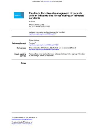
  • 11. but severe peak in cases will be more difficult for all services to cope with, compared with an identical number of cases distributed over a longer time course. For example, during the A/H3N2 pandemic a long first wave occurred in the winter of 1968/69 with morbidity and mortality approximately at the same level as the previous seasonal influenza; but in the following winter of 1969/ 70 a short and more severe epidemic occurred with a threefold higher peak in general practice consultation rates and a fourfold higher peak in mortality attributed to influenza, bronchitis and pneumonia. The high peak in consultation rates is well illustrated in figure 2.1. 2.5 Pandemic waves In 1918/19, the A/H1N1 pandemic occurred in three distinct epidemic waves: early spring 1918, autumn 1918 and late winter 1919. The second wave was by far the largest and case- fatality rates were also higher than in the first wave. The A/ H3N2 pandemic caused an epidemic wave in the winter of 1968/69 but a more severe one in 1969/70. In contrast, the second wave of the 1957/58 pandemic in the UK was very small in comparison to the first.7 Thus it should be considered a possibility that more than one wave of influenza will occur within a few months of the emergence of a pandemic virus and a subsequent wave could be worse than the first. 2.6 Health impact projections It is impossible to predict reliably with precision the level of excess mortality that will be experienced in the next pandemic. However, table 2.1 illustrates the broad range of excess mortality that it is reasonable to consider, based on various realistic combinations of case fatality rate and clinical attack rates derived from previous pandemics and epidemics. A case fatality rate of 0.37% corresponds to the aggregate rate observed in recent epidemic seasons (1989/90, 1991/92, 1993/ 94, 1995/96, 1996/97, 1997/98 and 1998/99) and the 1957 pandemic, although the overall case-fatality rate observed in the 1918–19 pandemic was in the region of 1–2%. A clinical attack rate of around 25% corresponds to the approximate clinical attack rate seen in all three previous pandemics of the twentieth century. Thus, a figure of at least 50,000 excess deaths is likely. Using mathematical projections, it is possible to illustrate the potential impact of the next pandemic, but these do not amount to accurate predictions. Table 2.2 summarises the number of events that might be expected by a GP with 1000 patients on his/her list and by a Primary Care Trust (PCT) serving a population of 100,000 persons. Using the same assumptions, table 2.3 illustrates the number of events by week over an assumed 15 week (single wave) pandemic period in a typical PCT population of 100 000. Most 0 100 200 300 400 500 600 700 800 900 1000 1100 1200 1300 1966/67 1967/68 1968/69 1969/70 1970/71 1971/72 1972/73 1973/74 1974/75 1975/76 1976/77 1977/78 1978/79 1979/80 1980/81 1981/82 1982/83 1983/84 1984/85 1985/86 1986/87 1987/88 1988/89 1989/90 1990/91 1991/92 1992/93 1993/94 1994/95 1995/96 1996/97 1997/98 1998/99 1999/00 2000/01 2001/02 2002/03 2003/04 2004/05 Year Rateper100000population 1969/70 1972/73 1975/76 1989/90 1967/68 Figure 2.1 Royal College of General Practitioners’ index for influenza and influenza-like illness, 1966 and 2005 (year marked at start of season, that is, week 40 (October)). Table 2.1 (A) Range of possible excess deaths based on various permutations of case-fatality rates and clinical attack rates for England and Wales Overall case fatality rate Clinical attack rate 10% 25% 50% 0.37% 19,300 48,400* 96,700 1.00% 51,700 129,200 258,400 1.5% 77,100 192,700 385,400 2.5% 129,200 323,000 645,900 *Corresponds to aggregate for recent epidemics (see text). Table 2.1 (B) Range of possible excess deaths based on various permutations of case-fatality rates and clinical attack rates for the UK Overall case fatality rate Clinical attack rate 10% 25% 50% 0.37% 21,500 53,700* 107,500 1.00% 56,700 141,800 283,700 1.5% 85,100 212,800 425,500 2.5% 141,800 354,600 709,300 *Corresponds to aggregate for recent epidemics (see text). Pandemic flu: clinical management of patients with an influenza-like illness during an influenza pandemic i7 www.thoraxjnl.com
  • 12. major acute trusts receive patients from a catchment area spanning several PCTs and the figures below require pro-rata adjustment before applying to individual hospitals. 3 CLINICAL FEATURES IN ADULTS Summary 1. Influenza is clinically defined as the presence of fever and new (or, in those with chronic lung disease, worsening) cough of acute onset in the context of influenza circulating in the community. This clinical definition may be modified once a pandemic occurs. 2. The spectrum of clinical disease associated with a pandemic strain cannot be forecast. 3. Pneumonia, either primary viral or secondary bacterial, is the commonest complication of influenza in adults. 4. Neurological complications are rare in adults. 3.1 How reliable is a clinical diagnosis of influenza infection during a pandemic? The clinical manifestations of infection by influenza viruses are diverse, ranging from asymptomatic infection to fulminant respiratory distress leading to respiratory failure and death. Furthermore, the presence of an ILI comprising of a combina- tion of fever, cough, sore throat, myalgia and headache is not specific for influenza infection. Other respiratory pathogens that may present with an ILI include viruses such as respiratory syncytial virus (RSV), adenovirus, rhinovirus and parainfluenza virus, as well as bacterial pathogens such as Chlamydia pneumoniae, Legionella sp, Mycoplasma pneumoniae and S pneumo- niae.14–16 Studies that have examined the value of a clinical definition of ILI in the diagnosis of influenza infection have not always used the same clinical definition for an ILI and have included different study populations, making comparison between studies complicated. A systematic review of the literature in this area identified the threefold combination of the presence of fever, cough and acute onset to be the most predictive clinical features. The accuracy of this clinical definition was higher in persons aged 60 years and above compared to patient groups without age restrictions (positive likelihood ratio (95% CI) 5.4 (3.8 to 7.7) v 2.0 (1.8 to 2.1)).17 The probability of influenza infection also increases with increasing level of fever.18 19 Importantly, the predictive value of clinical definitions based on an ILI increases when influenza virus is known to be Table 2.2 Estimated burden of illness attributable to pandemic influenza over the entire pandemic based on a 25% clinical attack rate and illustrative case hopitalisation and case- fatality rates of 0.55% and 0.37% respectively Population People with clinical symptoms/Health Care Contacts GP consultations A&E presentations Minimum excess hospitalisations Minimum excess deaths Population of 1000 250 (100–500) 25 (10–50) 13 (5–25) 1 (0–3) 1 (0–2) Population of 100,000 25,000 (10,000– 50,000) 2500 (1000–5000) 1250 (500–2500) 140 (50–300) 90 (40–180) Health Care Contacts represent the equivalent of GP consultations outside the pandemic period. It is envisaged that individuals experiencing symptoms will be diverted away from GPs in a pandemic. General practitioner consultations represent the remaining contacts required to deal with complications and with young children (see text for explanation). Figures are rounded and represent work additional to normal background health service activity. (Figures in parentheses illustrate the range from 10% (lower limit) to 50% (upper limit) attack rates.) Table 2.3 Demand for Health Care Contacts by primary care unit: weekly totals for the number of new clinical cases, and thus potential demand for Heath Care Contacts, per 100 000 population, and per Primary Care Trust (PCT), community pharmacy, GP practice or GP list of various sizes (see footnote for definition of ‘‘small’’, ‘‘medium’’ and ‘‘large’’ as they are used in the table) Period Clinical cases Cases per 100,000 % of total cases Cases per PCT Cases per pharmacy Cases per GP practice Cases per GP Small Medium Large Small Medium Large Small Medium Large Small Medium Large Week 1 21,367 36 0.1% 28 54 109 1 2 3 1 2 3 0 1 1 Week 2 30,400 51 0.2% 40 77 155 2 3 4 2 3 5 1 1 1 Week 3 121,886 205 0.8% 162 310 620 7 11 18 8 13 19 3 3 4 Week 4 464,219 780 3.1% 617 1181 2360 28 41 67 29 49 72 10 12 15 Week 5 1,569,434 2638 10.6% 2086 3992 7977 94 137 226 99 166 242 33 42 52 Week 6 3,206,013 5388 21.6% 4261 8155 16,295 192 280 462 203 339 494 67 85 106 Week 7 3,147,669 5290 21.2% 4183 8007 15,999 189 275 454 199 333 485 66 84 105 Week 8 2,122,779 3568 14.3% 2821 5400 10,790 127 185 306 134 224 327 44 56 70 Week 9 1,444,925 2428 9.7% 1920 3676 7344 87 126 208 91 153 223 30 38 48 Week 10 1,122,055 1886 7.5% 1491 2854 5703 67 98 162 71 119 173 23 30 37 Week 11 778,167 1308 5.2% 1034 1980 3955 47 68 112 49 82 120 16 21 26 Week 12 387,404 651 2.6% 515 985 1969 23 34 56 25 41 60 8 10 13 Week 13 232,944 392 1.6% 310 593 1184 14 20 34 15 25 36 5 6 8 Week 14 128,240 216 0.9% 170 326 652 8 11 18 8 14 20 3 3 4 Week 15 97,498 164 0.7% 130 248 496 6 9 14 6 10 15 2 3 3 All weeks 14,875,000 25,000 100% 19,770 37,839 75,606 891 1299 2145 942 1572 2292 311 396 494 ‘‘Small’’, ‘‘medium’’ and ‘‘large’’ refer to the 2.5th, 50th and 97.5th percentiles for the population served by a PCT, community pharmacy, GP practice or GP list, as follows. Small: PCT, 80,000; Pharmacy, 3600; GP practice, 3800; GP list, 1200. Medium: PCT, 150,000; Pharmacy, 5200; GP practice, 6300; GP list, 1600. Large: PCT, 300,000; Pharmacy, 8600; GP practice 9200; GP list, 2000. i8 Provisional guidelines from the BIS/BTS/HPA with the Department of Health www.thoraxjnl.com
  • 13. circulating in the community.15 17 20 In cohort studies, correla- tion of ILI with laboratory-confirmed influenza infection ranges from 25–45%, while in clinical trials rates of 70% have been consistently reported.15 21–23 These findings relate to influenza infections during inter- pandemic periods. During a global influenza pandemic, when a pandemic strain is known to be circulating locally in an immunologically susceptible population, the presence of an ILI would be expected to be highly predictive for influenza infection. (However, the extent to which a clinical diagnosis of ILI becomes predictive during a pandemic will also be determined by the behaviour of the public. If many, who would not normally present to a health professional, are prompted to present, then the predictive value of a clinical diagnosis of ILI will be reduced.) 3.2 What are the clinical features of uncomplicated influenza? The following description will relate mainly to interpandemic influenza A infections. Influenza B and C are not considered pandemic threats. Different strains may be associated with different clinical presentations and disease severity. For instance, there is evidence to suggest that the H3N2 subtype causes more severe disease than H1N1 subtype.24 The spectrum of clinical disease associated with a new influenza A subtype (for example, a pandemic strain) cannot be determined currently and may differ from that described for interpandemic influenza. The incubation period prior to the onset of symptoms is commonly 2–4 days (range 1–7 days). In adults, the illness typically presents as an abrupt onset of fever accompanied by a range of other symptoms as listed in Box 3.2.25–29 Fever is the paramount symptom and may reach 41˚C although more usually it ranges between 38–40˚C. The peak occurs within 24 hours of onset and lasts typically for three days (range 1–5 days).25–29 The cough is generally dry although in up to 40% of cases it may be productive. A productive cough together with chest tightness and substernal soreness is more common in patients with underlying chronic lung disease. Myalgia affects mainly the back and limbs. Gastrointestinal symptoms such as vomiting and diarrhoea are uncommon (,10%) in adults. Abdominal pain is rare. Clinical findings include a toxic appearance in the initial stages, hot and moist skin, a flushed face, injected eyes and hyperaemic mucous membranes around the nose and pharynx. Tender cervical lymphadenopathy is found in a minority (,10%) of cases. Wheezing or lung crackles are recognised findings (,10%) more commonly noted in patients with coexisting chronic lung disease. Although the overall clinical picture of uncomplicated influenza in any specific age group is similar for different influenza A subtypes, the frequency of certain symptoms may vary. For instance, during the ‘‘Asian’’ pandemic of 1957 (H2N2), headache and sore throat were frequent initial symptoms.30 In uncomplicated infection, the illness usually resolves in seven days although cough, malaise and lassitude may persist for weeks. Box 3.1 Clinical case definition of influenza (March 2006) The presence of fever and new (or, in those with chronic lung disease, worsening) cough of acute onset in the context of influenza circulating in the community. (Important note: this definition may be modified once a pandemic occurs.) Box 3.2 Range of symptoms associated with uncomplicated influenza infection N Cough (,85%) N Malaise (,80%) N Chills (,70%) N Headache (,65%) N Anorexia (,60%) N Coryzal symptoms (,60%) N Myalgia (,53%) and N Sore throat (,50%). Table 3.1 Complications associated with influenza infection in adults Complication Incidence Comments Respiratory Acute bronchitis Common More common in elderly and those with chronic medical conditions Primary viral pneumonia Uncommon Onset within 48 hours of start of fever Secondary bacterial pneumonia Common Typically occurs four to five days after onset of illness Cardiovascular ECG abnormalities Common Non-specific T wave and rhythm changes, ST segment deviation. Mostly not associated with cardiac symptoms Myocarditis Rare Pericarditis Rare Muscle Myositis Uncommon Occurs during early convalescence Myoglobinuria and renal failure Rare Central nervous system Encephalitis/encephalopathy Rare Occurs within first week of illness. More common in children and in Japan Transverse myelitis Very rare Guillain-Barre´ syndrome Very rare Others Otitis media Common Much more common in children Toxic shock syndrome Rare Parotitis Very rare Pandemic flu: clinical management of patients with an influenza-like illness during an influenza pandemic i9 www.thoraxjnl.com
  • 14. 3.3 What complications are associated with influenza infection? Influenza virus infection has been associated with worsening in the clinical condition of patients with a range of existing medical conditions, such as heart failure, diabetes, coronary heart disease, asthma and COPD. In addition, specific complications associated with influenza infection regardless of coexisting medical conditions are recognised (table 3.1). Based on data from interpandemic influenza, certain persons are identified as being at high risk from influenza-related complications. Such patients are similar to the group currently recommended for influenza vaccination by the Department of Health. These include those of all ages with chronic respiratory disease including asthma, chronic heart disease, chronic renal disease, chronic liver disease, immunosuppression due to disease or treatment, or diabetes mellitus, and all those aged 65 years or older, or those in long stay residential care (see Appendix 2). In the course of a pandemic, it may emerge that the patient group at high risk of complications differs from the group currently identified. In such circumstance, details of the ‘‘high risk’’ patient group will be altered according to relevant clinico- epidemiological data. 3.3.1 Influenza-related pneumonia The incidence of pneumonia (defined as a combination of respiratory symptoms and signs supported by chest radio- graphic changes consistent with infection) complicating influ- enza infection varies widely, from 2% to 38%, and is dependent on viral and host factors.25–27 Pneumonia generally occurs more frequently and with greater severity in patients with pre- existing chronic cardiac and respiratory conditions. Patients who develop pneumonia may present with symp- toms and signs indistinguishable from pneumonia related to other viral and bacterial pathogens. In the context of an influenza pandemic, the presence of an ILI and new or worsening dyspnoea should prompt a careful examination for the presence of complicating pneumonia. Two main types of influenza-related pneumonia are recognised: primary viral pneumonia and secondary bacterial pneumonia.25–28 Primary viral pneumonia Patients with primary viral pneumonia typically become breathless within the first 48 hours of onset of fever. An initially dry cough may become productive of blood-stained sputum. Cyanosis, tachypnoea, bilateral crepitations and wheeze on chest examination and leucocytosis are usual. The commonest chest radiographic abnormality is of bilateral interstitial infiltrates predominantly in the mid-zones, although focal consolidation is also well recognised. Rapid clinical deterioration with respiratory failure may ensue.31 The mortal- ity in hospitalised patients is high (.40%) despite maximum supportive treatment on intensive care.25–28 In the majority of fatal cases, death occurs within seven days of hospital admission. Secondary bacterial pneumonia Secondary bacterial pneumonia is more common (up to four times) than primary viral pneumonia. Typically, symptoms and signs of pneumonia develop during the early convalescent period (4–5 days from onset of initial symptoms). In others, symptoms of pneumonia blend in with the initial symptoms of influenza. Chest radiography usually demonstrates a lobar pattern of consolidation. Mortality rate ranges from 7% to 24%,25–29 32 although some small studies report higher mortality rates. The spectrum of pathogens implicated is similar to that observed in CAP and includes S pneumoniae, S aureus, H influenzae and Groups A, C and G beta-haemolytic streptococci.27 28 33–35 Different pathogens have predominated at different times. For instance, in the 1918 pandemic, H influenzae, beta-haemolytic streptococci and S pneumoniae were the predominant pathogens isolated. In 1968, S pneumoniae was the predominant pathogen (48%) followed by S aureus (26%) and non-typeable H influenzae (11%).34 Notably, S aureus was identified two and a half times more frequently during the 1968 pandemic compared to pneumonia occurring in the interpandemic period.34 36 Secondary staphylococcal pneumonia is associated with a higher incidence of lung abscess formation (14% v 2%) and carries a poorer prognosis compared to non-staphylococcal pneumonias (mortality 47% v 16%).25 29 32 37 During the 1957 pandemic, S aureus was the predominant bacterial pathogen isolated in fatal cases of influenza-related pneumonia (up to 69% of cases in some series).25 Mixed viral-bacterial pneumonia Bacterial and viral pneumonia can occur concurrently. In these instances, the chest radiograph may demonstrate lobar consolidation superimposed on bilateral diffuse lung infiltrates. The mortality rate in mixed viral-bacterial pneumonia is high (.40%), as for primary viral pneumonia.25–28 3.3.2 Cardiovascular Minor abnormalities on ECG such as ST segment deviation, T wave changes and rhythm disturbances have been described in uncomplicated influenza illness. They have been reported in up to 81% of patients hospitalised with influenza.25 Most do not have cardiac symptoms. Myocarditis and pericarditis are occasionally encountered in severe illness.38 39 Postmortem evidence of necrotising myocarditis has been reported in patients without clinically significant myocarditis in the antemortem period. 3.3.3 Myositis In contrast with myalgia affecting the back and limbs, which is common on initial presentation, myositis generally develops after the subsidence of the acute upper respiratory tract symptoms. The gastrocnemius and soleus muscles are typically involved with pain and tenderness to palpation. Complete recovery usually occurs in three days. Elevation in serum creatine phophokinase is recognised.40 41 Rarely, this is asso- ciated with myoglobinuria and renal failure.42 43 Myositis is more commonly described in children than adults. 3.3.4 Central nervous system Central nervous system (CNS) involvement in adults is uncommon. Most reports originate from Japan and occur in children.44 45 The main clinical syndrome is an encephalitis or encephalopathy manifesting in the form of decreased con- sciousness and seizures about three days (range 0–7 days) following the onset of upper respiratory tract symptoms. Focal neurological signs such as paresis, aphasia, choreoathetosis and cranial nerve palsies are less common. Cerebrospinal fluid (CSF) examination may be normal or reveal an elevation in protein or white cell count. Imaging by CT or MRI may be normal and if so, is indicative of a good prognosis and full recovery may be anticipated.46 Young age and abnormal CT/MRI findings are associated with a poor outcome including death or recovery with severe neurological sequelae (a fuller description is given in Section 4.2.6). Acute necrotising encephalopathy is a rare fulminant syndrome associated with multifocal brain lesions that is described mainly in Japan.46 Other rare manifestations include transverse myelitis and Guillain-Barre´ syndrome.47 48 Reye’s syndrome, characterised by an encephalopathy, acute fatty liver, association with aspirin use and high mortality i10 Provisional guidelines from the BIS/BTS/HPA with the Department of Health www.thoraxjnl.com
  • 15. (,40%), is a special situation that is almost exclusively seen in children and adolescents.46 Nevertheless, physicians managing adults are advised to be aware of this complication (a fuller description is given in Section 4.2.6.1.1). 3.3.5 Others Other complications rarely encountered in adults with influ- enza A infection include toxic shock syndrome in conjunction with secondary S aureus infection49 50 and parotitis.51 Otitis media is more commonly encountered in children than adults. 3.4 Avian influenza A (H5N1) infection in humans Human infections have been caused by different avian influenza A viruses in the past, including H9N2, H7N7, H7N3 and H7N2. In recent years, outbreaks of human infections by a novel strain of avian influenza A (H5N1) have raised particular concerns globally regarding the risk of a human pandemic.52 These concerns have been due in part to recognition that (a) avian influenza A (H5 N1) can pass directly from birds to humans and that (b) once in humans, avian influenza A (H5N1) causes severe disease with a high mortality. The full spectrum of human illness associated with avian influenza A (H5N1) infection is not completely known. Descriptions of the clinical features of influenza A (H5N1) infection in humans are based largely on case series of hospitalised patients. Subclinical infections, mild illnesses and atypical presentations of influenza A (H5N1) infections in humans have been reported, but the frequency of such infections is difficult to determine.53–55 In hospitalised patients, an ILI similar to that associated with seasonal influenza A (H1N1 or H3N2) infection is recognised. Gastrointestinal symptoms are present in a relatively large proportion of both adult and paediatric cases, in contrast to the relatively low incidence of gastrointestinal symptoms in seasonal influenza. The majority of patients develop a severe primary viral pneumonia usually associated with lymphopenia, thrombocytopenia and deranged liver function tests. Renal failure and multi-organ failure may develop subsequently. Mortality is high. A more detailed description is given in Appendix 10. Should influenza A (H5N1) acquire efficient human-to- human transmission capabilities, it may result in an influenza pandemic. In such an event, the clinical features of human H5N1 disease may alter. 4 CLINICAL FEATURES IN CHILDREN Summary 1. The commonest presenting features of influenza during an epidemic are fever, cough and rhinorrhoea. In infants, fever with non-specific symptoms or diarrhoea and vomiting is common; in older children pharyngitis and headache are frequent. 2. The clinical features of influenza in children during a pandemic cannot be forecast. 3. Children with underlying respiratory or cardiac disease, immune compromise or who are non-ambulant are more likely to be severely affected. 4. The younger the child the more likely hospital admission will be needed. 5. The severe and life-threatening complications of influenza are likely to be – bacterial pneumonia – acute respiratory distress syndrome – encephalopathy or encephalitis presenting as seizures or altered mental status. 4.1 What are the clinical features of uncomplicated influenza in children? The clinical features of influenza presenting in a pandemic cannot be predicted as they appear to be dependent on the strain of influenza and, in some respects, the host. A new strain of influenza A responsible for an epidemic or pandemic may result in a different spectrum of clinical features than previous strains.56 57 Common features during previous epidemics have been described and depend on the age of the child. The studies of clinical features are hospital based and are therefore likely to reflect more severe illness. These are nevertheless informative as one of the main issues in a pandemic is which patients require hospital admission. In young children presenting to primary care in a non-pandemic influenza season there are no specific clinical features that distinguish influenza from other winter viruses.58 Previously healthy infants and children 4.1.1 Neonates may present with non-specific signs of sepsis such as pallor, floppiness, (poor peripheral circulation, poor tone), lethargy, poor feeding, episodes of apnoea.59 Fever may be the only presenting feature. A North American study identified influenza as the most common reason for children aged 0–60 days being admitted to hospital during an epidemic with fever as the only clinical feature.60 4.1.2 Infants and very young children (under two years). Fever may be the only presenting feature in this age group too. They may also be irritable and toxic and are more likely than older children to present with gastrointestinal symptoms such as diarrhoea and vomiting. Febrile convulsions, particularly repeated convulsions, are positively associated with influenza A.61 Otitis media is also a common complication in children.62 Admission rates for under 2 year olds are 12 times higher than children aged 5–17 years.63 4.1.3 Older children. The presentation does not differ significantly from adults. Common features are sudden onset of high fever, chills (76–100%), cough, headache, sore throat, fatigue (51–75%), nasal stuffiness and conjunctivitis (26– 50%). Fever tends to settle 2–4 days later though a dry cough and clear nasal discharge last for 1–2 weeks.59 A clinical prediction model from North America for influenza in children has shown that the triad of cough, headache and pharyngitis had a sensitivity of 80% and a specificity of 78% for a positive viral culture for influenza.64 The subjects, mean age 6 years, presented during an epidemic to a suburban emergency department with a febrile respiratory illness and one or more symptoms of influenza. A Finnish retrospective study of children referred to hospital from 1980–99 with influenza confirmed by antigen testing reported that the median age for those with influenza A was 2 years. The most common features were cough, fever and rhinorrhoea.62 These were also the commonest features reported in a Chinese study where the mean age of the subjects with influenza A was 4 years.65 Children with underlying medical conditions 4.1.4 Children with asthma and other chronic medical conditions66 (table 4.1) and those who are not ambulant67 experience substantial morbidity during influenza seasons with a disproportionate number requiring inpatient care and ventilatory support. Of the 22% of previously healthy children who were hospitalised with influenza in Texas during the winter of 1998–99, 75% were under 1 year old. Of the 60% hospitalised who had underlying conditions, only 27% were under 1 year.68 Pandemic flu: clinical management of patients with an influenza-like illness during an influenza pandemic i11 www.thoraxjnl.com
  • 16. 4.2 Complications and rarer clinical features (table 4.2) 4.2.1 Pneumonia As in adults, influenza can present with either primary viral pneumonia or bacterial pneumonia most commonly caused by S pneumoniae or S aureus. There is much less published about pneumonia complicating influenza in children. An outbreak of severe pneumococcal pneumonia in children occurred in Iowa in the winter of 1995–96. This was coincident with an epidemic of influenza (H1N1). Compared with controls, patients were 12 times more likely to have experienced a recent ILI. There were also more likely to have family members with the illness and to have positive serology in the convalescent period. Many of these patients required chest drainage.69 Another study in 2002 of 202 children with proven influenza reported that 78 who had chest radiographs had either radiographic evidence of viral pneumonia or normal radio- graphs. No child had lobar pneumonia reported.70 Evidence from recent outbreaks of avian influenza (H5N1) in Hong Kong and Vietnam suggests that while some children had mild disease,71 others appeared to have multi-organ disease including acute respiratory distress syndrome (ARDS).57 All children who developed progressive pneumonia with ARDS died. There were no reports of bacterial pneumonia. There is no reason to believe that, apart from ARDS, pneumonia complicating influenza presents differently from CAP in children.72 The general clinical indicators for severity assessment of lower respiratory tract infection are summarised in the British Thoracic Society guidelines72 (Appendix 8). Failure to improve following 48 hours of antibiotics, or deterioration including a new, distinct spike of fever, should also be treated as severe and further complicating factors sought. 4.2.2 Croup The clinical course of croup caused by influenza appears to be more severe than croup caused by the more common parain- fluenza virus.73 It is more likely to be complicated by bacterial tracheitis.62 4.2.3 Otitis media Influenza is a well recognised cause of otitis media.74 It is the commonest bacterial superinfection of influenza and is reported in approximately 25% patients aged ,5 years.75 4.2.4 Bronchiolitis Influenza ranks second only to respiratory syncytial virus as a cause of bronchiolitis.76 The clinical features are the same.77 4.2.5 Febrile convulsions Children with influenza may present with febrile convulsions. In a community study in the Netherlands, recurrent febrile seizures were positively related to influenza A. It was recommended that children who have had a previous febrile convulsion should be immunised against influenza A.61 4.2.6 Encephalopathy and encephalitis These complications are described in small case series. 4.2.6.1 Encephalopathy This is defined as depressed or altered level of consciousness including lethargy and/or extreme irritability in younger children or significant change in personality or behaviour persisting beyond 24 hours, or confusion (older children). Encephalopathy usually presents as seizures within several days of the onset of fever.78 Seizures at this point are usually the first symptom of involvement of the CNS. Febrile convulsions, which are more likely to be repeated with influenza than with other causes of fever, generally occur with the onset of fever. Disturbances of behaviour and neurological deficit have been reported. A rapid and severe clinical course is usual with encephalopathy and is thought to be due to brain oedema mediated by cytokines rather than by direct invasion of the brain. Steroids are therefore considered. 202 children with encephalopathy were recognised in Japan between 1997 and 2001. Death occurred in 31%, residual neurological deficit in 26% and full recovery in 43%.79 4.2.6.1.1 Reye’s syndrome This is a rare childhood acute encephalopathy associated with liver dysfunction. The cause is unknown but it typically follows Table 4.2 Complications of influenza in children Complication Incidence Comments Respiratory Otitis media Very common Lung Common (,10%) The younger, the more likely to require hospital admissionBronchiolitis Primary viral pneumonia Secondary bacterial pneumonia Croup Presenting feature in ,5% Worse clinically than with para- influenza Central nervous system Febrile convulsions Common May be repeated Encephalopathy Rare Includes acute necrotising encephalopathy, Reye’s syndrome Encephalitis Rare Guillain-Barre´ Rare Others Myositis Rare Myocarditis Rare Pericarditis Rare Table 4.1 The most common underlying conditions in children admitted to hospital, Texas 1998–99.68 Asthma (42%) Congenital anomalies mostly cardiac (28.5%) Chronic lung disease of prematurity Immunodeficiencies Malignancies Renal disease Haemaglobinopathies Diabetes (and other metabolic conditions) i12 Provisional guidelines from the BIS/BTS/HPA with the Department of Health www.thoraxjnl.com
  • 17. viral illness and there is a clear association with aspirin therapy: thus an innate susceptibility coupled with aspirin taken for relief of viral symptoms. Influenza (particularly influenza B) is commonly implicated.80 There was a dramatic fall in incidence following warnings about aspirin use in children.81 It is possible that children on long term aspirin treatment for medical conditions may be at increased risk if they develop influenza infection. Reye’s syndrome is characterised by protracted vomiting and encephalopathy in afebrile patients with minimal or absent jaundice, and hepatomegaly in 50% of patients. It comprises: N acute non-inflammatory encephalopathy with an altered level of consciousness N elevation of ammonia levels 24–48 hours after the onset of mental status changes (the most frequent laboratory abnormality) N hepatic dysfunction with a liver biopsy showing fatty metamorphosis or a more than threefold increase in alanine aminotransferase (ALT), aspartate aminotransferase (AST). Neurological symptoms usually occur 24–48 hours after the onset of vomiting. Lethargy is usually the first neurological manifestation. Diarrhoea and hyperventilation may be the first signs in children younger than two years. Other investigations. Head CT scanning may reveal cerebral oedema but results are usually normal. An electro- encephalogram (EEG) may reveal slow wave activity in the early stages and flattened waves in advanced stages. Cerebrospinal fluid may or may not have increased opening pressure with white blood cells (WBCs) fewer than 9/ml3 (usually lymphocytes). There is no specific treatment for Reye’s syndrome. Key aspects of management are correction of metabolic imbalance and reduction of intracranial pressure. Advice should be requested from a specialist in metabolic medicine. Many children have an underlying inborn error of metabolism. Mortality has fallen from 50% to less than 20% as a result of earlier diagnosis and more aggressive therapy. 4.2.6.1.2 Acute necrotising encephalopathy Acute necrotising encephalopathy occurs mainly in Japan where it was first described in 1995. An estimated 100 deaths per annum are related to CNS complications of influenza in Japan.82 This suggests either a genetic predisposition for this complication or a variation in the strains of influenza circulating in Japan. Acute necrotising encephalopathy is characterised by high fever, convulsions and coma in children aged 1–5 years. The onset is 2–4 days after the respiratory symptoms and fewer than 10% of patients survive.83 There are no specific markers although some patients have raised liver transaminases. In many, the CSF is normal. Symmetrical multifocal brain lesions are seen and bilateral thalamic involvement is characteristic and may be demonstrated on MRI.83 4.2.6.2 Encephalitis This is defined as encephalopathy plus two of the following: fever of 38˚C or higher, seizures, focal neurological findings, WBC .5 cells/ml in CSF, electroencephalogram findings consistent with encephalitis, abnormal neuro-imaging.84 4.2.6.3 Differential diagnoses These must be considered when a child presents with altered level of consciousness or irritability. There is good evidence of an increased risk of meningococcal disease following influenza infection.85 During a pandemic, the focus will be on diagnosing influenza-related illness. Other neurological conditions or drug toxicity, for example, may be missed. 4.2.7 Myositis A literature review of 316 cases of myositis86 suggested that this was a complication mainly of schoolchildren. The calf muscles are predominantly affected. Rhabdomyolysis and renal failure are rare. 4.2.8 Myocarditis and pericarditis These are also rare complications but have been described in children with underlying medical conditions.62 Pandemic flu: clinical management of patients with an influenza-like illness during an influenza pandemic i13 www.thoraxjnl.com
  • 18. PART 1 Clinical management in primary care . . . . . . . . . . . . . . . . . . . . . . . . . . . . . . . . . . . . . . . . . . . . . . . . . . . . . . . . . . . . . . . . . . . . . . . . . . . . . . . . . . . . . . . . . . . . . . . . . . . . . . . . . . . . . . . . . . . . . . . . . . . . . . . . . . . Thorax 2007;62(Suppl I):i14–i18. doi: 10.1136/thx.2006.073080 5 GENERAL MANAGEMENT AND INVESTIGATIONS IN PRIMARY CARE 5.1 Triage With widespread concern during a pandemic, a significantly increased demand for advice and consultation should be anticipated. There are likely to be significantly higher consulta- tion rates for all types of respiratory tract infections including those which are normally managed well at home using over the counter remedies (for example, febrile colds, sore throat with temperatures). Consequently, demand management in both the practice and the Primary Care Trust (PCT) will be crucial to avoid the service’s capacity to triage care being overwhelmed. Guidance on demand management and health service delivery is given in the Primary Care Operational Plan (see Section 1.4).87 Management decisions of patients with influenza should be based primarily on: N an assessment of illness severity N identification of whether the individual is in an ‘‘at risk’’ group N current advice from Department of Health/local public health officials based on the epidemiology of the pandemic. Patients who are not considered to be at high risk and who have no features suggesting severe disease or complications may not need to be seen in face-to-face consultations by a primary care clinician. 5.2 General advice and symptomatic treatment in adults All patients presenting in general practice with symptoms suggestive of influenza (except perhaps those in whom urgent admission is required) should be given both general advice and advice on symptomatic treatment. It is important that clinicians identify and address individual concerns and expectations, provide information about the illness, and provide information about what patients can do to help themselves and when they should seek further help. Some useful facts that can be provided to the patient are included in Box 5.1. There is little scientific evidence for most symptomatic and self-help treatment, but experience suggests that some of the following may help, and are unlikely to cause harm: N treatment of fever, myalgias and headache with paracetamol or ibuprofen N rest N drinking plenty of fluids N avoiding smoking N consider: short course of topical decongestants, throat lozenges, saline nose drops. 5.3 General management in children Many infants and children will have coughs and mild fevers which may be due to other infections such as respiratory syncytial virus, especially over the winter months. These children should be managed in the usual way at home by parents with antipyretics and fluids. Note: aspirin should not be used in children. 5.3.1 Children with high fever (.38.5˚C) and cough or influenza-like symptoms Management of these children is determined by disease severity (see Appendix 5). The principles of symptomatic management are similar to those for adults. Recommendations N Children under one year of age year and those at high risk of complications (see Appendix 2) should be seen and assessed by a general practioner (GP) or at the A&E department. N Children age over 1 but under 7 years of age may be seen by a nurse or a GP and those aged 7 years and above may be seen by a member of the community health team (for example, community pharmacist). N All children (and parents) should be given advice on antipyretics and fluids. N Aspirin is contraindicated in children (aged under 16 years). 5.4 When should patients re-consult? Examples of what should prompt a patient to re-consult are given in Box 5.2. Patients who are started on antiviral agents (see Section 7 for indications for antiviral use) would be expected to begin to improve within 48 hours of starting treatment. Failure to improve two days after starting an Abbreviations: BTS, British Thoracic Society; CAP, community acquired pneumonia; COPD, chronic obstructive pulmonary disease; CRP, C-reactive protein; GP, general practitioner; HDU, high dependency unit; HPA, Health Protection Agency; ICU, intensive care unit; ILI, influenza-like illness; PCR, polymerase chain reaction; PCT, Primary Care Trust Box 5.1 Information about influenza to provide to patients N Influenza is caused by a number different types of ‘‘influenza’’ viruses. N The incubation period is typically 1–4 days and infected adults are usually contagious from the day of illness onset to five days after. Children are typically contagious for seven days, although sometimes for longer. N Fever usually declines after 2–3 days and normally disappears by the sixth day. N Cough, weakness and fatigue can persist for 1–2 weeks and up to six weeks. N Antibiotics do not benefit most people with influenza but are sometimes needed to treat secondary infections. Important note: this information may be modified once a pandemic occurs. i14 www.thoraxjnl.com
  • 19. antiviral agent is an indication to re-consult. At the time of re- consultation, an alternative diagnosis should be considered as well as the occurrence of any influenza-related complications. Recommendations N Any rapid deterioration following first consultation should prompt a patient to re-consult. N Failure to improve two days after starting an antiviral agent is an indication to re-consult. N If the first consultation did not involve contact with a physician, re-consultation should preferably involve a physician, usually a GP. 5.5 What general investigations should be done in the community? Recommendation N General investigations, including a chest x ray, are not necessary for the majority of patients managed in the community. 5.6 What microbiological investigations should be undertaken for patients in the community? The aim of microbiological investigations early in a pandemic (UK alert levels 1, 2 and 3) will be to confirm that influenza A is circulating in the local community. Once a pandemic is established (UK alert level 4), microbiological investigations are not recommended routinely or likely to be available readily. Routine testing for bacterial pathogens is not recommended at any stage. Recommendations N Where possible, early in a pandemic (UK alert levels 1, 2 and 3), nose and throat swabs, or nasopharyngeal swabs (in children), in virus transport medium should be submitted to the local laboratory. N Once a pandemic is established (UK alert level 4), micro- biological investigations are not recommended. 6 CRITERIA FOR HOSPITAL REFERRAL 6.1 Which adults require hospital referral? Adults with uncomplicated influenza infection usually do not require hospital referral. Patients who might require hospital admission fall into two main groups; those with worsening of a pre-existing medical condition and those with an influenza- related complication. 6.1.1 Worsening of pre-existing medical condition Patients who experience a worsening or clinical deterioration of pre-existing medical problems due to influenza infection should be managed according to recommended best practice for the medical condition in question. For instance, a patient with an acute exacerbation of chronic obstructive pulmonary disease (COPD) triggered by influenza infection should be managed according to current NICE Guidelines for COPD.88 Those with worsening of a pre-existing condition are likely to be in a group at ‘‘high risk’’ of influenza-related respiratory complications and consequently at risk of hospitalisation or death (Appendix 2). This group should be promptly reassessed if the illness is getting worse and considered for hospital referral. 6.1.2 Influenza-related pneumonia Pneumonia is the commonest influenza-related complication requiring hospital admission. Patients complaining of new or worsening dyspnoea should be carefully assessed for signs of pneumonia. If pneumonia is diagnosed, disease severity assessment is recommended and hospital referral made accordingly. There is no validated severity assessment tool developed specifically for influenza-related pneumonia. The CRB-65 score (table 6.1) is a well validated severity assessment tool developed for patients with community acquired pneumonia (CAP)89 90 and recommended in the British Thoracic Society (BTS) CAP Guidelines 2004 for use in the community setting.72 It is offered as an example of an assessment tool for influenza- related pneumonia. The use of any severity assessment tool does not replace clinical judgement. A patient’s social circumstances should also always be taken into account. In view of the rapid and fulminant course of primary viral pneumonia, patients with pneumonia who have bilateral chest signs (crackles) should be considered for hospital referral. 6.1.3 Other complications Other influenza-related complications are uncommon. There are no specific recommendations relating to criteria for hospital admission or disease severity assessment in these cases. Recommendations N Patients with clinically defined uncomplicated influenza infection would be expected to make a full recovery. They Box 5.2 Examples of what should prompt patients to re-consult N Shortness of breath at rest or while doing very little N Painful or difficult breathing N Coughing up bloody sputum N Drowsiness, disorientation or confusion N Fever for 4–5 days and not starting to get better (or getting worse) N Started to feel better then developing high fever and feeling unwell again N If taking antiviral drugs (for example, oseltamivir), symptoms should start to improve within two days. Lack of any improvement after two days from starting antiviral drugs is an indication to re-consult. Important note: this information may be modified once a pandemic occurs. Table 6.1 Severity assessment used to determine the management of influenza-related pneumonia in patients in the community (CRB-65 score) CRB-65 score* Recommended action 0 Likely suitable for home treatment 1 or 2 Consider hospital referral, particularly with score 2 3 or 4 Urgent hospital referral Any (0 to 4), in the presence of bilateral chest signs of pneumonia Consider hospital referral Score 1 point for each feature present: Confusion (Mental Test Score of (8, or new disorientation in person, place or time); Respiratory rate >30/min; Blood pressure (SBP ,90 mmHg or DBP (60 mmHg); age >65 years. Pandemic flu: clinical management of patients with an influenza-like illness during an influenza pandemic i15 www.thoraxjnl.com
  • 20. require good symptomatic management, access to antiviral treatment, information about the natural history, and advice as to when to re-consult. N Patients with new or worsening symptoms—particularly shortness or breath or recrudescent fever not responding to treatment—should be examined to assess the presence and severity of influenza-related pneumonia. N Patients with worsening of pre-existing comorbid medical conditions should be managed according to best practice for that condition with reference to published disease-specific guidelines, if available. N In patients with influenza-related pneumonia clinically, hospital referral and assessment should be considered for patients with a CRB-65 score of 1 or 2 (particularly score 2) and urgent admission for those with CRB-65 score of 3 or more. N Patients with bilateral chest signs of pneumonia should be referred to hospital for further assessment regardless of CRB-65 score. N The CRB-65 score does not replace clinical judgment. 6.2 Which children require hospital referral? (see Appendix 5) Recommendation N Children who are severely ill should be referred for assessment for admission. Indicators of severe disease are: (1) cyanosis (2) severe dehydration (3) altered conscious level (4) complicated or prolonged seizures (5) signs of sepsis such as extreme pallor, hypotension, a floppy infant (6) signs of respiratory distress such as markedly raised respiratory rate, grunting, intercostal recession or breathlessness with chest signs (a useful severity assessment tool for respiratory distress taken from the BTS pneumonia guidelines is given in Appendix 8). 7 ANTIVIRAL USE IN PRIMARY CARE 7.1 Introduction The guidance given in this Section summarises the key recommendations relevant to primary care. Full details relating to the principles and practice of antiviral use in adults and children are provided in Sections 13 and 19 respectively. Guidance relating to the delivery of antivirals is laid out in detail in the UK Operational Framework for stockpiling, distributing and using of antiviral drugs in the event of pandemic influenza5 and in the Primary Care Operational Plan. 7.2 What drugs should be used for antiviral treatment during a pandemic? Recommendations (see Appendix 9) N The antiviral treatment of choice is oseltamivir (TamifluH). This is given as a five-day course of oral tablets; 75 mg twice daily for adults. Liquid suspension is available for children from the age of 1 year upwards (see table 7.1). 7.3 What are the anticipated benefits of antiviral treatment? From clinical trial data accrued to date and based on seasonal, interpandemic influenza, the anticipated positive effect of antivirals in a pandemic will be: (a) reduction of illness duration by 24 hours, and therefore more rapid mobilisation of affected individuals including essential workers (b) a possible reduction in hospitalisation of infected indivi- duals (c) a reduction of subsequent antibiotic use by infected individuals. The evidence accrued to date does not suggest there will be a reduction of overall mortality, nor does it rule it out. 7.4 Who should receive antiviral drugs? Recommendations N Ideally, antiviral treatment should be offered to every patient who is over one year of age who (a) has an acute influenza-like illness (b) fever (>38˚C in adults, or >38.5˚C in children) and (c) presents within 48 hours of the onset of symptoms. N Exceptions: (i) Patients who are unable to mount an adequate febrile response—for example, the immunocompromised or very elderly—may still be eligible for antiviral treat- ment despite the lack of documented fever. (ii) Immunosuppressed patients, including those on long term corticosteroid therapy, may suffer more prolonged viraemia, and could possibly benefit from antiviral therapy commenced later than 48 hours after the onset of influenza-like illness (ILI). (iii) Patients who are severely ill, but who have not been hospitalised due to non-clinical reasons, may benefit from antiviral therapy commenced later than 48 hours after the onset of ILI. There is no strong evidence to support antiviral use in these exceptional situations. 7.5 What are the adverse effects of oseltamivir? The commonest adverse effect of oseltamivir is nausea in about 10% of patients. This can be managed with mild anti-emetic medication. Other side effects are listed in Appendix 9. 7.6 Delivery of antivirals in primary care National distribution arrangements are laid out in the UK Operational Framework for stockpiling, distributing and using antiviral drugs in the event of pandemic influenza5 and the Primary Care Operational Plan. The drug will be made available through these arrangements to pharmacies, PCTs and/or GP surgeries. Table 7.1 Adult and child dosages of oseltamivir Child aged .1 year; body weight 15 kg or lower 30 mg 12-hourly Child .15–23 kg 45 mg 12-hourly Adult, and child >24 kg 75 mg 12-hourly Dose to be reduced by 50% if creatinine clearance is less than 30 ml/ minute. i16 Provisional guidelines from the BIS/BTS/HPA with the Department of Health www.thoraxjnl.com