Male reproductive system
•Download as PPTX, PDF•
4 likes•852 views

Most species have two sexes: male and female. Each sex has its own unique reproductive system. They are different in shape and structure, but both are specifically designed to produce, nourish, and transport either the egg or sperm. Unlike the female, whose sex organs are located entirely within the pelvis, the male has reproductive organs, or genitals, that are both inside and outside the pelvis. The male genitals include: the testicles the duct system, which is made up of the epididymis and the vas deferens the accessory glands, which include the seminal vesicles and prostate gland the penis

Recommended
More Related Content
What's hot
What's hot (20)
Similar to Male reproductive system
Similar to Male reproductive system (20)
More from christenashantaram
More from christenashantaram (20)
Recently uploaded
Recently uploaded (20)
Male reproductive system
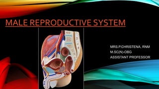
- 1. MALE REPRODUCTIVE SYSTEM MRS.P.CHRISTENA, RNM M.SC(N)-OBG ASSISTANT PROFESSOR
- 2. INTRODUCTION • Most species have two sexes: male and female. Each sex has its own unique reproductive system. They are different in shape and structure, but both are specifically designed to produce, nourish, and transport either the egg or sperm. • Unlike the female, whose sex organs are located entirely within the pelvis, the male has reproductive organs, or genitals, that are both inside and outside the pelvis. The male genitals include: • the testicles • the duct system, which is made up of the epididymis and the vas deferens • the accessory glands, which include the seminal vesicles and prostate gland • the penis
- 3. PURPOSES • The purpose of the organs of the male reproductive system is to perform the following functions: • To produce, maintain, and transport sperm (the male reproductive cells) and protective fluid (semen) • To discharge sperm within the female reproductive tract during sex • To produce and secrete male sex hormones responsible for maintaining the male reproductive system
- 4. EXTERNAL MALE REPRODUCTIVE STRUCTURES • Most of the male reproductive system is located outside of your abdominal cavity or pelvis. The external parts of the male reproductive system include • the penis, • the scrotum and • the testicles.
- 5. THE PENIS
- 6. • The penis is an external organ of the male reproductive system. It has two main functions: • Sexual intercourse – During erotic stimulation, the penis undergoes erection, becoming engorged with blood. Following emission, (mixing of the components of semen in the prostatic urethra) ejaculation can occur, whereby semen moves out of the urethra through the external urethral orifice. Finally, the penis undergoes remission, returning to a flaccid state. • Micturition – The penis also has an important urinary role. It contains the urethra, which carries urine from the bladder to the external urethral orifice, where it is expelled from the body.
- 7. STRUCTURE OF THE PENIS • The penis can be anatomically divided into three parts: • Root – the most proximal, fixed part of the penis. It is located in the superficial perineal pouch of the pelvic floor, and is not visible externally. The root contains three erectile tissues (two crura and bulb of the penis), and two muscles (ischiocavernosus and bulbospongiosus). • Body – the free part of the penis, located between the root and glans. It is suspended from the pubic symphysis. It is composed of three cylinders of erectile tissue – two corpora cavernosa, and the corpus spongiosum. • Glans – the most distal part of the of penis. It is conical in shape, and is formed by the distal expansion of the corpus spongiosum. This contains the opening of the urethra, termed the external urethral orifice.
- 8. Erectile Tissues • The erectile tissues fill with blood during sexual arousal, producing an erection. The root and body of the penis are spanned by three masses of erectile tissue. • In the root, these tissues are known as the left and right crura, and the bulb of the penis. • The erectile tissues continue into the body of the penis. • The left and right crura continue anteriorly into the dorsal part of the penis – they form the two corpora cavernosa. • They are separated by the septum of the penis, although often incompletely. The bulb forms the corpus spongiosum, which lies ventrally. • The male urethra runs through the corpus spongiosum – to prevent it becoming occluded during erection the corpus spongiosum fills to a reduced pressure. • Distally, the corpus spongiosum expands to form the glans penis.
- 10. • The prepuce (foreskin) is a double layer of skin and fascia, located at the neck of the glans. It covers the glans to a variable extent. • The prepuce is connected to the surface of the glans by the frenulum, a median fold of skin on the ventral surface of the penis. • The potential space between the glans and prepuce is termed the preputial sac. • Vasculature- The penis receives arterial supply from three sources: • Dorsal arteries of the penis • Deep arteries of the penis • Bulbourethral artery • Innervation • Sensory and sympathetic innervation to the skin and glans penis is supplied by the dorsal nerve of the penis, a branch of the pudendal nerve.
- 11. THE TESTES AND EPIDIDYMIS • The testes and epididymis are paired structures, located within the scrotum. • The testes are the site of sperm production and hormone synthesis, while the epididymis has a role in the storage of sperm. • The testes have an ellipsoid shape. • They consist of a series of lobules, each containing seminiferous tubules supported by interstitial tissue. • The seminiferous tubules are lined by Sertoli cells that aid the maturation process of the spermatozoa. • In the interstitial tissue lie the Leydig cells that are responsible for testosterone production. • Spermatozoa are produced in the seminiferous tubules. The developing sperm travels through the tubules, collecting in the rete testes. • Ducts known as efferent tubules transport the sperm from the rete testes to the epididymis for storage and maturation.
- 12. • Inside the scrotum, the testes are covered almost entirely by the tunica vaginalis, a closed sac of parietal peritoneal origin that contains a small amount of viscous fluid. This sac covers the anterior surface and sides of each testicle and works much like the peritoneal sac, lubricating the surfaces of the testes and allowing for friction-free movement. • The testicular parenchyma is protected by the tunica albuginea, a fibrous capsule that encloses the testes. It penetrates into the parenchyma of each testicle with diaphragms, dividing it into lobules. • The epididymis consists of a single heavily coiled duct. It can be divided into three parts; head, body and tail. • Head – The most proximal part of the epididymis. It is formed by the efferent tubules of the testes, which transport sperm from the testes to the epididymis. • Body – Formed by the heavily coiled duct of the epididymis. • Tail – The most distal part of the epididymis. It marks the origin of the vas deferens, which transports sperm to the prostatic portion of the urethra for ejaculation.
- 14. Innervation • The testes and epididymis receive innervation from the testicular plexus Vascular Supply • The main arterial supply to the testes and epididymis is via the paired testicular arteries, • However, the testes are also supplied by branches of the cremasteric artery (from the inferior epigastric artery) and the artery of the vas deferens (from the inferior vesical artery). • Venous drainage is achieved via the paired testicular veins Lymphatics • Since the testes are originally retroperitoneal organs, the lymphatic drainage is to the lumbar and para-aortic nodes, along the lumbar vertebrae. • This is in contrast to the scrotum, which drains into the nearby superficial inguinal nodes.
- 15. THE SCROTUM • The scrotum contains three major (paired) structures: • Testis – the site of sperm production. • Epididymis – situated at the head of each testicle. It functions as a storage reservoir for sperm. • Spermatic cord – a collection of muscle fibres, vessels, nerves and ducts that run to and from the testes. • There are also muscle fibres located within the scrotum.
- 16. Neurovascular Supply • The scrotum receives neurovascular supply from the nearby vessels and nerves. This is in contrast to the testes – which carry their vessels, nerves and lymph drainage from the abdomen during their development. Vessels • The scrotum receives arterial supply from the anterior and posterior scrotal arteries. The anterior scrotal artery arises from the external pudendal artery, while the posterior is derived from the internal pudendal artery. • The scrotal veins follow the major arteries, draining into the external pudendal veins. Nerves • Cutaneous innervation to the scrotum is supplied via several nerves, according to the topography: • Anterior and anterolateral aspect – Anterior scrotal nerves derived from the genital branch of genitofemoral nerve and ilioinguinal nerve • Posterior aspect – Posterior scrotal nerves derived from the perineal branches of the pudendal nerve and posterior femoral cutaneous nerve. Lymphatics • The lymphatic fluid from the scrotum drains to the nearby superficial inguinal nodes.
- 17. THE SPERMATIC CORD • The anatomical course of the spermatic cord is relatively short, beginning in the inferior abdomen and ending in the scrotum. • The spermatic cord is formed at the opening of the inguinal canal, known as the deep inguinal ring. This opening is located laterally to the inferior epigastric vessels. • The cord passes through the inguinal canal, entering the scrotum via the superficial inguinal ring. It continues into the scrotum, ending at the posterior border of the testes. Here, its contents disperse to supply the various structures of the testes and scrotum.
- 18. The spermatic cord conveys several important structures that run to and from the testis. • Blood vessels: • Testicular artery – branch of the aorta that arises just inferiorly to the renal arteries. • Cremasteric artery and vein – supplies the cremasteric fascia and muscle. • Artery to the vas deferens – branch of the inferior vesicle artery, which arises from the internal iliac. • Pampiniform plexus of testicular veins – drains venous blood from the testes into the testicular vein. • Nerves: • Genital branch of the genitofemoral nerve – supplies the cremaster muscle. • Autonomic nerves • Other structures: • Vas deferens – the duct that transports sperm from the epididymis to the ampulla (a dilated terminal part of the duct), ready for ejaculation. • Processus vaginalis – projection of peritoneum that forms the pathway of descent for the testes during embryonic development. In the adult, it is fused shut. • Lymph vessels – these drain into the para-aortic nodes, located in the lumbar region.
- 20. THE PROSTATE GLAND • The prostate is positioned inferiorly to the neck of the bladder and superiorly to the external urethral sphincter, with the levator ani muscle lying inferolaterally to the gland. • Most importantly, posteriorly to the prostate lies the ampulla of the rectum – this anatomical arrangement is utilised during Digital Rectal Examinations (DRE), allowing physicians to examine the gland. • The proteolytic enzymes leave the prostate via the prostatic ducts. These open into the prostatic portion of the urethra, through 10-12 openings at each side of the seminal colliculus (or verumontanum); secreting the enzymes into the semen immediately before ejaculation. Vasculature • The arterial supply to the prostate comes from the prostatic arteries • Venous drainage of the prostate is via the prostatic venous plexus Innervation • The prostate receives sympathetic, parasympathetic and sensory innervation from the inferior hypogastric plexus
- 22. THE BULBOURETHRAL GLANDS • The bulbourethral glands (also known as Cowper’s glands) are a pair of pea shaped exocrine glands located posterolateral to the membranous urethra. • They contribute to the final volume of semen by producing a lubricating mucus secretion. Embryology • Embryologically the bulbourethral glands are derived from the urogenital sinus, along with the bladder, prostate and urethra. Their development is greatly influenced by DHT (dihydrotestosterone). • The bulbourethral glands (also known as Cowper’s glands) are a pair of pea shaped exocrine glands located posterolateral to the membranous urethra. They contribute to the final volume of semen by producing a lubricating mucus secretion. • In this article, we shall look at the anatomy of the bulbourethral glands – their structure, vasculature and innervation. Function • During sexual arousal, the bulbourethral glands produce a mucus secretion containing glycoproteins. This substance has three main purposes: • Serves as lubrication medium for the urethra and the tip of the penis. • Expels any residue of urine, dead cells or mucous through the urethral meatus, preparing a clean and lubricated pathway for ejaculation. • Helps to neutralise residual acidity in the male urethra (secretions are alkaline).
- 24. Vasculature • The arterial supply of the bulbourethral glands is derived from the arteries to the bulb of the penis. Innervation • In a mammal study (in pigs), neurons projecting to the bulbourethral glands were found in pelvic ganglia (PG), sympathetic chain ganglia (L2–S3), the caudal mesenteric ganglion and dorsal root ganglia (L1–L3, S1–S3); • They reach the bulbourethral glands via the the hypogastric nerve and the pelvic nerve or pelvic branch of the pudendal nerve. Lymphatics • Like the seminal vesicles the bulbourethral glands drain into the internal and external iliac lymph nodes
- 25. THE SEMINAL VESICLES • The seminal vesicles (also known as the vesicular or seminal glands) are a pair of glands found in the male pelvis, which function to produce many of the constituent ingredients of semen. • They ultimately provide around 70% of the total volume of semen. Embryology • The Seminal glands, along with the Ejaculatory ducts, Epididymis and Ductus (vas) deferens, are derived from the mesonephric ducts, the precursor structure of male internal genitalia. • These structures can easily be remembered using the acronym SEED.
- 26. Function • The secretions of the seminal gland have a key role in the normal functioning of semen, making up 70% of its total volume. • It is notable however that the first fractions of expelled semen contain mainly spermatozoa and prostatic secretions; the fluids from the seminal vesicles are included in the late ejaculate fractions. These fluids contain: • Alkaline fluid – neutralises the acidity of the male urethra and vagina in order to facilitate the survival of spermatozoa. • Fructose – provides an energy source for spermatozoa. • Prostaglandins – have a role in suppressing the female immune response to foreign semen. • Clotting factors – designed to keep semen in the female reproductive tract post- ejaculation. • The remaining volume of semen is made up of testicular spermatozoa, prostatic secretions and mucus from the bulbourethral gland.
- 27. Vasculature • The arteries to the seminal gland are derived from the inferior vesicle, internal pudendal and middle rectal arteries, all of which stem from the internal iliac artery. Innervation • The innervation of the gland, like much of the male internal genitalia, is mainly sympathetic in origin. Lymphatic Drainage • The lymphatic drainage of the gland is the external and internal iliac lymph nodes.
- 30. REFERNCES • Dutta, D. C., Hiralal, K., & Konar, H. (2018). DC Dutta’s Textbook of Obstetrics: Including Perinatology and Contraception (9th ed.). Jaypee Brothers Medical Pub. • Fhea, W. A. M. C. S. R., & Rgn, G. A. B. P. (2018). Ross & Wilson Anatomy and Physiology in Health and Illness (13th ed.). Elsevier. • Male Reproductive System. (2021). RCHSD. https://www.rchsd.org/health-articles/male-reproductive-system/ • The Male Reproductive System. (2002, February 5). WebMD. https://www.webmd.com/sex-relationships/guide/male-reproductive- system • The Male Reproductive System. (2020, June 18). TeachMeAnatomy. https://teachmeanatomy.info/pelvis/the-male-reproductive-system/