
Recommended
More Related Content
What's hot
What's hot (19)
Viewers also liked
Viewers also liked (9)
Similar to DIPA 2000 Particle Size & Shape Analyzer
Similar to DIPA 2000 Particle Size & Shape Analyzer (20)
Recently uploaded
Recently uploaded (20)
DIPA 2000 Particle Size & Shape Analyzer
- 1. Particle Size & Shape Analyzer
- 2. Slide 3/43 “Equivalent Sphere” Attitude These diameters are fundamentally different for the same particle. The equivalent sphere diameters differ for any non-spherical material. Every PSA technique yields different results for non-spherical particles.
- 3. Slide 5/43 DIPA-2000 Measuring Channels Single Instrument ; Dual Measurement Channels
- 4. Slide 6/43 DIPA-2000 Laser Channel Main Advantages: Quick & Straight Forward Measurement High Resolution Detection Capabilities Time Domain Analysis, No “Pre-Knowledge”
- 5. Slide 7/43 DIPA-2000 Laser Channel The rotating laser beam A B C D E F scans single particles Scanning Laser within its focus. The diameter of the particle is directly correlated to the duration of the obscuration. V The measurement principle is a time interval, not an intensity measurement. The time domain is independent of particles Ta Tb Tc Td Te Tf Time optical or physical properties.
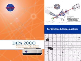
- 6. Slide 8/43 DIPA-2000 Laser Channel From the duration of the obscuration (T), and the known rotation velocity of the laser beam (V), the particle diameter (D) can be calculated: D=V·T In relation to the high speed of the laser rotating laser, the particles are stationary, thus will not effect particle size measurement. No requirements for particles pre-knowledge of: 1. Refractive index 2. Temperature 3. Viscosity variation 4. Electrical conductivity 5. Etc…
- 7. Slide 9/43 DIPA-2000 Laser Channel Summery Data is collected directly on each analyzed particle. Detection of minor fractions, high resolution. Correct measurement of particle size, not influenced by physical or optical properties. Operation in Liquid, Dry and Aerosol mediums. No calibration/alignment required by user. Robust measurement, on-line indication for statistical significance. Concentration measurement. FDA 21 CFR Part 11 S/W security compliance.
- 8. Slide 10/43 DIPA-2000 Measuring Channels Single Instrument ; Dual Measurement Channels
- 9. DIPA-2000 Video Channel Main Advantages: Sample Visualization Dynamic Shape Analysis Advanced Particles Size and Shape information
- 10. Slide 14/43 Dynamic Shape Analysis (DSA) Principle of Operation Real time images of particles in motion are collected Images are converted into a grid containing a collection of picture elements (pixels) 420 µm Each individual pixel has a value for brightness Image (grey level): 0 = Dark; 255 = White Conversion All images are analyzed according to a pre-defined set of Image Analysis characteristics Measurement is finished when the analysis end conditions are met (# of particles/images, confidence level, time, etc) 18 µm
- 11. Slide 15/43 DIPA-2000 Video Channel DIPA-2000 DSA Main Features: • Easy setup and measurement using software wizards • Advanced pre-processing algorithms • Size and Shape Filter/Grouping • Automated Report Generation • Storage of real raw date (un-processed images) • Re-processing of raw data • Internal optical calibration and verification tools available • Interface with 3rd party hardware (Microscopy)
- 12. Slide 16/43 DIPA-2000 Video Channel • For each individual particle, all size and shape parameters are determined. • The DIPA-2000 Image Analysis S/W automates set-up and measurement. • DIPA-2000 IA software includes many procedures such as: – Pre-Processing Procedures – Image Quality Filters – Region of Interest Determination – Out-Of-Focus Rejection – Morphology Operations – Grouping according to size/shape – Re-Processing of stored images and movies – Manual Lens Calibration (Microscopy)
- 13. Slide 20/43 DIPA-2000 Video Channel Summery Seeing is believing, visualize your sampled particles. Better and advanced characterization of materials. High resolution data (down to pixel size data). Detection and quantification of shape parameters. Validation technique for the Laser Channel or other particle size methods. Detection capability of minor fractions for fine particles or agglomerated materials. Quantification of material homogenity. Advanced tool for shape differences characterisation. FDA 21 CFR Part 11 S/W security compliance.
- 14. Slide 22/43 One System = Variety of Applications “Adapting the technology to the application”, rather than - “adapting the application to the technology”
- 15. Slide 23/43 Some Global Applications Geology Soil, clay, sand, kaolin Ocean water, tap water, waste water, membrane filtration, Environmental dust, flocculation Powders, suspensions, syrups, emulsions, pastes, micro- Pharma/Bio-Tech carriers, injectable solutions, collagen, microcapsules Pesticides, dispersants, catalysts, resins, emulsions, Chemicals preservatives Alumina, silica, magnetic powders, tungsten, strontium, Ceramics & Metals sintered products, stainless steel, cobalt Energy Coal, fuels, slurries, shale-oil emulsions, fly ash Emulsions, fine powders, beer, coffee, chocolate, flour, Food Products ground products, agglomerated crystals, peanut butter Polymers, oil droplets, wear particles, calk, fillers, toners, Heavy Industry pulp & paper, coatings, pigments, PVC, paint Bacteria, smears, inhalation toxicology, cell research, Life Science algae growth, yeast, blood analysis
- 16. Slide 24/43 DIPA 2000 Particle Size & Shape Analyzer Elementary and Advanced S/W Features
- 17. Slide 26/43
- 18. Slide 27/43
- 19. Slide 31/43
- 20. Slide 33/43
- 21. Slide 35/45
- 22. Slide 36/43
- 23. Slide 37/43
- 24. Slide 38/43
- 25. Slide 39/43
- 26. Slide 41/43
- 27. Slide 43/43 DIPA 2000 S/W Features Summery • Easy setup and measurement using software wizards • Advanced pre-processing algorithmes • Size and Shape Grouping/Filtering • Automated report generator • Storage of real-time raw data (un-processed images) • Re-processing of raw data (images & movie files) • Interface with 3rd party hardware (e.g. Microscopy) • Manual calibration and verification tools avaible • Integrated laser and video software • Simultaneous laser and video results • FDA 21 CFR Part 11 S/W security compliance
Editor's Notes
- Now that we have measured the duration of the obscuration, it is simple to calculate the size of the particle:Since the rotation velocity of the laser beam (by the wedge prism) is known and strictly controlled by the DIPA-2000, the obscuration time is used for calculating the particle’s size with a simple formula: Particle Diameter equals velocity multiplied by obscuration time.This formula holds true under the condition that the particles are stationary relative to the scanning motion of the laser beam. Since the laser beam is rotating at a much higher speed than the sample flow, this is automatically achieved.A strong advantage of this principle is that the measurement does not require any pre-knowledge of the material under investigation.