SlideShare a Scribd company logo
1 of 13
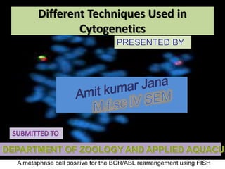
A metaphase cell positive for the BCR/ABL rearrangement using FISH
Different Techniques Used in
Cytogenetics
Introduction & Definition
• Cytogenetics is a branch of genetics that is
concerned with the study of the structure and
function of the cell, especially the
chromosomes. It includes routine analysis of G-
banded chromosomes, other cytogenetic
banding techniques, as well as molecular
cytogenetics such as Fluorescent In
Situ Hybridization (FISH) and Comparative
Genomic Hybridization (CGH).
History
• Chromosomes were first observed in plant cells by Karl Wilhelm von Nägeli in
1842. Their behavior in animal (salamander) cells was described by Walther
Flemming, the discoverer of mitosis, in 1882. The name was coined by another
German anatomist, von Waldeyer in 1888.
• In science books, the number of human chromosomes remained at 48 for over
thirty years. New techniques were needed to correct this error. Joe Hin Tjio
working in Albert Levan’s lab was responsible for finding the approach:
• Using cells in culture
• Pre-treating cells in a hypotonic solution, which swells them and spreads the
chromosomes
• Arresting mitosis in metaphase by a solution of colchicine
• Squashing the preparation on the slide forcing the chromosomes into a single
plane
• Cutting up a photomicrograph and arranging the result into an indisputable
karyogram.It took until 1956 until it became generally accepted that the
karyotype of man included only 46 chromosomes.Rather interestingly,
the greatapes have 48 chromosomes. Human chromosome 2was formed by a
merger of ancestral chromosomes, reducing the number.
Techniques
1. Karyotyping
1. Slide preparation
2. Analysis
2. Fluorescent in situ hybridization
1. Slide preparation
2. Analysis
Techniques Karyotyping
• Routine chromosome analysis (Karyotyping) refers to analysis
of metaphase chromosomes which have been banded
using trypsin followed by Giemsa, Leishmanns, or a mixture of the
two.
Several chromosome-banding techniques are used in cytogenetics
laboratories.
Quinacrine banding (Q-banding) was the first staining method used to
produce specific banding patterns. This method requires a
fluorescence microscope and is no longer as widely used
as Giemsa banding (G-banding). Reverse banding, or R-banding,
requires heat treatment and reverses the usual black-and-white
pattern that is seen in G-bands and Q-bands. This method is
particularly helpful for staining the distal ends of chromosomes.
Other staining techniques include C-banding.
Slide Preparation
• Cells from bone marrow, blood, amniotic fluid, cord blood, tumor, and
tissues (including skin, umbilical cord, chorionic villi, liver, and many
other organs) can be cultured using standard cell culture techniques in
order to increase their number.
• A mitotic inhibitor (colchicine, colcemid) is then added to the culture.
This stops cell division at mitosis which allows an increased yield of
mitotic cells for analysis. The cells are then centrifuged and media and
mitotic inhibitor are removed, and replaced with a hypotonic solution.
This causes the white blood cells or fibroblasts to swell so that the
chromosomes will spread when added to a slide as well as lyses the
red blood cells. After the cells have been allowed to sit in hypotonic
solution, Carnoy's fixative (3:1 methanol to glacial acetic acid) is
added. This kills the cells and hardens the nuclei of the remaining
white blood cells. The cells are generally fixed repeatedly to remove
any debris or remaining red blood cells. The cell suspension is then
dropped onto specimen slides. After aging the slides in an oven or
waiting a few days they are ready for banding and analysis.
Analysis
Analysis of banded chromosomes is done at
a microscope by a clinical laboratory specialist in
cytogenetics (CLSp(CG)). Generally 20 cells are
analyzed which is enough to rule out mosaicism to
an acceptable level. The results are summarized and
given to a board-certified cytogeneticist for review,
and to write an interpretation taking into account
the patients previous history and other clinical
findings.
Fluorescent in situ hybridization
Fluorescent in situ hybridization refers to using
fluorescently labeled probe to hybridize to cytogenetic
cell preparations.
In addition to standard preparations FISH can also be
performed on:
1. bone marrow smears
2. blood smears
3. paraffin embedded tissue preparations
4. enzymatically dissociated tissue samples
5. uncultured bone marrow
6. uncultured amniocytes
7. cytospin preparations Fig of FSH
Slide preparation
• The slide is aged using a salt solution usually
consisting of 2X SSC (salt, sodium citrate). The slides
are then dehydrated in ethanol, and the probe
mixture is added. The sample DNA and the probe
DNA are then co-denatured using a heated plate
and allowed to re-anneal for at least 4 hours. The
slides are then washed to remove excess unbound
probe, and counterstained with 4',6-Diamidino-2-
phenylindole (DAPI) or propidium iodide.
Analysis
• Analysis of FISH specimens is done
by fluorescence microscopy by a clinical
laboratory specialist in cytogenetics. For oncology
generally a large number of interphase cells are
scored in order to rule out low-level residual
disease, generally between 200 and 1,000 cells
are counted and scored. For congenital problems
usually 20 metaphase cells are scored.
Future of cytogenetics
• Advances now focus on molecular cytogenetics
including automated systems for counting the
results of standard FISH preparations and
techniques for virtual karyotyping, such as
comparative genomic hybridization arrays, CGH
and Single nucleotide polymorphism arrays.
Conclusion
• So far, no system can classify banded chromosomes as
robustly and accurately as a skilled cytogeneticist,
despite the millions of dollars that have been invested
in automated karyotype analysis since 1968. Currently
cytogenetics is paving its way into the molecular
approaches in deciphering the structure, function and
evolution of chromosomes. Still,conventional
cytogenetics where routine banding techniques are
employed remains a simple and popular technique to
get an overview of the human genome. Routine
banded karyotype analysiscan now be combined with
M-FISH and other molecular techniques leading to
moreprecise detection of various syndromes in
children.
Different techniques used in cytogenetics

More Related Content

What's hot

Fluorescent in situ hybridization (FISH)
Fluorescent in situ hybridization (FISH)Fluorescent in situ hybridization (FISH)
Fluorescent in situ hybridization (FISH)Gauravrajsinh Vaghela
 
Insuite hybridization
Insuite hybridizationInsuite hybridization
Insuite hybridizationNoman Ch
 
Hormonal cytology
Hormonal cytologyHormonal cytology
Hormonal cytologyAnkita072
 
Comparative genomic hybridization
Comparative genomic hybridizationComparative genomic hybridization
Comparative genomic hybridizationvlmawia
 
Advanced Techniques In Molecular Cytogenetics Karyotyping, FISH, CGH, SKY
Advanced Techniques In Molecular Cytogenetics Karyotyping, FISH, CGH, SKYAdvanced Techniques In Molecular Cytogenetics Karyotyping, FISH, CGH, SKY
Advanced Techniques In Molecular Cytogenetics Karyotyping, FISH, CGH, SKYDr. Akshay Joshi
 
Cytotechniques
Cytotechniques  Cytotechniques
Cytotechniques drtousif
 
Basics of Immunohistochemistry (IHC)
Basics of Immunohistochemistry (IHC)Basics of Immunohistochemistry (IHC)
Basics of Immunohistochemistry (IHC)BioGenex
 
Special stains bmlt
Special stains bmltSpecial stains bmlt
Special stains bmltSizan Thapa
 
fish- Fluorescence in situ hybridization
fish- Fluorescence in situ hybridization fish- Fluorescence in situ hybridization
fish- Fluorescence in situ hybridization gaurav raja
 
Frozen section and cryostat
Frozen section and cryostatFrozen section and cryostat
Frozen section and cryostatMAHMOUD IBRAHIM
 
Chromosomal banding technique
Chromosomal banding techniqueChromosomal banding technique
Chromosomal banding techniquekishoreGupta17
 
Immunohistochemistry METHODS
Immunohistochemistry  METHODSImmunohistochemistry  METHODS
Immunohistochemistry METHODSK BHATTACHARJEE
 
Cytogenetics 1
Cytogenetics 1Cytogenetics 1
Cytogenetics 1137156
 
Enzyme histochemistry
Enzyme histochemistryEnzyme histochemistry
Enzyme histochemistryShabab Ali
 

What's hot (20)

Fluorescent in situ hybridization (FISH)
Fluorescent in situ hybridization (FISH)Fluorescent in situ hybridization (FISH)
Fluorescent in situ hybridization (FISH)
 
Insuite hybridization
Insuite hybridizationInsuite hybridization
Insuite hybridization
 
Hormonal cytology
Hormonal cytologyHormonal cytology
Hormonal cytology
 
Comparative genomic hybridization
Comparative genomic hybridizationComparative genomic hybridization
Comparative genomic hybridization
 
Advanced Techniques In Molecular Cytogenetics Karyotyping, FISH, CGH, SKY
Advanced Techniques In Molecular Cytogenetics Karyotyping, FISH, CGH, SKYAdvanced Techniques In Molecular Cytogenetics Karyotyping, FISH, CGH, SKY
Advanced Techniques In Molecular Cytogenetics Karyotyping, FISH, CGH, SKY
 
Cytotechniques
Cytotechniques  Cytotechniques
Cytotechniques
 
Immunohistochemistry
Immunohistochemistry Immunohistochemistry
Immunohistochemistry
 
Basics of Immunohistochemistry (IHC)
Basics of Immunohistochemistry (IHC)Basics of Immunohistochemistry (IHC)
Basics of Immunohistochemistry (IHC)
 
Special stains bmlt
Special stains bmltSpecial stains bmlt
Special stains bmlt
 
CYTOGENETICS.pptx
CYTOGENETICS.pptxCYTOGENETICS.pptx
CYTOGENETICS.pptx
 
fish- Fluorescence in situ hybridization
fish- Fluorescence in situ hybridization fish- Fluorescence in situ hybridization
fish- Fluorescence in situ hybridization
 
Karyotyping.pptx
Karyotyping.pptxKaryotyping.pptx
Karyotyping.pptx
 
Frozen section and cryostat
Frozen section and cryostatFrozen section and cryostat
Frozen section and cryostat
 
Chromosomal banding technique
Chromosomal banding techniqueChromosomal banding technique
Chromosomal banding technique
 
Immunohistochemistry METHODS
Immunohistochemistry  METHODSImmunohistochemistry  METHODS
Immunohistochemistry METHODS
 
Cytogenetics 1
Cytogenetics 1Cytogenetics 1
Cytogenetics 1
 
Flow cytometry
Flow cytometryFlow cytometry
Flow cytometry
 
In situ hybridization
In situ hybridizationIn situ hybridization
In situ hybridization
 
Immunocytochemistry Protocol
Immunocytochemistry ProtocolImmunocytochemistry Protocol
Immunocytochemistry Protocol
 
Enzyme histochemistry
Enzyme histochemistryEnzyme histochemistry
Enzyme histochemistry
 

Similar to Different techniques used in cytogenetics

Intorduction to Cytogenetics
Intorduction to CytogeneticsIntorduction to Cytogenetics
Intorduction to Cytogeneticsmohammadjamil37
 
Genetic Series Cytogenetics.pptx
Genetic Series Cytogenetics.pptxGenetic Series Cytogenetics.pptx
Genetic Series Cytogenetics.pptxMathew Joseph
 
MOLECULAR AND CYTOGENETIC ANALYSIS -BMLS GENERAL &HBT-1.pptx
MOLECULAR AND CYTOGENETIC ANALYSIS -BMLS GENERAL &HBT-1.pptxMOLECULAR AND CYTOGENETIC ANALYSIS -BMLS GENERAL &HBT-1.pptx
MOLECULAR AND CYTOGENETIC ANALYSIS -BMLS GENERAL &HBT-1.pptxAmosiRichard
 
Chromosome analysis
Chromosome analysisChromosome analysis
Chromosome analysisnaren
 
Cytogenetics in diagnosis of disease
Cytogenetics in diagnosis of diseaseCytogenetics in diagnosis of disease
Cytogenetics in diagnosis of diseasejatadhar123
 
Flow cytometry for cell componenet analysis
Flow cytometry for cell componenet analysisFlow cytometry for cell componenet analysis
Flow cytometry for cell componenet analysisRAJA GOPAL
 
Molecular testing techniques in cytology specimens
Molecular testing techniques in cytology specimensMolecular testing techniques in cytology specimens
Molecular testing techniques in cytology specimensSudipta Naskar
 
Ezhil Final. Ppt
Ezhil Final. PptEzhil Final. Ppt
Ezhil Final. Pptguest094207
 
Laboratory diagnosis of neoplasm
Laboratory diagnosis of neoplasmLaboratory diagnosis of neoplasm
Laboratory diagnosis of neoplasmSunita Patil
 
HLA typing.ppt
HLA typing.pptHLA typing.ppt
HLA typing.pptTilak Saha
 
CYTOGENETICS cytogenetics abnormality.pptx
CYTOGENETICS cytogenetics abnormality.pptxCYTOGENETICS cytogenetics abnormality.pptx
CYTOGENETICS cytogenetics abnormality.pptxmfasna35
 
Applications of Flow Cytometry | Cell Analysis
Applications of Flow Cytometry | Cell AnalysisApplications of Flow Cytometry | Cell Analysis
Applications of Flow Cytometry | Cell AnalysisUniversity of The Punjab
 
immunohistochemically and in situ hybridizati.pptx
immunohistochemically and in situ hybridizati.pptximmunohistochemically and in situ hybridizati.pptx
immunohistochemically and in situ hybridizati.pptxMarwa972783
 
immunohistochemically and in situ hybridizati.pptx
immunohistochemically and in situ hybridizati.pptximmunohistochemically and in situ hybridizati.pptx
immunohistochemically and in situ hybridizati.pptxMarwa972783
 
Internship - FMRI, Gurgaon (Dec '18 - Jan '19)
Internship - FMRI, Gurgaon (Dec '18 - Jan '19) Internship - FMRI, Gurgaon (Dec '18 - Jan '19)
Internship - FMRI, Gurgaon (Dec '18 - Jan '19) AryanDugar
 
Tool And Technology In Diagnostic Cycle.pptx
Tool And Technology In Diagnostic Cycle.pptxTool And Technology In Diagnostic Cycle.pptx
Tool And Technology In Diagnostic Cycle.pptxGunaliChaudhari
 

Similar to Different techniques used in cytogenetics (20)

Intorduction to Cytogenetics
Intorduction to CytogeneticsIntorduction to Cytogenetics
Intorduction to Cytogenetics
 
Genetic Series Cytogenetics.pptx
Genetic Series Cytogenetics.pptxGenetic Series Cytogenetics.pptx
Genetic Series Cytogenetics.pptx
 
Flow cytometry
Flow cytometryFlow cytometry
Flow cytometry
 
MOLECULAR AND CYTOGENETIC ANALYSIS -BMLS GENERAL &HBT-1.pptx
MOLECULAR AND CYTOGENETIC ANALYSIS -BMLS GENERAL &HBT-1.pptxMOLECULAR AND CYTOGENETIC ANALYSIS -BMLS GENERAL &HBT-1.pptx
MOLECULAR AND CYTOGENETIC ANALYSIS -BMLS GENERAL &HBT-1.pptx
 
Chromosome analysis
Chromosome analysisChromosome analysis
Chromosome analysis
 
Cytogenetics in diagnosis of disease
Cytogenetics in diagnosis of diseaseCytogenetics in diagnosis of disease
Cytogenetics in diagnosis of disease
 
GENETICS.pptx
GENETICS.pptxGENETICS.pptx
GENETICS.pptx
 
Flow cytometry for cell componenet analysis
Flow cytometry for cell componenet analysisFlow cytometry for cell componenet analysis
Flow cytometry for cell componenet analysis
 
Molecular testing techniques in cytology specimens
Molecular testing techniques in cytology specimensMolecular testing techniques in cytology specimens
Molecular testing techniques in cytology specimens
 
Ezhil Final. Ppt
Ezhil Final. PptEzhil Final. Ppt
Ezhil Final. Ppt
 
Laboratory diagnosis of neoplasm
Laboratory diagnosis of neoplasmLaboratory diagnosis of neoplasm
Laboratory diagnosis of neoplasm
 
HLA typing.ppt
HLA typing.pptHLA typing.ppt
HLA typing.ppt
 
CYTOGENETICS cytogenetics abnormality.pptx
CYTOGENETICS cytogenetics abnormality.pptxCYTOGENETICS cytogenetics abnormality.pptx
CYTOGENETICS cytogenetics abnormality.pptx
 
Cytogenetics iscn
Cytogenetics iscnCytogenetics iscn
Cytogenetics iscn
 
Applications of Flow Cytometry | Cell Analysis
Applications of Flow Cytometry | Cell AnalysisApplications of Flow Cytometry | Cell Analysis
Applications of Flow Cytometry | Cell Analysis
 
immunohistochemically and in situ hybridizati.pptx
immunohistochemically and in situ hybridizati.pptximmunohistochemically and in situ hybridizati.pptx
immunohistochemically and in situ hybridizati.pptx
 
immunohistochemically and in situ hybridizati.pptx
immunohistochemically and in situ hybridizati.pptximmunohistochemically and in situ hybridizati.pptx
immunohistochemically and in situ hybridizati.pptx
 
Liquid biopsy
Liquid biopsyLiquid biopsy
Liquid biopsy
 
Internship - FMRI, Gurgaon (Dec '18 - Jan '19)
Internship - FMRI, Gurgaon (Dec '18 - Jan '19) Internship - FMRI, Gurgaon (Dec '18 - Jan '19)
Internship - FMRI, Gurgaon (Dec '18 - Jan '19)
 
Tool And Technology In Diagnostic Cycle.pptx
Tool And Technology In Diagnostic Cycle.pptxTool And Technology In Diagnostic Cycle.pptx
Tool And Technology In Diagnostic Cycle.pptx
 

Recently uploaded

Animal Communication- Auditory and Visual.pptx
Animal Communication- Auditory and Visual.pptxAnimal Communication- Auditory and Visual.pptx
Animal Communication- Auditory and Visual.pptxUmerFayaz5
 
Nightside clouds and disequilibrium chemistry on the hot Jupiter WASP-43b
Nightside clouds and disequilibrium chemistry on the hot Jupiter WASP-43bNightside clouds and disequilibrium chemistry on the hot Jupiter WASP-43b
Nightside clouds and disequilibrium chemistry on the hot Jupiter WASP-43bSérgio Sacani
 
Botany 4th semester series (krishna).pdf
Botany 4th semester series (krishna).pdfBotany 4th semester series (krishna).pdf
Botany 4th semester series (krishna).pdfSumit Kumar yadav
 
Unlocking the Potential: Deep dive into ocean of Ceramic Magnets.pptx
Unlocking  the Potential: Deep dive into ocean of Ceramic Magnets.pptxUnlocking  the Potential: Deep dive into ocean of Ceramic Magnets.pptx
Unlocking the Potential: Deep dive into ocean of Ceramic Magnets.pptxanandsmhk
 
GBSN - Microbiology (Unit 2)
GBSN - Microbiology (Unit 2)GBSN - Microbiology (Unit 2)
GBSN - Microbiology (Unit 2)Areesha Ahmad
 
Pests of cotton_Borer_Pests_Binomics_Dr.UPR.pdf
Pests of cotton_Borer_Pests_Binomics_Dr.UPR.pdfPests of cotton_Borer_Pests_Binomics_Dr.UPR.pdf
Pests of cotton_Borer_Pests_Binomics_Dr.UPR.pdfPirithiRaju
 
Presentation Vikram Lander by Vedansh Gupta.pptx
Presentation Vikram Lander by Vedansh Gupta.pptxPresentation Vikram Lander by Vedansh Gupta.pptx
Presentation Vikram Lander by Vedansh Gupta.pptxgindu3009
 
Broad bean, Lima Bean, Jack bean, Ullucus.pptx
Broad bean, Lima Bean, Jack bean, Ullucus.pptxBroad bean, Lima Bean, Jack bean, Ullucus.pptx
Broad bean, Lima Bean, Jack bean, Ullucus.pptxjana861314
 
Recombinant DNA technology (Immunological screening)
Recombinant DNA technology (Immunological screening)Recombinant DNA technology (Immunological screening)
Recombinant DNA technology (Immunological screening)PraveenaKalaiselvan1
 
Nanoparticles synthesis and characterization​ ​
Nanoparticles synthesis and characterization​  ​Nanoparticles synthesis and characterization​  ​
Nanoparticles synthesis and characterization​ ​kaibalyasahoo82800
 
Traditional Agroforestry System in India- Shifting Cultivation, Taungya, Home...
Traditional Agroforestry System in India- Shifting Cultivation, Taungya, Home...Traditional Agroforestry System in India- Shifting Cultivation, Taungya, Home...
Traditional Agroforestry System in India- Shifting Cultivation, Taungya, Home...jana861314
 
Pests of cotton_Sucking_Pests_Dr.UPR.pdf
Pests of cotton_Sucking_Pests_Dr.UPR.pdfPests of cotton_Sucking_Pests_Dr.UPR.pdf
Pests of cotton_Sucking_Pests_Dr.UPR.pdfPirithiRaju
 
Isotopic evidence of long-lived volcanism on Io
Isotopic evidence of long-lived volcanism on IoIsotopic evidence of long-lived volcanism on Io
Isotopic evidence of long-lived volcanism on IoSérgio Sacani
 
VIRUSES structure and classification ppt by Dr.Prince C P
VIRUSES structure and classification ppt by Dr.Prince C PVIRUSES structure and classification ppt by Dr.Prince C P
VIRUSES structure and classification ppt by Dr.Prince C PPRINCE C P
 
Spermiogenesis or Spermateleosis or metamorphosis of spermatid
Spermiogenesis or Spermateleosis or metamorphosis of spermatidSpermiogenesis or Spermateleosis or metamorphosis of spermatid
Spermiogenesis or Spermateleosis or metamorphosis of spermatidSarthak Sekhar Mondal
 
GFP in rDNA Technology (Biotechnology).pptx
GFP in rDNA Technology (Biotechnology).pptxGFP in rDNA Technology (Biotechnology).pptx
GFP in rDNA Technology (Biotechnology).pptxAleenaTreesaSaji
 
Disentangling the origin of chemical differences using GHOST
Disentangling the origin of chemical differences using GHOSTDisentangling the origin of chemical differences using GHOST
Disentangling the origin of chemical differences using GHOSTSérgio Sacani
 
DIFFERENCE IN BACK CROSS AND TEST CROSS
DIFFERENCE IN  BACK CROSS AND TEST CROSSDIFFERENCE IN  BACK CROSS AND TEST CROSS
DIFFERENCE IN BACK CROSS AND TEST CROSSLeenakshiTyagi
 
Discovery of an Accretion Streamer and a Slow Wide-angle Outflow around FUOri...
Discovery of an Accretion Streamer and a Slow Wide-angle Outflow around FUOri...Discovery of an Accretion Streamer and a Slow Wide-angle Outflow around FUOri...
Discovery of an Accretion Streamer and a Slow Wide-angle Outflow around FUOri...Sérgio Sacani
 

Recently uploaded (20)

Animal Communication- Auditory and Visual.pptx
Animal Communication- Auditory and Visual.pptxAnimal Communication- Auditory and Visual.pptx
Animal Communication- Auditory and Visual.pptx
 
Nightside clouds and disequilibrium chemistry on the hot Jupiter WASP-43b
Nightside clouds and disequilibrium chemistry on the hot Jupiter WASP-43bNightside clouds and disequilibrium chemistry on the hot Jupiter WASP-43b
Nightside clouds and disequilibrium chemistry on the hot Jupiter WASP-43b
 
Botany 4th semester series (krishna).pdf
Botany 4th semester series (krishna).pdfBotany 4th semester series (krishna).pdf
Botany 4th semester series (krishna).pdf
 
Unlocking the Potential: Deep dive into ocean of Ceramic Magnets.pptx
Unlocking  the Potential: Deep dive into ocean of Ceramic Magnets.pptxUnlocking  the Potential: Deep dive into ocean of Ceramic Magnets.pptx
Unlocking the Potential: Deep dive into ocean of Ceramic Magnets.pptx
 
GBSN - Microbiology (Unit 2)
GBSN - Microbiology (Unit 2)GBSN - Microbiology (Unit 2)
GBSN - Microbiology (Unit 2)
 
Pests of cotton_Borer_Pests_Binomics_Dr.UPR.pdf
Pests of cotton_Borer_Pests_Binomics_Dr.UPR.pdfPests of cotton_Borer_Pests_Binomics_Dr.UPR.pdf
Pests of cotton_Borer_Pests_Binomics_Dr.UPR.pdf
 
Presentation Vikram Lander by Vedansh Gupta.pptx
Presentation Vikram Lander by Vedansh Gupta.pptxPresentation Vikram Lander by Vedansh Gupta.pptx
Presentation Vikram Lander by Vedansh Gupta.pptx
 
Broad bean, Lima Bean, Jack bean, Ullucus.pptx
Broad bean, Lima Bean, Jack bean, Ullucus.pptxBroad bean, Lima Bean, Jack bean, Ullucus.pptx
Broad bean, Lima Bean, Jack bean, Ullucus.pptx
 
Recombinant DNA technology (Immunological screening)
Recombinant DNA technology (Immunological screening)Recombinant DNA technology (Immunological screening)
Recombinant DNA technology (Immunological screening)
 
The Philosophy of Science
The Philosophy of ScienceThe Philosophy of Science
The Philosophy of Science
 
Nanoparticles synthesis and characterization​ ​
Nanoparticles synthesis and characterization​  ​Nanoparticles synthesis and characterization​  ​
Nanoparticles synthesis and characterization​ ​
 
Traditional Agroforestry System in India- Shifting Cultivation, Taungya, Home...
Traditional Agroforestry System in India- Shifting Cultivation, Taungya, Home...Traditional Agroforestry System in India- Shifting Cultivation, Taungya, Home...
Traditional Agroforestry System in India- Shifting Cultivation, Taungya, Home...
 
Pests of cotton_Sucking_Pests_Dr.UPR.pdf
Pests of cotton_Sucking_Pests_Dr.UPR.pdfPests of cotton_Sucking_Pests_Dr.UPR.pdf
Pests of cotton_Sucking_Pests_Dr.UPR.pdf
 
Isotopic evidence of long-lived volcanism on Io
Isotopic evidence of long-lived volcanism on IoIsotopic evidence of long-lived volcanism on Io
Isotopic evidence of long-lived volcanism on Io
 
VIRUSES structure and classification ppt by Dr.Prince C P
VIRUSES structure and classification ppt by Dr.Prince C PVIRUSES structure and classification ppt by Dr.Prince C P
VIRUSES structure and classification ppt by Dr.Prince C P
 
Spermiogenesis or Spermateleosis or metamorphosis of spermatid
Spermiogenesis or Spermateleosis or metamorphosis of spermatidSpermiogenesis or Spermateleosis or metamorphosis of spermatid
Spermiogenesis or Spermateleosis or metamorphosis of spermatid
 
GFP in rDNA Technology (Biotechnology).pptx
GFP in rDNA Technology (Biotechnology).pptxGFP in rDNA Technology (Biotechnology).pptx
GFP in rDNA Technology (Biotechnology).pptx
 
Disentangling the origin of chemical differences using GHOST
Disentangling the origin of chemical differences using GHOSTDisentangling the origin of chemical differences using GHOST
Disentangling the origin of chemical differences using GHOST
 
DIFFERENCE IN BACK CROSS AND TEST CROSS
DIFFERENCE IN  BACK CROSS AND TEST CROSSDIFFERENCE IN  BACK CROSS AND TEST CROSS
DIFFERENCE IN BACK CROSS AND TEST CROSS
 
Discovery of an Accretion Streamer and a Slow Wide-angle Outflow around FUOri...
Discovery of an Accretion Streamer and a Slow Wide-angle Outflow around FUOri...Discovery of an Accretion Streamer and a Slow Wide-angle Outflow around FUOri...
Discovery of an Accretion Streamer and a Slow Wide-angle Outflow around FUOri...
 

Different techniques used in cytogenetics

  • 1. A metaphase cell positive for the BCR/ABL rearrangement using FISH Different Techniques Used in Cytogenetics
  • 2. Introduction & Definition • Cytogenetics is a branch of genetics that is concerned with the study of the structure and function of the cell, especially the chromosomes. It includes routine analysis of G- banded chromosomes, other cytogenetic banding techniques, as well as molecular cytogenetics such as Fluorescent In Situ Hybridization (FISH) and Comparative Genomic Hybridization (CGH).
  • 3. History • Chromosomes were first observed in plant cells by Karl Wilhelm von Nägeli in 1842. Their behavior in animal (salamander) cells was described by Walther Flemming, the discoverer of mitosis, in 1882. The name was coined by another German anatomist, von Waldeyer in 1888. • In science books, the number of human chromosomes remained at 48 for over thirty years. New techniques were needed to correct this error. Joe Hin Tjio working in Albert Levan’s lab was responsible for finding the approach: • Using cells in culture • Pre-treating cells in a hypotonic solution, which swells them and spreads the chromosomes • Arresting mitosis in metaphase by a solution of colchicine • Squashing the preparation on the slide forcing the chromosomes into a single plane • Cutting up a photomicrograph and arranging the result into an indisputable karyogram.It took until 1956 until it became generally accepted that the karyotype of man included only 46 chromosomes.Rather interestingly, the greatapes have 48 chromosomes. Human chromosome 2was formed by a merger of ancestral chromosomes, reducing the number.
  • 4. Techniques 1. Karyotyping 1. Slide preparation 2. Analysis 2. Fluorescent in situ hybridization 1. Slide preparation 2. Analysis
  • 5. Techniques Karyotyping • Routine chromosome analysis (Karyotyping) refers to analysis of metaphase chromosomes which have been banded using trypsin followed by Giemsa, Leishmanns, or a mixture of the two. Several chromosome-banding techniques are used in cytogenetics laboratories. Quinacrine banding (Q-banding) was the first staining method used to produce specific banding patterns. This method requires a fluorescence microscope and is no longer as widely used as Giemsa banding (G-banding). Reverse banding, or R-banding, requires heat treatment and reverses the usual black-and-white pattern that is seen in G-bands and Q-bands. This method is particularly helpful for staining the distal ends of chromosomes. Other staining techniques include C-banding.
  • 6. Slide Preparation • Cells from bone marrow, blood, amniotic fluid, cord blood, tumor, and tissues (including skin, umbilical cord, chorionic villi, liver, and many other organs) can be cultured using standard cell culture techniques in order to increase their number. • A mitotic inhibitor (colchicine, colcemid) is then added to the culture. This stops cell division at mitosis which allows an increased yield of mitotic cells for analysis. The cells are then centrifuged and media and mitotic inhibitor are removed, and replaced with a hypotonic solution. This causes the white blood cells or fibroblasts to swell so that the chromosomes will spread when added to a slide as well as lyses the red blood cells. After the cells have been allowed to sit in hypotonic solution, Carnoy's fixative (3:1 methanol to glacial acetic acid) is added. This kills the cells and hardens the nuclei of the remaining white blood cells. The cells are generally fixed repeatedly to remove any debris or remaining red blood cells. The cell suspension is then dropped onto specimen slides. After aging the slides in an oven or waiting a few days they are ready for banding and analysis.
  • 7. Analysis Analysis of banded chromosomes is done at a microscope by a clinical laboratory specialist in cytogenetics (CLSp(CG)). Generally 20 cells are analyzed which is enough to rule out mosaicism to an acceptable level. The results are summarized and given to a board-certified cytogeneticist for review, and to write an interpretation taking into account the patients previous history and other clinical findings.
  • 8. Fluorescent in situ hybridization Fluorescent in situ hybridization refers to using fluorescently labeled probe to hybridize to cytogenetic cell preparations. In addition to standard preparations FISH can also be performed on: 1. bone marrow smears 2. blood smears 3. paraffin embedded tissue preparations 4. enzymatically dissociated tissue samples 5. uncultured bone marrow 6. uncultured amniocytes 7. cytospin preparations Fig of FSH
  • 9. Slide preparation • The slide is aged using a salt solution usually consisting of 2X SSC (salt, sodium citrate). The slides are then dehydrated in ethanol, and the probe mixture is added. The sample DNA and the probe DNA are then co-denatured using a heated plate and allowed to re-anneal for at least 4 hours. The slides are then washed to remove excess unbound probe, and counterstained with 4',6-Diamidino-2- phenylindole (DAPI) or propidium iodide.
  • 10. Analysis • Analysis of FISH specimens is done by fluorescence microscopy by a clinical laboratory specialist in cytogenetics. For oncology generally a large number of interphase cells are scored in order to rule out low-level residual disease, generally between 200 and 1,000 cells are counted and scored. For congenital problems usually 20 metaphase cells are scored.
  • 11. Future of cytogenetics • Advances now focus on molecular cytogenetics including automated systems for counting the results of standard FISH preparations and techniques for virtual karyotyping, such as comparative genomic hybridization arrays, CGH and Single nucleotide polymorphism arrays.
  • 12. Conclusion • So far, no system can classify banded chromosomes as robustly and accurately as a skilled cytogeneticist, despite the millions of dollars that have been invested in automated karyotype analysis since 1968. Currently cytogenetics is paving its way into the molecular approaches in deciphering the structure, function and evolution of chromosomes. Still,conventional cytogenetics where routine banding techniques are employed remains a simple and popular technique to get an overview of the human genome. Routine banded karyotype analysiscan now be combined with M-FISH and other molecular techniques leading to moreprecise detection of various syndromes in children.