Cardiovascular ppt. fall 08 web v1
•Download as PPT, PDF•
5 likes•2,724 views

This document discusses the cardiovascular system and heart failure. It provides details on diagnostic tests related to the cardiovascular system including complete blood count, coagulation tests, and chest x-rays. It then covers symptoms of heart failure such as dyspnea, edema, fatigue, and chest pain. The causes, pathophysiology, and clinical manifestations of left and right heart failure are summarized.
![Case Study Intro… ,[object Object]](data:image/gif;base64,R0lGODlhAQABAIAAAAAAAP///yH5BAEAAAAALAAAAAABAAEAAAIBRAA7)
Recommended
More Related Content
What's hot
What's hot (18)
Viewers also liked
Viewers also liked (20)
Similar to Cardiovascular ppt. fall 08 web v1
Similar to Cardiovascular ppt. fall 08 web v1 (20)
Recently uploaded
Recently uploaded (20)
Cardiovascular ppt. fall 08 web v1
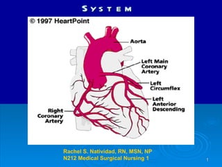
- 1. The Cardiovascular System Rachel S. Natividad, RN, MSN, NP N212 Medical Surgical Nursing 1
- 3. Circulation through the Heart
- 5. Diagnostic Studies: Blood Components
- 7. WBC with Differential 30-40% 55-70% 5-6% 1-2% <1%
- 8. Diagnostic Studies: WBC Differential–Neutrophils Segmented Neutrophils (Mature) Band Neutrophils (Immature) NEUTROPHIL MATURATION Bands Segs: Mature Bands (immature neutrophils-released into circulation In response to severe infection ( Left Shift or “ Bandemia ”)
- 12. Diagnostic Studies: Case Study #1 #2 POD PE: Incision site appears pink and slightly edematous with moderate amt. serosanguinous drainage, JP drain intact draining reddish colored drainage. VS: Temp 99.8 F, Resp 20/min, P 98 BPM, BP 138/88. CBC results 2 day post-op reveal ->->->->
- 16. Diagnostic Studies: Case Study #2
- 25. Chest Pain: What’s the difference? ANGINA MI PERICARDITIS Cause Ischemia Onset; Precipitating factors Sudden No precip. Factors Often early am Quality Severity squeezing stabbing pain or pressure Severe Sharp stabbing Moderate to severe Location Region Substernal May spread to chest, arms, back, Substernal May spread to ant. Chest, arms, back, jaw, neck Substernal Usually spreads to left side or back Duration, Relieving Factors < 15 min Rest, Nitro, O2 30 min or longer Not relived by rest Relieved with opioids
- 31. Pulmonary Edema The most severe manifestation of Left Heart Failure Fluid leak into the pulmonary interstitial spaces (Pulmonary congestion/edema) Hypoxia and poor 02 exchange
- 33. CXR: Left Heart Failure Pulmonary edema
- 34. Systemic Edema Unresolved Left failure : eventually leads to right sided failure by venous congestion in the systemic circulation Also other causes …
- 36. Heart Failure Clinical manifestations : Pulmonary Congestion (L) and Systemic Congestion (R) Right Heart Failure Left Heart Failure Pulmonary fluid overload Peripheral fluid overload
Editor's Notes
- Elevation in LVEDP (left ventricular end-diastolic pressure) Increases left atrial pressure Backs into the pulmonary vascular bed &quot;Pulmonary edema&quot; is water on the lungs. Fluid is not only in the lung tissues, but actually in the air spaces as well. This is a severe degree of heart failure, and requires immediate and aggressive management. When the heart's output decreases, the body does many things to try and compensate for it. It will release hormones to make the heart beat stronger. The heart will beat faster. Many of these reflexes however, only create a short term gain, and may ultimately hurt the heart's function. When the kidneys sense a decrease in flow, they release hormones which cause the body to hold sodium and water. In the short term, this will lead to an increase in the volume of blood which is circulating, and provide the kidneys with the blood volume they are looking for. However, this extra volume of fluid is more than can be held in the blood vessels, and it will start to exude out into the tissues of the bo Develops when the imbalance in pump function causes an increase in lung fluid secondary to leakage from pulmonary capillaries into the interstitium and alveoli of the lung. Life threatening situation in which the lung alveoli become filled with serosanguinous fluid. Most common cause is acut L venricular failure secondary to CAD –thus producing the cymptom of pink frothy sputum---
- Fluid in pulmonary vessels - infiltrates