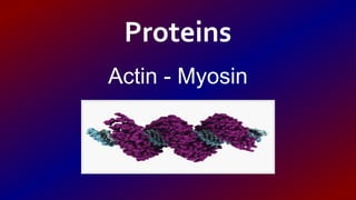
Actin & Myosin
•Download as ODP, PDF•
13 likes•13,689 views

The structure of actin and myosin proteins and their role in muscular contraction.

Recommended
More Related Content
What's hot
What's hot (20)
Similar to Actin & Myosin
Similar to Actin & Myosin (20)
More from Aglaia Koutra
Recently uploaded
Recently uploaded (20)
Actin & Myosin
- 2. The proteins v Proteins are large biomolecules having huge importance for living organisms. v Proteins account for 50% of dry cell mass and play a crucial role in all cell functions.
- 3. The role of proteins v Structural elements of cells and tissues v Storage of useful substances. v They transfer molecules. v They take part in cellular communication and signaling. v They help in cellular movement. v They participate in cell defence.
- 4. Myosin Myosin is a muscle protein constisting of head, neck and tail domains The head domain binds the filamentous actin, and uses ATP hydrolysis to generate force and to "walk" along the filament towards the barbed (+) . The neck domain acts as a linker and as a lever arm for transducing force generated by the catalytic motor domain. The neck domain can also serve as a binding site for myosin light chains which are distinct proteins that form part of a macromolecular complex and generally have regulatory functions The tail domain generally mediates interaction with cargo molecules and/or other myosin subunits. In some cases, the tail domain may play a role in regulating
- 5. Actin An actin protein is the monomeric subunit of two types of filaments in cells: microfilaments, one of the three major components of the cytoskeleton, and thin filaments, part of the contractile apparatus in muscle cells. It can be present as either a free monomer called G-actin (globular) or as part of a linear polymer microfilament called F-actin (filamentous), both of which are essential for such important cellular functions as the mobility and contraction of cells during cell division. Actin participates in many important cellular processes, including muscle contraction, cell motility, cell division and cytokinesis, vesicle and organelle movement, cell signaling, and the establishment and maintenance of cell junctions and cell shape.
- 6. Actin filaments Cellular actin has two forms: monomeric globules called G-actin and polymeric filaments called F- actin (that is, as filaments made up of many G- actin monomers). F-actin can also be described as a microfilament. Two parallel F-actin strands must rotate 166 degrees to lie correctly on top of each other. This creates the double helix structure of the microfilaments found in the cytoskeleton. Microfilaments measure approximately 7 nm in diameter with the helix repeating every 37 nm. Each molecule of actin is bound to a molecule of adenosine triphosphate (ATP) or adenosine diphosphate (ADP) that is associated with a Mg2+ cation. The most commonly found forms of actin, compared to all the possible combinations, are ATP-G-Actin and ADP-F-actin.
- 7. Muscle contraction Muscle contraction is the activation of tension-generating sites within muscle fibers. Muscle contractions can be described based on two variables: length and tension. A muscle contraction is described as isometric, if the muscle tension changes, but the muscle length remains the same. In contrast, a muscle contraction is isotonic, if muscle length changes, but the muscle tension remains the same. If the muscle length shortens, the contraction is concentric. If the muscle length lengthens, the contraction is eccentric.
- 8. The mechanism of muscle contraction In muscle cells, actomyosin myofibrils make up much of the cytoplasmic material. These myofibrils are made of thin filaments of actin (typically around 7 nm in diameter), and thick filaments of the motor-protein myosin (typically around 15 nm in diameter). Using the hydrolysis of ATP for energy, myosin heads undergo a cycle during which they attach to thin filaments, exert a tension, and then, depending on the load, perform a power stroke that causes the thin filaments to slide past, shortening the muscle.
- 9. Muscular Spasms Various kinds of involuntary muscle activity may be referred to as a "spasm". A spasm may be a muscle contraction caused by abnormal nerve stimulation or by abnormal activity of the muscle itself. A spasm may lead to muscle strains or tears in tendons and ligaments if the force of the spasm exceeds the tensile strength of the underlying connective tissue. This can occur with a particularly strong spasm or with weakened connective tissue.
- 10. Thank you for your attention!!! Work done by the students of C2 Class: • Atha Kournouti • Alexandros Kostas • Giorgos Marmaras • Anastasia Michioti • Argyris Bazos • Panagiotis Babatsias