hallux valgus & hallux rigidus
•Download as PPTX, PDF•
74 likes•18,924 views

a useful presentation .. dedicated for orthopedic residents

Recommended
More Related Content
What's hot
What's hot (20)
Similar to hallux valgus & hallux rigidus
Similar to hallux valgus & hallux rigidus (20)
Recently uploaded
Recently uploaded (20)
hallux valgus & hallux rigidus
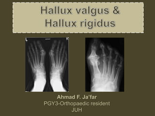
- 1. Ahmad F. Ja’far PGY3-Orthopaedic resident JUH
- 2. Hallux Valgus • Complex deformity. • Often accompanied by deformities of lesser toes. • Associated conditions : hammer toe, calluses • Definition : Lateral deviation of great toe with medial deviation of first metatarsal. • More common in women. • 70% of pts with hallux valgus have family history genetic predisposition with anatomic anomalies. • Adult type. • Adolescent & juvenile hallux valgus.
- 3. Risk factors Intrinsic • Genetic predisposition. • Ligamentous laxity. • Convex metatarsal head. • Pes planus. • Rheumatoid arthritis. • Cerebral palsy. Extrinsic • Shoes with high heel. • Shoes with narrow toe box.
- 4. Pathoanatomy • Valgus deviation promotes varus position of metatarsal • Sesamoid… complex becomes lateral to the metatarsal head, which moves medially • Medial Capsule… joint capsule becomes stretched and attenuated. • Lateral capsule… becomes contracted • Adductor tendon becomes deforming force – inserts on fibular sesamoid • Lateral deviation of EHL • Plantar and lateral migration of the abductor hallucis causes muscle to plantar flex and pronate phalanx • Windlass mechanism becomes less effective – leads to transfer metatarsalgia
- 5. Pathoanatomy
- 6. Presentation-symptoms • Difficulty with shoe wear due to medial eminence (80%) • Pain over prominence at MTP joint (70%) • Cosmetic concerns in (60%) • Pain underneath the second metatarsal head in 40%. • Compression of digital nerve may cause symptoms.
- 7. Physical Exam • Hallux rests in valgus and pronated due to deforming forces illustrated above • Examine entire first ray for – 1st MTP ROM – 1st tarsometatarsal mobility • Evaluate associated deformities – Pes planus – Lesser toe deformities – Midfoot and hindfoot conditions – Corns, calluses, warts, interdigital neuromas, bunionettes, hammer toes, and claw toes. – Generalized ligamentous laxity
- 9. Radiographs • Views – Standard series should include weight bearing AP and Lat. – Sesamoid view can be useful. • Findings – Radiographic parameters ….. guide treatment – Displacement of sesamoids • often displaced laterally – Joint congruency. – Presence of first MTP joint and first metatarso- cuneiform joint degenerative changes should be noted .
- 11. Hallux valgus angle Normal < 15°
- 12. 1st/2nd Intermetatarsal angle (IMA) Normal < 9°
- 13. Distal metatarsal articular angle (DMMA) Normal < 10° Joint line congruency Moderately severe hallux valgus w/ a significantly increased DMMA will be associated w/ a congruent bunion
- 14. Halluv valgus interphalangeus (HVI) Normal < 10° Joint line congruency
- 16. Classification
- 17. Management • Conservative • Shoe modification/ pads/ orthoses – First line treatment • Exercises, and activity adjustments. • Orthoses more helpful in patients with pes planus or metatarsalgia.
- 18. Surgical options • surgical correction ---- Indications – when symptoms present with 2ry deformities and shoe modification fails – do not perform for cosmetic reasons alone • General rules …. • More than 130 operations … – Soft tissue procedure • indicated in very mild disease in young female (almost never alone) – Distal MTB osteotomy • indicated in mild disease – Proximal or combined MTB osteotomy • indicated in more moderate disease – Fusion procedures • indicated in severe disease with 2ry degenerative changes. – MTP resection arthroplasty • only indicated in elderly patients with low functional demand
- 19. Soft tissue: Modified Mcbride Distal MTB osteotomy HVA ≤ 40, IMA < 13: Chaveron.. biplanar Chevron Mitchel Proximal MTB osteotomy: HVA >40°, IMA >13° Scarf Crescentric Ludloff Broomstick Combined MTB osteotomy: severe disease (HVA 41-50°, IMA 16-20°) Proximal phalanx osteotomy Akin Arthrodesis 1st MTJ Lapidus-1st metatarsocuneiform
- 20. Soft Tissue Procedure • Modified McBride • Indications • 30- to 50-year-old woman with clinical • HVA --- 15 to 25 degrees • IMA --- less than 13 degrees. • HVI --- less than 15 degrees • No degenerative changes at the metatarsophalangeal joint – never appropriate in isolation – in conjunction with medial eminence resection and osteotomy.
- 21. Modified McBride
- 22. Distal metatarsal osteotomy-Mitchel • Distal 1st MT osteotomy (extra-articular). • More proximal than Chevron. • Problems : metatarsalgia, attributable to dorsiflexion malunion of the distal fragment, excessive shortening of the metatarsal or Both…. recurrence
- 23. Distal metatarsal osteotomy-Chaveron • V-shaped lateral translational osteotomy – sagittal plane. • non-congruent deformity with a normal DMAA. • More distal than Mitches (Cancellous bone) • less shortening of the metatarsal, lessa metatarsalgia more stability.
- 24. Combined Chaveron-Aiken osteotomy • Moderate to sever deformity • Increased HVI • Proximal phalanx medial close wedge osteotomy
- 25. Osteotomy of the proximal first metatarsal and shaft osteotomy • Severe deformity IMA > 20 HVA > 50 • have high corrective power due to their proximal location providing a long lever arm. • More demanding. • Accompanied by soft tissue procedure Crescentic Proximal chaveron Ludloff Scarf
- 26. Combined proximal and distal osteotomies • Increased intermetatarsal angle (IMA) and distal metatarsal articular angle (DMAA).
- 27. Scarf osteotomy • Added stability • +/- soft tissue procedure .. +/- pahalangeal osteotomy
- 29. 1st metatarsophalangeal joint arthrodesis Indications • cerebral palsy • Down's syndrome • Rheumatoid arthritis • Gout • Severe DJD • Ehler-Danlos • Recurrent hallux valgus.
- 30. 1st metatarsophalangeal joint arthrodesis • 15 to 20 degrees of valgus • 30 degrees of dorsiflexion in relation to metatarsal shaft or • 10 to 15 degrees of dorsiflexion in relation to floor
- 31. Lapidus procedure (1st metatarsocuneiform arthrodesis) Indication • Severe deformity • Metatarsus primus varus • Hypermobile 1st tarsometatarsal joint
- 32. Resection arthroplasty (Keller’s) • Largely abandoned • Still indicated in some elderly patient with reduced function demands • Include medial eminence removal and resection of base of proximal phalanx
- 33. Complications of surgery Recurrence • Most common cause of failure is insufficient preoperative assessment and failure to follow indications – e.g., failure to recognize DMAA > 15° – e.g., failure to do adequate distal soft tissue realignment • More common in juvenile/adolescent population • Noncompliant patient that bears weight.
- 34. Complications of surgery Avascular necrosis • Medial capsulotomy is primary insult to blood flow to metatarsal head • Distal metatarsal oseotomy and lateral soft tissue release inconjuction do not increase risk for AVN.
- 35. Complications of surgery Dorsal malunion with transfer metatarsalgiadue to overload of lesser metatarsal heads • Risk associated with – Lapidus fusion. – Proximal crescentric osteotomy. • 2nd MT transfer metatarsalgia often seen concomitant with hallux valgus • Shortening metatarsal osteotomy (Weil) indicated with extensor tendon and capsular release
- 36. Complications of surgery Hallux Varus caused by – Overcorrection of 1st IMA – Excessive lateral capsular release with overtightening of medial capsule – Overresection of medial first metatarsal head – Fibular sesamoidectomy
- 37. Complications of surgery Cock up toe deformity - Due to injury of FHL - Most severe complication with Keller resection Neuropraxia - Painful incisional neuromas----involve the dorsomedial cutaneous branch of the superficial peroneal nerve. - Branches of the deep peroneal nerve to this area are rare.
- 38. Juvenile / adolescent hallux valgus • How much different ?? – Often bilateral and familial – Pain usually not primary complaint (cosmetic) – Varus of first MT with widened IMA usually present – DMAA usually increased – Often associated with flexible flatfoot • complications – recurrence is most common complication and hallux varus
- 39. • Nonoperative – Shoe modification • indications – pursue nonoperative management until physis closes • Operative – Surgical correction • indications – best to wait until skeletal maturity to operate » Medial cuneiform osteotomy. » Surgery indicated in symptomatic patients with an IMA > 10° and HVA of > 20° – Severe deformity with a DMAA > 20 perform a double MT osteotomy Juvenile / adolescent hallux valgus
- 41. Hallux Rigidus and DJD
- 42. Hallux Rigidus • A condition characterized by loss of motion of first MTP joint in adults due to degenerative arthritis . • Osteophyte formation leads to dorsal impingement Pathoanatomy …..primary etiology unknown • acute trauma and repetitive microtrauma predispose to arthritic changes • anatomic variations of first metatarsal may play a yet unproven role in arthritic predisposition
- 43. Classification Exam Findings Radiographic Findings Grade 0 Stiffness Normal Grade 1 mild pain at extremes of motion mild dorsal osteophyte, normal joint space Grade 2 moderate pain with range of motion increasingly more constant moderate dorsal osteophyte, <50% joint space narrowing Grade 3 significant stiffness, pain at extreme ROM, no pain at mid-range severe dorsal osteophyte, >50% joint space narrowing Grade 4 significant stiffness, pain at extreme ROM, pain at mid- range of motion same as grade III
- 44. Presentation Symptoms – First ray and 1st MTP pain and swelling worse with push off or forced dorsiflexion of great toe – Shoe irritation due to dorsal osteophytes and compression of dorsal cutaneous nerve may lead to paresthesias – Pain becomes less severe as the disease progresses Physical exam – Limited dorsiflexion – Pain with grind test
- 45. Radiographs recommended views – AP, lateral, and oblique views Findings – osteophytes, especially dorsal – joint space narrowing – subchondral sclerosis and cysts
- 46. Treatment • Nonoperative • NSAIDS, activity modification & orthotics – Indications • grade 0 and 1 disease – Activity modifications • avoid activities that lead to excessive great toe dorsiflexion – Types of orthotics • Morton's extension with stiff foot plate is the mainstay of treatment • stiff sole shoe and shoe box stretching may also be used
- 47. Operative • Joint debridement and synovectomy • patients with acute osteochondral or chondral defects • Dorsal cheilectomy • Grade 1 and 2 disease. • Pain with dorsiflexion is an indicator of good results with dorsal cheilectomy • Shoe wear irritation from dorsal prominence and pain (ideal candidate) • contraindicated when pain located in the mid-range of the joint during passive motion – Technique • remove 25-30% of the dorsal aspect of the metatarsal head along with dorsal osteophyte resection • the goal of surgery is to obtain 70% to 90% dorsiflexion intraoperatively.
- 49. Moberg procedure • Dorsal closing wedge osteotomy of the proximal phalanx). – runners with reduced dorsiflexion (60° is needed to run) – Failure of cheilectomy to provide at least 30 to 40 degrees of motion
- 50. Keller Procedure • Resection arthroplasty – elderly, low demand patients with significant joint degeneration and loss of motion • Technique – involves removing the base of the first proximal phalanx – risk of hyperextension (cock-up deformity), weakness with push-off, and transfer metatarsalgia (decreased with capsular interposition)
- 51. MTP arthroplasty ((controversial)) • Technique – capsular interpositonal arthroplasty gaining popularity – Silicone implants are not recommended due to poor long-term results • Outcomes – silicone implants may have a good short term satisfaction rate – osteolysis and synovitis cause mid to long term pain and joint destruction
- 52. MTP joint arthrodesis • Indications – grade 3 and 4 disease (significant joint arthritis) – most common procedure for hallux rigidus • Outcomes – 70% to 100% fusion rate – 15% of patients experience degeneration of IP joint after surgery (mostly asymptomatic)
- 53. Summary
- 54. Thank you
Editor's Notes
- Not a single deformity rather a complex deormity of 1st ray
- Pronation of hallux. A, Normal. B, Note plantar shift of abductor hallucislateral shift of sesamoids with associated intrinsic muscles of hallux
- pronation of the great toe is easily noted by comparing the angulation of the nail of the great toe relative to the floor.Hammer toe deformity. Note severe crossover toe deformity of second toe associated with severe hallux valgus. Primary complaint frequently is not severe hallux valgus deformity but pain beneath second metatarsal head.
- Plantar surface of first metatarsal head.Entire sesamoid sling with attached intrinsic musculature has been rotated distally off metatarsal head to present schematicallyrelationships of muscle, tendon, capsule, ligaments, and articular configuration of first metatarsophalangeal joint. B, As metatarsalhead moves medially, sesamoid sling apparatus becomes valgus deforming force, and metatarsal rotates (pronates) on its longitudinalaxis.
- Marks are placed in the mid-diaphyseal region of the proximal phalanx and the first metatarsal at an equal distance from the medial and lateral cortices. The longitudinal axis of the proximal phalanx is determined by an axis drawn though points A and B, and the longitudinal axis of the first metatarsal is determined by a line drawn through points C and D. The hallux valgus angle is formed by the intersection of the diaphyseal axes of the first metatarsal (line CD) and the proximal phalanx (line AB). the valgus angle of the first metatarsophalangeal joint exceeds 30 to 35 degrees,
- first-second intermetatarsal angle is formed by the intersection of these two axes
- e DMAA defines the relationship of the articular surface of the distal first metatarsal with the longitudinal axis of the first metatarsal. Points are placed on the most medial and lateral extent of the distal metatarsal articular surface (X′, Y′). A line drawn to connect these two points defines the “slope laterally of the articular surface.” Another line through points W and Z is drawn perpendicular to the first line X′ Y′. A third line through points C and D defines the longitudinal axis of the first metatarsal. The angle subtended by the perpendicular line (W, Z) and the longitudinal axis of the first metatarsal (C, D) defines the DMAA.
- . Marks are placed in the mid-diaphyseal region of the proximal phalanx and the first metatarsal at an equal distance from the medial and lateral cortices. The longitudinal axis of the proximal phalanx is determined by an axis drawn though points A and B, and the longitudinal axis of the first metatarsal is determined by a line drawn through points C and D. The hallux valgus angle is formed by the intersection of the diaphyseal axes of the first metatarsal (line CD) and the proximal phalanx (line AB).
- ux valgus deformity with subluxation (noncongruent joint) is characterized by lateral deviation of the articular surface of the proximal phalanx in relation to the articular surface of the distal first metatarsal. B, Hallux valgus deformity with a nonsubluxated (congruent) metatarsophalangeal joint is caused most often by lateral inclination of the distal metatarsal articular surface. Points X and Y determine the medial and lateral extent of the articular surface of the proximal phalanx; points X′ and Y′ determine the medial and lateral extent of the metatarsal articular surface. Note the lateral slope of the distal metatarsal articular surface.
- Medial capsular imbrication ,, A, Preoperative deformity in 30-year-old patient. B, Correction obtained at surgery. C, Preoperative and postoperative radiographs (note fibular sesamoid was not removed). More deformity can be corrected by fibular sesamoidectomy, but overcorrection (hallux varus) is risk. techniquemedial capsule of 2nd MTP is sutured to lateral capsule of 1st. adductor hallucis is released and interposed.
- (1)removal of the medial eminence, (2) an close wedge osteotomy of the distal portion of the first metatarsal shaft, (3) lateral displacement and angulation of the capital fragment, and (4) medial capsulorrhaphythrough excessive shortening and dorsally angulated malunion, has resulted in transfer metatarsalgia. This biplanar deformity is most difficult to correct.
- (1) medial eminence removal, (2) a V-shaped intracapsular osteotomy through the first metatarsal head, (3) lateral displacement of the capitalfragment, (4) removal of the resulting projection of the first metatarsal, and (5) medial capsulorrhaphy.
- A, Resection of medial eminence parallel to medial border of foot. B, Chevronosteotomy cut is made, and metatarsal head is shifted laterally 2.5 to 3.0 mm. C, Osteotomy is fixed with 0.045-inch smoothpin, and protruding medial border of metatarsal is osteotomized flush with metatarsal head. D, Akin cut parallels concavity at baseof proximal phalanx, and 1-mm wedge of bone is removed. E, Suture closure of Akin osteotomy corrects residual valgus of hallux. (From
- Correction of the IMA with a proximal osteotomy causes the proximal phalanx to override the second toe secondary to the DMAA. C, A distal closed-wedge osteotomy is performed. D,With closure of the osteotomy, the phalanx is now directed parallel to the metatarsal shaft, correcting the overall deformity.
- osteotomy. A, Placement of Kirschner wires at corner point of planned osteotomy. Standard orientation forlateral translation is 90 degrees to longitudinal axis of second metatarsal (A) and in approximately 20 degrees horizontal inclinationto plantar surface (XY). B, Osteotomy cuts. C, Lateral displacement is obtained if short osteotomy cuts are perpendicular to longitudinalaxis of foot. D, Proximally oriented inclination of short osteotomy cuts caused shortening depending on angle of inclinationand amount of translation. E, After lateral displacement, osteotomy is fixed with two minifragment screws.
- Patient with moderatemetatarsus primus varus and hallux valgusdeformity. B, Both the intermetatarsal angleand the hallux valgus are corrected; anAkin osteotomy also was done to obtain completecorrection of the hallux valgus
- A, Hallux valgus in 46-year-old woman. B, Oneyear after arthrodesis
- Twenty to 25 percent of metatarsal head is removed dorsally.