Signs of manipulations of physical rx negatives seen from the aspect of production
•
0 likes•127 views

Proof of deliberate Conspiracy of Silence, obstruction of TX CT scan investigation. Medical crime is allowed when kept silent is the conclusion with my study about the praxis in the Netherlands.

Recommended
More Related Content
Similar to Signs of manipulations of physical rx negatives seen from the aspect of production
Similar to Signs of manipulations of physical rx negatives seen from the aspect of production (10)
More from siegfried van hoek
More from siegfried van hoek (20)
Recently uploaded
Recently uploaded (20)
Signs of manipulations of physical rx negatives seen from the aspect of production
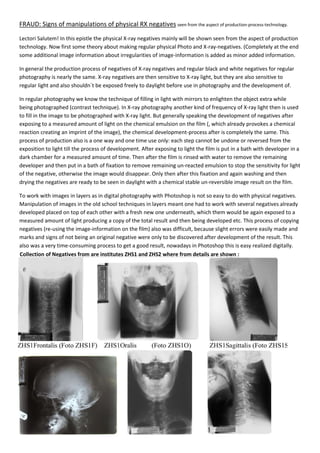
- 1. FRAUD: Signs of manipulations of physical RX negatives seen from the aspect of production-process-technology. Lectori Salutem! In this epistle the physical X-ray negatives mainly will be shown seen from the aspect of production technology. Now first some theory about making regular physical Photo and X-ray-negatives. (Completely at the end some additional image information about irregularities of image-information is added as minor added information. In general the production process of negatives of X-ray negatives and regular black and white negatives for regular photography is nearly the same. X-ray negatives are then sensitive to X-ray light, but they are also sensitive to regular light and also shouldn´t be exposed freely to daylight before use in photography and the development of. In regular photography we know the technique of filling in light with mirrors to enlighten the object extra while being photographed (contrast technique). In X-ray photography another kind of frequency of X-ray light then is used to fill in the image to be photographed with X-ray light. But generally speaking the development of negatives after exposing to a measured amount of light on the chemical emulsion on the film (, which already provokes a chemical reaction creating an imprint of the image), the chemical development-process after is completely the same. This process of production also is a one way and one time use only: each step cannot be undone or reversed from the exposition to light till the process of development. After exposing to light the film is put in a bath with developer in a dark chamber for a measured amount of time. Then after the film is rinsed with water to remove the remaining developer and then put in a bath of fixation to remove remaining un-reacted emulsion to stop the sensitivity for light of the negative, otherwise the image would disappear. Only then after this fixation and again washing and then drying the negatives are ready to be seen in daylight with a chemical stable un-reversible image result on the film. To work with images in layers as in digital photography with Photoshop is not so easy to do with physical negatives. Manipulation of images in the old school techniques in layers meant one had to work with several negatives already developed placed on top of each other with a fresh new one underneath, which them would be again exposed to a measured amount of light producing a copy of the total result and then being developed etc. This process of copying negatives (re-using the image-information on the film) also was difficult, because slight errors were easily made and marks and signs of not being an original negative were only to be discovered after development of the result. This also was a very time-consuming process to get a good result, nowadays in Photoshop this is easy realized digitally. Collection of Negatives from are institutes ZHS1 and ZHS2 where from details are shown :
- 2. ZHS1 Cervical Frontalis extraction from phot SR-REGULAR 007406 ZHS1 Cervical Oralis extraction from ph ZHS1 Cervical Sagittalis extraction from ZHS1H Head Sagittalis extraction from ZHS1 Cervical Frontalis extraction from photo ZHS1F REGULAR 007406 KN / NG- SR 97490717 extraction from photo ZHS1O KN / MG-SR 97490717 KN / MG-SR 97490717 extraction from photo ZHS1S KN / MG - KN / MG – extraction from photo ZHS1H KN / MG - On all ZHS1 negatives the same nametag with exactly the sane handwriting and even the s scotch for attaching is being used. There is difference in series registration numbers: from from …369 to 371 and then jumps 907. This is odd for old fashion regular X made right one after another with the equipment by one person This nametag shows a remain of other information underneath. Inside the ‘six’ there originally should be this is the first sign of re of image information The X-rays were made right one after another, so I cannot explain the jump of registration numbers of X made and also the largest scan ZHS1H only had just one registration number. SR 97490717 – 2 22207371 SR 97490717-2 22207369 SR 97490717-2 222073(70?) SP 08190762-1 13200309 – SP 08190762-1 13200308 SP 08190763 - 1 43300907 On all ZHS1 negatives the same exactly the sane handwriting and even the same cotch for attaching is being used. in series and a leap in digital registration numbers: from …308 to 309 and then and then jumps all the way to … fashion regular X-rays being made right one after another with the same X-ray by one person in that same institute. This nametag shows a remain of other information underneath. Inside the ‘six’ there be no emulsion meaning this is the first sign of re-use (manipulation) age information in layers. rays were made right one after not explain the jump of registration numbers of X-rays being also the largest scan ZHS1H only had just one registration number.
- 3. ZHS2 Cervical Oralis extraction from ZHS2 Cervical Sagittalis extraction from The object-rotation on sagittal to frontal scan ZHS1 extraction from photo ZHS2O extraction from photo ZHS2S digital number 0007770400939 This nametag shows a remain of other information underneath. This is a sign of (manipulation use of image information leading to questioning the integrity very X-ray negative. Brand information runs into dark, but on that spot there should not be any Photo white ciphers This nametag other information underneath too and was made within a minute time after the first one. Again this is a sign of of image information leading to questioning integrity of. Brand information runs dark, but also other brand information is mirrored: that is a very obvious mark of traces of image manipulation. digital number. 0007770400938 to frontal scan is around a diagonal axis and not 90o around a vertical ax The position of the object on ZHS1S and ZHS2S the vertebra equal in position and proportions, the object While the object is supposed to be locked up inside the vertebral canal it is also even reaching outside the vertebra ZHS2 This nametag shows a remain of other information underneath. This is a sign of (manipulation?) re- of image information leading to questioning the integrity of this ray negative. Brand information runs into dark, but on that spot there should not be any Photo-emulsion leaving ciphers as standard result. This nametag shows a remain of other information underneath and was made within a minute time after the first one. Again this is a sign of the re-use of image information leading to ing integrity of. Brand information runs away into dark, but also other brand information is mirrored: that is a very obvious mark of traces of image manipulation. digital number. 0007770400938 around a vertical axis as should be. The position of the object shown on ZHS1S and ZHS2S, although the vertebras are of shown here equal in position and proportions, the object however clearly is not. While the object is supposed to be locked up inside the vertebral canal it is also even reaching outside the vertebral body.
- 4. Before the second operation there was no metal substance not show any disturbances. Therefore the CT scan of Feb 15th 2000 also becomes questionable regarding integrity because that one does show metal present in the oral cavity, wh Supposed is a clip sagged down into the neck vertebral. Dimension and position differ between various scans Before the second operation there was no metal substance present inside the mouth or inside the neck; Therefore the CT scan of Feb 15th 2000 also becomes questionable regarding integrity because that one does show metal present in the oral cavity, while two MRI scans after before 2nd surgery do not neck vertebral. Dimension and position differ between various scans made after. or inside the neck; MRI does Therefore the CT scan of Feb 15th 2000 also becomes questionable regarding integrity, ile two MRI scans after before 2nd surgery do not.
- 5. After the second operation there are metal c mouth both MRI and CT scan show two pieces/locations of metal containing objects, however on MRI we see in the neck three parts/locations but on CT (and RX after) we only see one single metal containing object/location. On the 3D MRI resp. Sagittal Left and Posterior we clearly can see that that are more then one single metal containg parts. If there was only a single Michel Clip present them all the image disturbances were connected to eachother. The lower foreign body material parts also looks to surround the vertebral cord: is this the extinction artefact? MRI is not the most suitable scanner to define the exact form of metal, it only gives a vague shape of presence but for detecting metal containing parts it is t of a metal containing object, but being single photo they appeared more easily to be manipulated then a MRI result. Traces of ferro (blood) from skin towards vertebral canal in the middle image also indicates surgery being performed. front After the second operation there are metal containing substances present inside the nec mouth both MRI and CT scan show two pieces/locations of metal containing objects, however on MRI we see in the k three parts/locations but on CT (and RX after) we only see one single metal containing object/location. On the 3D MRI resp. Sagittal Left and Posterior we clearly can see that that are more then one single metal containg parts. If there was only a single Michel Clip present them all the image disturbances were connected to eachother. material parts also looks to surround the vertebral cord: is this the extinction artefact? MRI is not the most suitable scanner to define the exact form of metal, it only gives a vague shape of presence but for detecting metal containing parts it is the strongest scanner. CT/RX is the suitable scanner for defining the shape of a metal containing object, but being single photo they appeared more easily to be manipulated then a MRI result. races of ferro (blood) from skin towards vertebral canal in the middle image also indicates surgery being performed. Sagittal scan L Posterior L ck and the mouth. In the mouth both MRI and CT scan show two pieces/locations of metal containing objects, however on MRI we see in the k three parts/locations but on CT (and RX after) we only see one single metal containing object/location. On the 3D MRI resp. Sagittal Left and Posterior we clearly can see that that are more then one single metal containg parts. If there was only a single Michel Clip present them all the image disturbances were connected to eachother. material parts also looks to surround the vertebral cord: is this the extinction artefact? MRI is not the most suitable scanner to define the exact form of metal, it only gives a vague shape of presence but he strongest scanner. CT/RX is the suitable scanner for defining the shape of a metal containing object, but being single photo they appeared more easily to be manipulated then a MRI result. races of ferro (blood) from skin towards vertebral canal in the middle image also indicates surgery being performed. WHY THE SCAR IS IN THE NECK? WHAT IS THERE TO HIDE BY RX MANIPULA- TION? Posterior R
- 6. ZHS2O in full page in order to show the evidence with brand information being the left and right border of the upper half of the image. This is the negatives placed on top of each other for producing an new one with negative underneath. This is a mark of proof of a complex old school dark The clip ‘R’ under to the left also should be white show the evidence with brand information being really mirrored the left and right border of the upper half of the image. This is the most evident sign of manipulation with multiple negatives placed on top of each other for producing an new one with by photo-copying the two towards a fresh new tive underneath. This is a mark of proof of a complex old school dark-room technique for image under to the left also should be white caused by the metal clip that is used in really mirrored on one negative (on most evident sign of manipulation with multiple copying the two towards a fresh new room technique for image-manipulation. clip that is used in old-school RX-technology.