SlideShare a Scribd company logo
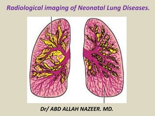
Radiological imaging of Neonatal Lung Diseases.
Dr/ ABD ALLAH NAZEER. MD.
Neonatal ICU chest radiographs are one of the
most common pediatric radiology examinations
performed. As modern medicine has advanced, the
lower age limit of viability has continued to
decrease. Nowadays, it is not uncommon for 23-
week-old infants to survive. Although there are
many complications associated with prematurity,
to include necrotizing enterocolitis, intracranial
hemorrhage, and sepsis, the most common cause
of neonatal morbidity and mortality remains lung
disease. This article describes the pathology and
radiographic findings of some of the most common
lung disease encountered in neonates.
Transient Tachypnea of the Newborn
Transient tachypnea of the newborn (TTN), also referred to as retained
fetal lung fluid, wet lung disease, or transient respiratory distress, is
caused by prolonged clearance of fetal lung fluid. Fetal lung fluid is not
the same as amniotic fluid-rather it represents an ultrafiltrate of the
fetal plasma. Symptoms of TTN include mild to moderate respiratory
distress which presents at birth but may be delayed up to 6 hours. The
symptoms typically peak within 36 hours after delivery and resolve by
72 hours.
Common risk factors of TTN include precipitous deliveries and cesarean
sections where it is thought that retained fluid is not fully expelled from
the neonate's lungs as would occur during a normal vaginal delivery.
Normally, 35% of fetal lung fluid is cleared in the first few days prior to
birth secondary to increased gene expression for an epithelial sodium
(Na+) channel. The rest is cleared by labor and postnatally during crying
and breathing.Other risk factors include prematurity, maternal diabetes,
hydrops and other forms of hypervolemia, maternal sedation, and a
history of maternal smoking.
In vivo experiments demonstrate that the poor fluid absorption may be
explained by a poorly developed epithelial Na+ transport protein. In
utero, the fetal lung epithelium secretes chloride (Cl) and fluid. Late in
gestation, the lung epithelium develops Na proteins to absorb the fetal
lung fluid by responding to increase catecholamines and
glucocorticoids. It is thought that infants with TTN have mature
surfactant and poorly developed respiratory epithelial transport
proteins, as opposed to neonatal respiratory distress syndrome (NRDS)
where both surfactant pathways and Na+ transport proteins are
deficient.
Radiographic findings of TTN often include hyperinflated lungs and
retained fluid within the alveoli and interstitium, to include pleural and
fissural fluid, as well as increased perihilar interstitial markings. Severe
cases may show alveolar opacities from the retained fluid.
Radiographic findings can be similar to heart failure, although without
marked cardiac silhouette enlargement. Imaging findings of TTN
typically improve within 24 hours as the excess fetal lung fluid is either
absorbed or expelled.
Transient tachypnea of the newborn (TTN). Portable chest radiograph reveals
perihilar interstitial markings and right fissural fluid, characteristic of TTN.
Transient tachypnea of the newborn (TTN). Portable chest radiograph demonstrates perihilar
interstitial markings, which can be seen with TTN from the retained fetal lung fluid.
Transient tachypnea of the newborn: Radiographic findings of diffuse
infiltrates and perihilar streaking, indicating retained lung fluid.
Meconium Aspiration Syndrome
Meconium aspiration syndrome (MAS) is the most common cause of
respiratory distress in the term or post-term neonate. Meconium is the
earliest stool in an infant, and its components include epithelial cells,
mucus, amniotic fluid, bile, blood, and lipids. Meconium was once
thought to be sterile, although research has shown that approximately
half of meconium is populated by Escherichia coli and the remaining
half by lactic acid producing bacteria, such as Lactobacillus.
Most cases of meconium aspiration occur during the distress of labor-
however, in utero meconium passage and aspiration can occur
secondary to fetal stressors, such as hypoxia and sepsis. These stressful
events cause a vagal response by the fetus, leading to defecation.
Clinically, infants may present with cyanosis, tachypnea, and
tachycardia. Signs of respiratory distress are invariably present,
including intercostal retractions and nasal flaring. If the neonate
defecated in utero, ingestion of meconium within the amniotic fluid can
result in a yellow or greenish appearance to the skin, nails, and urine.
Aspiration of meconium results in airway obstruction with a ball-valve
mechanism, chemical pneumonitis, and inactivation of surfactant by the
bile salts, causing secondary surfactant deficiency. This complex
pathophysiology results in a wide range of radiographic manifestations
of the disease.
The most common radiographic finding is pulmonary hyperinflation
secondary to the ball-valve mechanism of air trapping. Air trapping, in
combination with the chemical pneumonitis, results in barotrauma which
may cause pneumothoraces, pneumomediastinum, and pulmonary
interstitial emphysema. Other radiographic findings include perihilar
ropey opacities and interspersed areas of atelectasis. Pleural effusions
can be seen but are uncommon.
Meconium aspiration syndrome can result in persistent pulmonary
hypertension of the newborn. Treatment includes endotracheal
intubation to facilitate suctioning below the vocal cords, administration
of surfactant to replace the surfactant inactivated by bile salts, and
prophylactic antibiotics. The radiographic manifestations usually resolve
by 48 hours, although may take weeks if the meconium has a lower water
content.
Meconium aspiration. Portable chest radiograph demonstrates
hyperinflation with an anteromedial pneumothorax on the right.
Meconium aspiration with perihilar opacities. Portable chest radiograph in a newborn
infant with history of meconium aspiration shows rope-like perihilar opacities.
Chest radiograph of an infant with meconium aspiration syndrome.
Neonatal Respiratory Distress Syndrome
Neonatal respiratory distress syndrome (NRDS) is the clinical term used to describe
surfactant deficiency. It is also referred to as lung disease of prematurity. The term
hyaline membrane disease is a histologic term and describes a byproduct of the
disease.
The incidence of NRDS is approximately 6 in 1000 births.Risk factors include
prematurity, multiple gestations, oligohydramnios, and maternal diabetes.
Maternal diabetes is thought to cause fetal hyperinsulinemia which interferes
with surfactant biosynthesis, leading to NRDS. Boys and Caucasian babies are also
at increased risk, for unknown reasons.
Surfactant is produced in the endoplasmic reticulum of type II pneumocytes, which
are found in the alveolar walls. The surfactant is transported to the surface of the
pneumocyte where it is combined with surfactant apoproteins on the surface to
form a lipid monolayer. The surfactant layer reduces the surface tension and
allows the alveoli to more easily expand. If the type II pneumocytes are not
mature at the time of birth, surfactant deficiency occurs. The collapsed alveoli
result in decreased oxygenation, causing an increase in the pulmonary vascular
resistance. This in turn increases right to left shunting through a patent ductus
arteriosus, which usually does not close in the setting of prematurity and low
blood oxygenation. The increased shunting through the PDA exacerbates the
infant’s hypoxia.
The term hyaline membrane disease is derived from the appearance of
hyaline membranes in the bronchiole walls. The hyaline membranes, which
contain fibrin, mucin, and necrotic alveolar cells, are a byproduct of prolonged
alveolar collapse. The lecithin to sphingomyelin ratio in the amniotic fluid is
frequently used as a marker of fetal lung maturity. Fetal lung fluid flows into
the amniotic fluid throughout gestation. At approximately 32 to 33 weeks of
gestational age the lecithin content rapidly increases, indicating maturing
fetal lungs and production of surfactant by type II pneumocytes.
Radiographic findings in the setting of NRDS include stigmata of prematurity,
to include a bell-shaped thorax and absence of humeral head ossification
centers. The classic pattern of NRDS includes bilateral and symmetric granular
opacities, air bronchograms, effacement of the pulmonary vasculature, and
decreased lung volumes. The classic appearance of NRDS is less commonly
seen given the early administration of surfactant, frequently before baseline
imaging is obtained, and tendency for early intubation. This results in
appearances that can mimic meconium aspiration syndrome or neonatal
pneumonia with increased lung volumes and focal areas of consolidation.
Cystic lucencies from expanded parenchyma and asymmetric aeration can
resemble PIE. If only a single lung receives surfactant, it may asymmetrically
expand and cause mediastinal shift.
Classic appearance of NRDS. Portable chest radiograph demonstrate decreased lung volumes
and granular opacities which are more confluent centrally, resulting in air bronchograms and
effacement of the pulmonary vasculature. Patient is intubated with the ETT tip at the carina.
Mild RDS. Magnified
radiograph of the right
lung of a preterm neonate
shows effacement of
vascular definition by
diffuse reticulogranular
opacities. Peripheral air
bronchograms (arrows)
are visible at the medial
lung base. The minor
fissure (arrowheads) is
slightly thickened.
Diffuse bilateral reticulogranular opacities, and air bronchograms,
findings consistent with severe RDS. (b) Repeat radiograph, obtained 6
hours after endotracheal administration of one dose of surfactant, reveals
marked improvement in lung aeration and vascular definition.
CXR AP and lateral view in post-term neonate with RDS- note hyper
inflated lungs, scattered foci of ground glass opacities/ atelectasis and
collapse, complicated by air trapping and anterior pneumothorax
Neonate with respiratory distress syndrome, presents with
bilateral ground glass opacities. the lung volumes are low, air
bronchograms are seen, and there are no pleural effusions-
the most likely cause is surfactant deficiency.
Bronchopulmonary Dysplasia
Bronchopulmonary dysplasia (BPD), also known as chronic lung disease of infancy,
is a disease of unclear etiology, although it is likely multifactorial. The disease was
originally thought to be caused from NRDS and its treatment. The etiology of CPD
is now less clear given the advancements in neonatal treatment for NRDS, such as
surfactant, steroids, and low pressure algorithms of positive pressure ventilation.
Infectious organisms, such as Ureaplasma urealyticum, which is the most common
contaminant in amniotic fluid, have also been implicated as potential causes of
CPD. It has been postulated that inflammatory cytokines associated with
Ureaplasma infection injures the respiratory epithelium, which is then further
damaged by oxygen toxicity or barotrauma.6,8,9
The radiographic findings of CPD vary and have evolved over time from continuing
advances in medical therapy. Northway, et al. originally described four
radiographic stages of CPD. stage 1 (2-3 days after birth) resulted in the typically
granular opacities of NRDS- stage 2
(4-10 days) demonstrated granular opacities and with superimposed complete
pulmonary opacification in more severe cases-stage 3 (1030 days) revealed small
cystic lucencies alternating with small focal opacities-and stage 4 (greater than
one month) showing a “bubbly” appearance due to enlargement of cystic
lucencies and linear or ropy opacities.
Bronchopulmonary dysplasia (BPD). Portable chest radiograph of a
neonate with bronchopulmonary dysplasia demonstrates diffuse
granular opacities with more focal consolidation in the right lung.
“Classic” severe BPD in a 3-month-old premature infant. (a) Frontal radiograph shows
heterogeneous aeration, coarse strand-like areas of opacity, and intervening cystic
lucencies. (b) Axial CT scan demonstrates right upper lobe regional air trapping anteriorly,
architectural distortion with fibrotic subpleural parenchymal bands (arrows) and
subsegmental atelectasis posteriorly, and diffuse coarse reticular opacities in the left lung.
BPD in a 33-day-old preterm infant. Frontal chest radiograph demonstrates
uniform distribution of reticular opacities and small cystic lucencies.
Bronchopulmonary Dysplasia. The lungs are usually over aerated. There are diffuse
rope-like densities separated in some areas by zones of hyperlucency. The densities
may be coalescent in many areas. The heart borders can be completely obliterated.
Chest radiograph of infant with bronchopulmonary dysplasia.
Interstitial Air
The use of mechanical ventilation exposes the lungs to increased pressure, termed
barotrauma, and over distention, called volutrauma, resulting in lung injury. Injury
resulting in rupture at the junction of terminal bronchioles and alveoli, which
allows gas to infiltrate into the perivascular and peribronchial spaces. This is
referred to as pulmonary interstitial emphysema or PIE.
Interstitial gas may extend along numerous potential spaces in the interstitium
toward the peripheral lung. Subpleural blebs can form and rupture, resulting in a
pneumothorax. Interstitial gas may also extend centrally, resulting in
pneumomediastinum. The radiographic appearance of PIE can occasionally be
confused with aspiration pneumonia, pulmonary edema, and neonatal respiratory
distress syndrome.
There are two types of PIE, acute and persistent. acute PIE appears
radiographically as “bizarre tubular and cystic lucencies” which may be focal or
diffuse. The term persistent PIE is reserved for PIE that lasts longer than 1 week.
As with the acute form, it may be focal or diffuse. The cysts of persistent PIE have
been described as being lined with multinucleated giant cells.Persistent PIE may
be confused with other types of cystic thoracic chest masses in the infant.
However, PIE can usually be distinguished from other cystic lesions, since it arises,
occurs, and progresses in a ventilated patient.
The radiologic features of pneumothorax, pneumomediastinum,
pneumopericardium, and systemic air embolism have been extensively described.
In the supine infant, pleural air tends to collect anteriorly and may require cross-
table lateral or lateral decubitus views to confirm the diagnosis. Another feature
of pneumothorax peculiar to infants is the tendency of pleural air to “cloak”
diaphragmatic and mediastinal surfaces. Unlike in older children and adults, a
pleural line is often not discernible in infants with pneumothorax, and the
diagnosis may be suggested by an unusually well-defined costophrenic sulcus
(“deep sulcus sign”). The demonstration of the anterior junction line, which is not
normally seen on chest radiographs of healthy infants, can indicate bilateral
pneumothorax. Bilateral anterior pneumothoraces may also compress the
malleable lobes of the thymus, producing a bulging “figure 8” or “pseudomass”
configuration of the superior mediastinum. Mediastinal air may elevate the lobes
of the thymus (“angel wing” or “spinnaker sail sign”), track within the extrapleural
space and outline the inferior aspect of the heart (“continuous diaphragm sign”),
and dissect into the soft tissues of the neck or chest wall. Intrathoracic air leak
may also traverse the diaphragmatic hiatus to produce pneumoretroperitoneum
or Pneumoperitoneum, which may be under tension. Pericardial air outlines the
heart but is limited superiorly by the pericardial reflection about the great vessels.
Systemic air embolism manifests with striking findings of intracardiac, venous,
and arterial air
Pulmonary interstitial emphysema (PIE). Single frontal radiograph in a 7-month-old child
demonstrates the typical bizarre cystic lucencies of PIE in this tracheotomy dependent infant.
Development and resolution of PIE. Portable chest radiograph in a premature newborn infant (A) demonstrates an endotracheal tube tip near the carina,
diffuse granular opacities, and no interstitial air. Examination performed two days later (B) shows interval development of PIE within the left pulmonary
interstitium. Three days later, the right lung develops PIE (C). By two weeks from the initial radiograph, the pulmonary interstitial emphysema has resolved (D).
Diffuse persistent PIE in a 3-week-old girl with a history of positive-pressure mechanical
ventilation. (a) Frontal chest radiograph shows overexpansion of most of the left lung by
multiple cystic lucencies, producing contralateral mediastinal displacement and left retrocardiac
compressive atelectasis. (b) Axial CT scan shows multiple collections of interstitial gas in the left
upper lobe that surround lines (arrows) and dots (arrowheads) of soft-tissue attenuation
Localized persistent PIE in a 1-year-old boy delivered at 27 weeks gestational age.
(a) Frontal chest radiograph shows a circumscribed lucent lesion at the medial
right lung base with a smooth, lobulated, thin wall (arrows). (b) Axial CT scan
shows an aggregate of thin-walled, gas-containing cysts in the right lower lobe
that contains lines (arrows) and dots (arrowheads) of soft-tissue attenuation.
Bilateral pneumothoraces. (a) Anteroposterior chest radiograph shows juxta
mediastinal lucencies and bisagittal compression of the lobes of the thymus
(arrows), producing a “figure 8” configuration contour. A “deep sulcus sign”
(arrowhead) is seen in the right lung. (b) On a radiograph obtained after
spontaneous resolution of air leak, the mediastinal contour appears normal
Bilateral pneumothoraces. Frontal chest radiograph obtained with the infant rotated toward the
right reveals the anterior junction line (arrowheads) outlined by pleural gas. Both diaphragm
leaflets and the right aspect of the cardiothymic silhouette are abnormally well defined.
Pneumomediastinum. Frontal chest radiograph shows the lobes of the thymus
(arrows), which are displaced superolaterally by a large central lucent area.
Extensive air leak in a premature neonate who received positive-pressure assisted
ventilation for treatment of RDS. Frontal chest radiograph demonstrates a thin pericardial
membrane (straight white arrows), which is defined medially by intrapericardial gas and
laterally by pleural or mediastinal gas. Extrapleural gas outlines the medial aspect of the
left diaphragm (black arrows). Bilateral pneumothoraces produce deep sulcus signs (curved
arrows). Mediastinal gas tracks into the cervical soft tissues and right lateral chest wall
(arrowheads). The tip of the endotracheal tube enters the right mainstem bronchus.
Systemic air embolism in the setting of diffuse bilateral PIE. Frontal radiograph of the chest
and abdomen shows elevation and compression of the base of the heart (white arrowheads)
by tension pneumopericardium. The cardiac chambers are filled with gas, and intraluminal
gas is demonstrated in the inferior vena cava (straight arrow), hepatic veins (black
arrowhead), and abdominal aorta surrounding the tip of the umbilical artery catheter (curved
arrow). Both lungs are overexpanded by innumerable cystic lucencies representing PIE.
Summary
Neonatal lung disease remains one of the most
common causes of morbidity and mortality in this
patient population, especially in the setting of
prematurity. Neonatal chest radiographs play a critical
role in the diagnosis, categorization, and management
of the myriad of underlying neonatal lung diseases.
Therefore, it is critical that radiologists involved in
interpreting neonatal chest radiographs be familiar
with the imaging manifestations of common neonatal
lung pathologies. This will allow for prompt and
accurate characterization, as well as expedite and
guide treatment.
Thank You.

More Related Content

What's hot

CONGENITAL DISORDERS OF LUNG
CONGENITAL DISORDERS OF LUNGCONGENITAL DISORDERS OF LUNG
CONGENITAL DISORDERS OF LUNGAmeen Rageh
 
Ascites and Pleural Effusion
 Ascites and Pleural Effusion Ascites and Pleural Effusion
Ascites and Pleural EffusionMedia Genie
 
Presentation1.pptx, radiological signs in thoracic radiology.
Presentation1.pptx, radiological signs in thoracic radiology.Presentation1.pptx, radiological signs in thoracic radiology.
Presentation1.pptx, radiological signs in thoracic radiology.Abdellah Nazeer
 
Presentation1, radiological imaging of undescended testis.
Presentation1, radiological imaging of undescended testis.Presentation1, radiological imaging of undescended testis.
Presentation1, radiological imaging of undescended testis.Abdellah Nazeer
 
Neonatal Chest X-Ray
Neonatal Chest X-RayNeonatal Chest X-Ray
Neonatal Chest X-RayDrAzharZain
 
Neonatal neurosonography
Neonatal neurosonographyNeonatal neurosonography
Neonatal neurosonographyHarshita Saxena
 
Presentation1.pptx, radiological imaging of diffuse lung disease.
Presentation1.pptx, radiological imaging of diffuse lung disease.Presentation1.pptx, radiological imaging of diffuse lung disease.
Presentation1.pptx, radiological imaging of diffuse lung disease.Abdellah Nazeer
 
Diagnostic Imaging of Pulmonary infections
Diagnostic Imaging of Pulmonary infectionsDiagnostic Imaging of Pulmonary infections
Diagnostic Imaging of Pulmonary infectionsMohamed M.A. Zaitoun
 
Radiographic Presentation of Congenital Heart Disease
Radiographic Presentation of Congenital Heart DiseaseRadiographic Presentation of Congenital Heart Disease
Radiographic Presentation of Congenital Heart DiseaseTarique Ajij
 
Collapse consolidation
Collapse consolidationCollapse consolidation
Collapse consolidationairwave12
 
Congenital cystic adenomatoid malformation
Congenital cystic adenomatoid malformationCongenital cystic adenomatoid malformation
Congenital cystic adenomatoid malformationSneha Volvoikar
 
Diagnostic Imaging of Pulmonary Vasculature
Diagnostic Imaging of Pulmonary VasculatureDiagnostic Imaging of Pulmonary Vasculature
Diagnostic Imaging of Pulmonary VasculatureMohamed M.A. Zaitoun
 
A P L S Pediatric Emergency Radiology 1
A P L S  Pediatric  Emergency  Radiology 1A P L S  Pediatric  Emergency  Radiology 1
A P L S Pediatric Emergency Radiology 1Dang Thanh Tuan
 
Presentation1, radiological imaging of barium studies.
Presentation1, radiological imaging of barium studies.Presentation1, radiological imaging of barium studies.
Presentation1, radiological imaging of barium studies.Abdellah Nazeer
 

What's hot (20)

CONGENITAL DISORDERS OF LUNG
CONGENITAL DISORDERS OF LUNGCONGENITAL DISORDERS OF LUNG
CONGENITAL DISORDERS OF LUNG
 
Ascites and Pleural Effusion
 Ascites and Pleural Effusion Ascites and Pleural Effusion
Ascites and Pleural Effusion
 
Presentation1.pptx, radiological signs in thoracic radiology.
Presentation1.pptx, radiological signs in thoracic radiology.Presentation1.pptx, radiological signs in thoracic radiology.
Presentation1.pptx, radiological signs in thoracic radiology.
 
Neurosonogram.. Dr.Padmesh
Neurosonogram.. Dr.PadmeshNeurosonogram.. Dr.Padmesh
Neurosonogram.. Dr.Padmesh
 
Presentation1, radiological imaging of undescended testis.
Presentation1, radiological imaging of undescended testis.Presentation1, radiological imaging of undescended testis.
Presentation1, radiological imaging of undescended testis.
 
CXR: Lung Mass - Mediastinal Mass
CXR: Lung Mass - Mediastinal MassCXR: Lung Mass - Mediastinal Mass
CXR: Lung Mass - Mediastinal Mass
 
Neonatal Chest X-Ray
Neonatal Chest X-RayNeonatal Chest X-Ray
Neonatal Chest X-Ray
 
Neonatal neurosonography
Neonatal neurosonographyNeonatal neurosonography
Neonatal neurosonography
 
Presentation1.pptx, radiological imaging of diffuse lung disease.
Presentation1.pptx, radiological imaging of diffuse lung disease.Presentation1.pptx, radiological imaging of diffuse lung disease.
Presentation1.pptx, radiological imaging of diffuse lung disease.
 
Diagnostic Imaging of Pulmonary infections
Diagnostic Imaging of Pulmonary infectionsDiagnostic Imaging of Pulmonary infections
Diagnostic Imaging of Pulmonary infections
 
Radiographic Presentation of Congenital Heart Disease
Radiographic Presentation of Congenital Heart DiseaseRadiographic Presentation of Congenital Heart Disease
Radiographic Presentation of Congenital Heart Disease
 
Collapse consolidation
Collapse consolidationCollapse consolidation
Collapse consolidation
 
Pediatric radiology
Pediatric radiologyPediatric radiology
Pediatric radiology
 
Pneumomediastinum
PneumomediastinumPneumomediastinum
Pneumomediastinum
 
Congenital cystic adenomatoid malformation
Congenital cystic adenomatoid malformationCongenital cystic adenomatoid malformation
Congenital cystic adenomatoid malformation
 
Pediatric chest
Pediatric chestPediatric chest
Pediatric chest
 
Diagnostic Imaging of Pulmonary Vasculature
Diagnostic Imaging of Pulmonary VasculatureDiagnostic Imaging of Pulmonary Vasculature
Diagnostic Imaging of Pulmonary Vasculature
 
Normal chest ct
Normal chest ctNormal chest ct
Normal chest ct
 
A P L S Pediatric Emergency Radiology 1
A P L S  Pediatric  Emergency  Radiology 1A P L S  Pediatric  Emergency  Radiology 1
A P L S Pediatric Emergency Radiology 1
 
Presentation1, radiological imaging of barium studies.
Presentation1, radiological imaging of barium studies.Presentation1, radiological imaging of barium studies.
Presentation1, radiological imaging of barium studies.
 

Similar to Presentation1.pptx, radiological imaging of neonatal lung disease.

Respiratory distress syndrome^.^.pptx
Respiratory distress syndrome^.^.pptxRespiratory distress syndrome^.^.pptx
Respiratory distress syndrome^.^.pptxEADavidKoli
 
Weiying1新生儿
Weiying1新生儿Weiying1新生儿
Weiying1新生儿Deep Deep
 
Respiratory Distress Syndrome by DR FAITHFUL DANIEL.pptx
Respiratory Distress Syndrome  by DR FAITHFUL DANIEL.pptxRespiratory Distress Syndrome  by DR FAITHFUL DANIEL.pptx
Respiratory Distress Syndrome by DR FAITHFUL DANIEL.pptxDanielFaithful
 
Respiratory Distress Syndrome of the Newborn.pdf
Respiratory Distress Syndrome of the Newborn.pdfRespiratory Distress Syndrome of the Newborn.pdf
Respiratory Distress Syndrome of the Newborn.pdfShapi. MD
 
unilateral hyperlucent lung in children
unilateral hyperlucent lung in childrenunilateral hyperlucent lung in children
unilateral hyperlucent lung in childrenAnnie Agarwal
 
My presentation nicu
My presentation nicuMy presentation nicu
My presentation nicuAhmad Salama
 
USMLE RESP 04 dev of respiratory sys medical anatomy .pdf
USMLE   RESP 04 dev of respiratory sys medical anatomy .pdfUSMLE   RESP 04 dev of respiratory sys medical anatomy .pdf
USMLE RESP 04 dev of respiratory sys medical anatomy .pdfAHMED ASHOUR
 
Neonatal respiratory diseases
Neonatal respiratory diseasesNeonatal respiratory diseases
Neonatal respiratory diseasesTheva Thy
 
Respiratory distress
Respiratory distressRespiratory distress
Respiratory distressEric General
 
Respiratory disorder in neonate.pptx
Respiratory disorder in neonate.pptxRespiratory disorder in neonate.pptx
Respiratory disorder in neonate.pptxTashriiq_Ahmed
 
Lower & chronic respiratory disease in children
Lower & chronic respiratory disease in childrenLower & chronic respiratory disease in children
Lower & chronic respiratory disease in childrenRohit Tripathi
 
Respiratory distress in newborn
Respiratory distress in newborn Respiratory distress in newborn
Respiratory distress in newborn Aftab Siddiqui
 
Respiratory distress in newborn
Respiratory distress in newbornRespiratory distress in newborn
Respiratory distress in newbornNirav Dhinoja
 
The Neonatal chest dr.xay.pptx
The Neonatal chest dr.xay.pptxThe Neonatal chest dr.xay.pptx
The Neonatal chest dr.xay.pptxDr Xay Khant Ko
 
NEONATAL RESPIRATORY CARE FROM A PHYSIO POV.pptx
NEONATAL RESPIRATORY CARE FROM A PHYSIO POV.pptxNEONATAL RESPIRATORY CARE FROM A PHYSIO POV.pptx
NEONATAL RESPIRATORY CARE FROM A PHYSIO POV.pptxHanineHassan2
 

Similar to Presentation1.pptx, radiological imaging of neonatal lung disease. (20)

Respiratory distress syndrome^.^.pptx
Respiratory distress syndrome^.^.pptxRespiratory distress syndrome^.^.pptx
Respiratory distress syndrome^.^.pptx
 
Dffy
DffyDffy
Dffy
 
Nrds
NrdsNrds
Nrds
 
Weiying1新生儿
Weiying1新生儿Weiying1新生儿
Weiying1新生儿
 
Respiratory Distress Syndrome by DR FAITHFUL DANIEL.pptx
Respiratory Distress Syndrome  by DR FAITHFUL DANIEL.pptxRespiratory Distress Syndrome  by DR FAITHFUL DANIEL.pptx
Respiratory Distress Syndrome by DR FAITHFUL DANIEL.pptx
 
AIRDS
AIRDSAIRDS
AIRDS
 
Respiratory Distress in The Newborn
Respiratory Distress in The NewbornRespiratory Distress in The Newborn
Respiratory Distress in The Newborn
 
Respiratory Distress Syndrome of the Newborn.pdf
Respiratory Distress Syndrome of the Newborn.pdfRespiratory Distress Syndrome of the Newborn.pdf
Respiratory Distress Syndrome of the Newborn.pdf
 
unilateral hyperlucent lung in children
unilateral hyperlucent lung in childrenunilateral hyperlucent lung in children
unilateral hyperlucent lung in children
 
My presentation nicu
My presentation nicuMy presentation nicu
My presentation nicu
 
USMLE RESP 04 dev of respiratory sys medical anatomy .pdf
USMLE   RESP 04 dev of respiratory sys medical anatomy .pdfUSMLE   RESP 04 dev of respiratory sys medical anatomy .pdf
USMLE RESP 04 dev of respiratory sys medical anatomy .pdf
 
Neonatal respiratory diseases
Neonatal respiratory diseasesNeonatal respiratory diseases
Neonatal respiratory diseases
 
Respiratory distress
Respiratory distressRespiratory distress
Respiratory distress
 
Respiratory disorder in neonate.pptx
Respiratory disorder in neonate.pptxRespiratory disorder in neonate.pptx
Respiratory disorder in neonate.pptx
 
Lower & chronic respiratory disease in children
Lower & chronic respiratory disease in childrenLower & chronic respiratory disease in children
Lower & chronic respiratory disease in children
 
Neonatal respiratory sdr 2015
Neonatal respiratory sdr 2015Neonatal respiratory sdr 2015
Neonatal respiratory sdr 2015
 
Respiratory distress in newborn
Respiratory distress in newborn Respiratory distress in newborn
Respiratory distress in newborn
 
Respiratory distress in newborn
Respiratory distress in newbornRespiratory distress in newborn
Respiratory distress in newborn
 
The Neonatal chest dr.xay.pptx
The Neonatal chest dr.xay.pptxThe Neonatal chest dr.xay.pptx
The Neonatal chest dr.xay.pptx
 
NEONATAL RESPIRATORY CARE FROM A PHYSIO POV.pptx
NEONATAL RESPIRATORY CARE FROM A PHYSIO POV.pptxNEONATAL RESPIRATORY CARE FROM A PHYSIO POV.pptx
NEONATAL RESPIRATORY CARE FROM A PHYSIO POV.pptx
 

More from Abdellah Nazeer

Muculoskeletal Pediatic Imaging..pptx
Muculoskeletal Pediatic Imaging..pptxMuculoskeletal Pediatic Imaging..pptx
Muculoskeletal Pediatic Imaging..pptxAbdellah Nazeer
 
Presentation1, Ultrasound of the bowel loops and the lymph nodes..pptx
Presentation1, Ultrasound of the bowel loops and the lymph nodes..pptxPresentation1, Ultrasound of the bowel loops and the lymph nodes..pptx
Presentation1, Ultrasound of the bowel loops and the lymph nodes..pptxAbdellah Nazeer
 
Presentation1 Short cases MD..pptx
Presentation1 Short cases MD..pptxPresentation1 Short cases MD..pptx
Presentation1 Short cases MD..pptxAbdellah Nazeer
 
Presentation1, MD MCQ Cases..pptx
Presentation1, MD MCQ Cases..pptxPresentation1, MD MCQ Cases..pptx
Presentation1, MD MCQ Cases..pptxAbdellah Nazeer
 
Presentation1, Short Cases Quiz..pptx
Presentation1, Short Cases Quiz..pptxPresentation1, Short Cases Quiz..pptx
Presentation1, Short Cases Quiz..pptxAbdellah Nazeer
 
Presentation1, radiological imaging of lateral hindfoot impingement.
Presentation1, radiological imaging of lateral hindfoot impingement.Presentation1, radiological imaging of lateral hindfoot impingement.
Presentation1, radiological imaging of lateral hindfoot impingement.Abdellah Nazeer
 
Presentation2, radiological anatomy of the liver and spleen.
Presentation2, radiological anatomy of the liver and spleen.Presentation2, radiological anatomy of the liver and spleen.
Presentation2, radiological anatomy of the liver and spleen.Abdellah Nazeer
 
Presentation1, artifacts and pitfalls of the wrist and elbow joints.
Presentation1, artifacts and pitfalls of the wrist and elbow joints.Presentation1, artifacts and pitfalls of the wrist and elbow joints.
Presentation1, artifacts and pitfalls of the wrist and elbow joints.Abdellah Nazeer
 
Presentation1, artifact and pitfalls of the knee, hip and ankle joints.
Presentation1, artifact and pitfalls of the knee, hip and ankle joints.Presentation1, artifact and pitfalls of the knee, hip and ankle joints.
Presentation1, artifact and pitfalls of the knee, hip and ankle joints.Abdellah Nazeer
 
Presentation1, radiological imaging of artifact and pitfalls in shoulder join...
Presentation1, radiological imaging of artifact and pitfalls in shoulder join...Presentation1, radiological imaging of artifact and pitfalls in shoulder join...
Presentation1, radiological imaging of artifact and pitfalls in shoulder join...Abdellah Nazeer
 
Presentation1, radiological imaging of internal abdominal hernia.
Presentation1, radiological imaging of internal abdominal hernia.Presentation1, radiological imaging of internal abdominal hernia.
Presentation1, radiological imaging of internal abdominal hernia.Abdellah Nazeer
 
Presentation11, radiological imaging of ovarian torsion.
Presentation11, radiological imaging of ovarian torsion.Presentation11, radiological imaging of ovarian torsion.
Presentation11, radiological imaging of ovarian torsion.Abdellah Nazeer
 
Presentation1, musculoskeletal anatomy.
Presentation1, musculoskeletal anatomy.Presentation1, musculoskeletal anatomy.
Presentation1, musculoskeletal anatomy.Abdellah Nazeer
 
Presentation1, new mri techniques in the diagnosis and monitoring of multiple...
Presentation1, new mri techniques in the diagnosis and monitoring of multiple...Presentation1, new mri techniques in the diagnosis and monitoring of multiple...
Presentation1, new mri techniques in the diagnosis and monitoring of multiple...Abdellah Nazeer
 
Presentation1, radiological application of diffusion weighted mri in neck mas...
Presentation1, radiological application of diffusion weighted mri in neck mas...Presentation1, radiological application of diffusion weighted mri in neck mas...
Presentation1, radiological application of diffusion weighted mri in neck mas...Abdellah Nazeer
 
Presentation1, radiological application of diffusion weighted images in breas...
Presentation1, radiological application of diffusion weighted images in breas...Presentation1, radiological application of diffusion weighted images in breas...
Presentation1, radiological application of diffusion weighted images in breas...Abdellah Nazeer
 
Presentation1, radiological application of diffusion weighted images in abdom...
Presentation1, radiological application of diffusion weighted images in abdom...Presentation1, radiological application of diffusion weighted images in abdom...
Presentation1, radiological application of diffusion weighted images in abdom...Abdellah Nazeer
 
Presentation1, radiological application of diffusion weighted imges in neuror...
Presentation1, radiological application of diffusion weighted imges in neuror...Presentation1, radiological application of diffusion weighted imges in neuror...
Presentation1, radiological application of diffusion weighted imges in neuror...Abdellah Nazeer
 
Presentation1, mr physics.
Presentation1, mr physics.Presentation1, mr physics.
Presentation1, mr physics.Abdellah Nazeer
 
Presentation1. ct physics.
Presentation1. ct physics.Presentation1. ct physics.
Presentation1. ct physics.Abdellah Nazeer
 

More from Abdellah Nazeer (20)

Muculoskeletal Pediatic Imaging..pptx
Muculoskeletal Pediatic Imaging..pptxMuculoskeletal Pediatic Imaging..pptx
Muculoskeletal Pediatic Imaging..pptx
 
Presentation1, Ultrasound of the bowel loops and the lymph nodes..pptx
Presentation1, Ultrasound of the bowel loops and the lymph nodes..pptxPresentation1, Ultrasound of the bowel loops and the lymph nodes..pptx
Presentation1, Ultrasound of the bowel loops and the lymph nodes..pptx
 
Presentation1 Short cases MD..pptx
Presentation1 Short cases MD..pptxPresentation1 Short cases MD..pptx
Presentation1 Short cases MD..pptx
 
Presentation1, MD MCQ Cases..pptx
Presentation1, MD MCQ Cases..pptxPresentation1, MD MCQ Cases..pptx
Presentation1, MD MCQ Cases..pptx
 
Presentation1, Short Cases Quiz..pptx
Presentation1, Short Cases Quiz..pptxPresentation1, Short Cases Quiz..pptx
Presentation1, Short Cases Quiz..pptx
 
Presentation1, radiological imaging of lateral hindfoot impingement.
Presentation1, radiological imaging of lateral hindfoot impingement.Presentation1, radiological imaging of lateral hindfoot impingement.
Presentation1, radiological imaging of lateral hindfoot impingement.
 
Presentation2, radiological anatomy of the liver and spleen.
Presentation2, radiological anatomy of the liver and spleen.Presentation2, radiological anatomy of the liver and spleen.
Presentation2, radiological anatomy of the liver and spleen.
 
Presentation1, artifacts and pitfalls of the wrist and elbow joints.
Presentation1, artifacts and pitfalls of the wrist and elbow joints.Presentation1, artifacts and pitfalls of the wrist and elbow joints.
Presentation1, artifacts and pitfalls of the wrist and elbow joints.
 
Presentation1, artifact and pitfalls of the knee, hip and ankle joints.
Presentation1, artifact and pitfalls of the knee, hip and ankle joints.Presentation1, artifact and pitfalls of the knee, hip and ankle joints.
Presentation1, artifact and pitfalls of the knee, hip and ankle joints.
 
Presentation1, radiological imaging of artifact and pitfalls in shoulder join...
Presentation1, radiological imaging of artifact and pitfalls in shoulder join...Presentation1, radiological imaging of artifact and pitfalls in shoulder join...
Presentation1, radiological imaging of artifact and pitfalls in shoulder join...
 
Presentation1, radiological imaging of internal abdominal hernia.
Presentation1, radiological imaging of internal abdominal hernia.Presentation1, radiological imaging of internal abdominal hernia.
Presentation1, radiological imaging of internal abdominal hernia.
 
Presentation11, radiological imaging of ovarian torsion.
Presentation11, radiological imaging of ovarian torsion.Presentation11, radiological imaging of ovarian torsion.
Presentation11, radiological imaging of ovarian torsion.
 
Presentation1, musculoskeletal anatomy.
Presentation1, musculoskeletal anatomy.Presentation1, musculoskeletal anatomy.
Presentation1, musculoskeletal anatomy.
 
Presentation1, new mri techniques in the diagnosis and monitoring of multiple...
Presentation1, new mri techniques in the diagnosis and monitoring of multiple...Presentation1, new mri techniques in the diagnosis and monitoring of multiple...
Presentation1, new mri techniques in the diagnosis and monitoring of multiple...
 
Presentation1, radiological application of diffusion weighted mri in neck mas...
Presentation1, radiological application of diffusion weighted mri in neck mas...Presentation1, radiological application of diffusion weighted mri in neck mas...
Presentation1, radiological application of diffusion weighted mri in neck mas...
 
Presentation1, radiological application of diffusion weighted images in breas...
Presentation1, radiological application of diffusion weighted images in breas...Presentation1, radiological application of diffusion weighted images in breas...
Presentation1, radiological application of diffusion weighted images in breas...
 
Presentation1, radiological application of diffusion weighted images in abdom...
Presentation1, radiological application of diffusion weighted images in abdom...Presentation1, radiological application of diffusion weighted images in abdom...
Presentation1, radiological application of diffusion weighted images in abdom...
 
Presentation1, radiological application of diffusion weighted imges in neuror...
Presentation1, radiological application of diffusion weighted imges in neuror...Presentation1, radiological application of diffusion weighted imges in neuror...
Presentation1, radiological application of diffusion weighted imges in neuror...
 
Presentation1, mr physics.
Presentation1, mr physics.Presentation1, mr physics.
Presentation1, mr physics.
 
Presentation1. ct physics.
Presentation1. ct physics.Presentation1. ct physics.
Presentation1. ct physics.
 

Recently uploaded

Antiulcer drugs Advance Pharmacology .pptx
Antiulcer drugs Advance Pharmacology .pptxAntiulcer drugs Advance Pharmacology .pptx
Antiulcer drugs Advance Pharmacology .pptxRohit chaurpagar
 
Presentació "Advancing Emergency Medicine Education through Virtual Reality"
Presentació "Advancing Emergency Medicine Education through Virtual Reality"Presentació "Advancing Emergency Medicine Education through Virtual Reality"
Presentació "Advancing Emergency Medicine Education through Virtual Reality"Badalona Serveis Assistencials
 
Mastering Wealth: A Path to Financial Freedom
Mastering Wealth: A Path to Financial FreedomMastering Wealth: A Path to Financial Freedom
Mastering Wealth: A Path to Financial FreedomFatimaMary4
 
Young at heart: Cardiovascular health stations to empower healthy lifestyle b...
Young at heart: Cardiovascular health stations to empower healthy lifestyle b...Young at heart: Cardiovascular health stations to empower healthy lifestyle b...
Young at heart: Cardiovascular health stations to empower healthy lifestyle b...Catherine Liao
 
Effects of vaping e-cigarettes on arterial health
Effects of vaping e-cigarettes on arterial healthEffects of vaping e-cigarettes on arterial health
Effects of vaping e-cigarettes on arterial healthCatherine Liao
 
linearity concept of significance, standard deviation, chi square test, stude...
linearity concept of significance, standard deviation, chi square test, stude...linearity concept of significance, standard deviation, chi square test, stude...
linearity concept of significance, standard deviation, chi square test, stude...KavyasriPuttamreddy
 
TEST BANK For Williams' Essentials of Nutrition and Diet Therapy, 13th Editio...
TEST BANK For Williams' Essentials of Nutrition and Diet Therapy, 13th Editio...TEST BANK For Williams' Essentials of Nutrition and Diet Therapy, 13th Editio...
TEST BANK For Williams' Essentials of Nutrition and Diet Therapy, 13th Editio...kevinkariuki227
 
Final CAPNOCYTOPHAGA INFECTION by Gauri Gawande.pptx
Final CAPNOCYTOPHAGA INFECTION by Gauri Gawande.pptxFinal CAPNOCYTOPHAGA INFECTION by Gauri Gawande.pptx
Final CAPNOCYTOPHAGA INFECTION by Gauri Gawande.pptxgauripg8
 
"Central Hypertension"‚ in China: Towards the nation-wide use of SphygmoCor t...
"Central Hypertension"‚ in China: Towards the nation-wide use of SphygmoCor t..."Central Hypertension"‚ in China: Towards the nation-wide use of SphygmoCor t...
"Central Hypertension"‚ in China: Towards the nation-wide use of SphygmoCor t...Catherine Liao
 
Fundamental of Radiobiology -SABBU.pptx
Fundamental of Radiobiology  -SABBU.pptxFundamental of Radiobiology  -SABBU.pptx
Fundamental of Radiobiology -SABBU.pptxSabbu Khatoon
 
PT MANAGEMENT OF URINARY INCONTINENCE.pptx
PT MANAGEMENT OF URINARY INCONTINENCE.pptxPT MANAGEMENT OF URINARY INCONTINENCE.pptx
PT MANAGEMENT OF URINARY INCONTINENCE.pptxdrtabassum4
 
Retinal consideration in cataract surgery
Retinal consideration in cataract surgeryRetinal consideration in cataract surgery
Retinal consideration in cataract surgeryKafrELShiekh University
 
Multiple sclerosis diet.230524.ppt3.pptx
Multiple sclerosis diet.230524.ppt3.pptxMultiple sclerosis diet.230524.ppt3.pptx
Multiple sclerosis diet.230524.ppt3.pptxMeenakshiGursamy
 
Prix Galien International 2024 Forum Program
Prix Galien International 2024 Forum ProgramPrix Galien International 2024 Forum Program
Prix Galien International 2024 Forum ProgramLevi Shapiro
 
Couples presenting to the infertility clinic- Do they really have infertility...
Couples presenting to the infertility clinic- Do they really have infertility...Couples presenting to the infertility clinic- Do they really have infertility...
Couples presenting to the infertility clinic- Do they really have infertility...Sujoy Dasgupta
 
Is preeclampsia and spontaneous preterm delivery associate with vascular and ...
Is preeclampsia and spontaneous preterm delivery associate with vascular and ...Is preeclampsia and spontaneous preterm delivery associate with vascular and ...
Is preeclampsia and spontaneous preterm delivery associate with vascular and ...Catherine Liao
 
ANATOMY OF THE LOWER URINARY TRACT AND MALE [Autosaved] [Autosaved].pptx
ANATOMY OF THE LOWER URINARY TRACT AND MALE [Autosaved] [Autosaved].pptxANATOMY OF THE LOWER URINARY TRACT AND MALE [Autosaved] [Autosaved].pptx
ANATOMY OF THE LOWER URINARY TRACT AND MALE [Autosaved] [Autosaved].pptxBright Chipili
 
TEST BANK For Wong’s Essentials of Pediatric Nursing, 11th Edition by Marilyn...
TEST BANK For Wong’s Essentials of Pediatric Nursing, 11th Edition by Marilyn...TEST BANK For Wong’s Essentials of Pediatric Nursing, 11th Edition by Marilyn...
TEST BANK For Wong’s Essentials of Pediatric Nursing, 11th Edition by Marilyn...kevinkariuki227
 
US E-cigarette Summit: Taming the nicotine industrial complex
US E-cigarette Summit: Taming the nicotine industrial complexUS E-cigarette Summit: Taming the nicotine industrial complex
US E-cigarette Summit: Taming the nicotine industrial complexClive Bates
 

Recently uploaded (20)

Antiulcer drugs Advance Pharmacology .pptx
Antiulcer drugs Advance Pharmacology .pptxAntiulcer drugs Advance Pharmacology .pptx
Antiulcer drugs Advance Pharmacology .pptx
 
Presentació "Advancing Emergency Medicine Education through Virtual Reality"
Presentació "Advancing Emergency Medicine Education through Virtual Reality"Presentació "Advancing Emergency Medicine Education through Virtual Reality"
Presentació "Advancing Emergency Medicine Education through Virtual Reality"
 
Contact dermaititis (irritant and allergic).pdf
Contact dermaititis (irritant and allergic).pdfContact dermaititis (irritant and allergic).pdf
Contact dermaititis (irritant and allergic).pdf
 
Mastering Wealth: A Path to Financial Freedom
Mastering Wealth: A Path to Financial FreedomMastering Wealth: A Path to Financial Freedom
Mastering Wealth: A Path to Financial Freedom
 
Young at heart: Cardiovascular health stations to empower healthy lifestyle b...
Young at heart: Cardiovascular health stations to empower healthy lifestyle b...Young at heart: Cardiovascular health stations to empower healthy lifestyle b...
Young at heart: Cardiovascular health stations to empower healthy lifestyle b...
 
Effects of vaping e-cigarettes on arterial health
Effects of vaping e-cigarettes on arterial healthEffects of vaping e-cigarettes on arterial health
Effects of vaping e-cigarettes on arterial health
 
linearity concept of significance, standard deviation, chi square test, stude...
linearity concept of significance, standard deviation, chi square test, stude...linearity concept of significance, standard deviation, chi square test, stude...
linearity concept of significance, standard deviation, chi square test, stude...
 
TEST BANK For Williams' Essentials of Nutrition and Diet Therapy, 13th Editio...
TEST BANK For Williams' Essentials of Nutrition and Diet Therapy, 13th Editio...TEST BANK For Williams' Essentials of Nutrition and Diet Therapy, 13th Editio...
TEST BANK For Williams' Essentials of Nutrition and Diet Therapy, 13th Editio...
 
Final CAPNOCYTOPHAGA INFECTION by Gauri Gawande.pptx
Final CAPNOCYTOPHAGA INFECTION by Gauri Gawande.pptxFinal CAPNOCYTOPHAGA INFECTION by Gauri Gawande.pptx
Final CAPNOCYTOPHAGA INFECTION by Gauri Gawande.pptx
 
"Central Hypertension"‚ in China: Towards the nation-wide use of SphygmoCor t...
"Central Hypertension"‚ in China: Towards the nation-wide use of SphygmoCor t..."Central Hypertension"‚ in China: Towards the nation-wide use of SphygmoCor t...
"Central Hypertension"‚ in China: Towards the nation-wide use of SphygmoCor t...
 
Fundamental of Radiobiology -SABBU.pptx
Fundamental of Radiobiology  -SABBU.pptxFundamental of Radiobiology  -SABBU.pptx
Fundamental of Radiobiology -SABBU.pptx
 
PT MANAGEMENT OF URINARY INCONTINENCE.pptx
PT MANAGEMENT OF URINARY INCONTINENCE.pptxPT MANAGEMENT OF URINARY INCONTINENCE.pptx
PT MANAGEMENT OF URINARY INCONTINENCE.pptx
 
Retinal consideration in cataract surgery
Retinal consideration in cataract surgeryRetinal consideration in cataract surgery
Retinal consideration in cataract surgery
 
Multiple sclerosis diet.230524.ppt3.pptx
Multiple sclerosis diet.230524.ppt3.pptxMultiple sclerosis diet.230524.ppt3.pptx
Multiple sclerosis diet.230524.ppt3.pptx
 
Prix Galien International 2024 Forum Program
Prix Galien International 2024 Forum ProgramPrix Galien International 2024 Forum Program
Prix Galien International 2024 Forum Program
 
Couples presenting to the infertility clinic- Do they really have infertility...
Couples presenting to the infertility clinic- Do they really have infertility...Couples presenting to the infertility clinic- Do they really have infertility...
Couples presenting to the infertility clinic- Do they really have infertility...
 
Is preeclampsia and spontaneous preterm delivery associate with vascular and ...
Is preeclampsia and spontaneous preterm delivery associate with vascular and ...Is preeclampsia and spontaneous preterm delivery associate with vascular and ...
Is preeclampsia and spontaneous preterm delivery associate with vascular and ...
 
ANATOMY OF THE LOWER URINARY TRACT AND MALE [Autosaved] [Autosaved].pptx
ANATOMY OF THE LOWER URINARY TRACT AND MALE [Autosaved] [Autosaved].pptxANATOMY OF THE LOWER URINARY TRACT AND MALE [Autosaved] [Autosaved].pptx
ANATOMY OF THE LOWER URINARY TRACT AND MALE [Autosaved] [Autosaved].pptx
 
TEST BANK For Wong’s Essentials of Pediatric Nursing, 11th Edition by Marilyn...
TEST BANK For Wong’s Essentials of Pediatric Nursing, 11th Edition by Marilyn...TEST BANK For Wong’s Essentials of Pediatric Nursing, 11th Edition by Marilyn...
TEST BANK For Wong’s Essentials of Pediatric Nursing, 11th Edition by Marilyn...
 
US E-cigarette Summit: Taming the nicotine industrial complex
US E-cigarette Summit: Taming the nicotine industrial complexUS E-cigarette Summit: Taming the nicotine industrial complex
US E-cigarette Summit: Taming the nicotine industrial complex
 

Presentation1.pptx, radiological imaging of neonatal lung disease.

  • 1. Radiological imaging of Neonatal Lung Diseases. Dr/ ABD ALLAH NAZEER. MD.
  • 2. Neonatal ICU chest radiographs are one of the most common pediatric radiology examinations performed. As modern medicine has advanced, the lower age limit of viability has continued to decrease. Nowadays, it is not uncommon for 23- week-old infants to survive. Although there are many complications associated with prematurity, to include necrotizing enterocolitis, intracranial hemorrhage, and sepsis, the most common cause of neonatal morbidity and mortality remains lung disease. This article describes the pathology and radiographic findings of some of the most common lung disease encountered in neonates.
  • 3. Transient Tachypnea of the Newborn Transient tachypnea of the newborn (TTN), also referred to as retained fetal lung fluid, wet lung disease, or transient respiratory distress, is caused by prolonged clearance of fetal lung fluid. Fetal lung fluid is not the same as amniotic fluid-rather it represents an ultrafiltrate of the fetal plasma. Symptoms of TTN include mild to moderate respiratory distress which presents at birth but may be delayed up to 6 hours. The symptoms typically peak within 36 hours after delivery and resolve by 72 hours. Common risk factors of TTN include precipitous deliveries and cesarean sections where it is thought that retained fluid is not fully expelled from the neonate's lungs as would occur during a normal vaginal delivery. Normally, 35% of fetal lung fluid is cleared in the first few days prior to birth secondary to increased gene expression for an epithelial sodium (Na+) channel. The rest is cleared by labor and postnatally during crying and breathing.Other risk factors include prematurity, maternal diabetes, hydrops and other forms of hypervolemia, maternal sedation, and a history of maternal smoking.
  • 4. In vivo experiments demonstrate that the poor fluid absorption may be explained by a poorly developed epithelial Na+ transport protein. In utero, the fetal lung epithelium secretes chloride (Cl) and fluid. Late in gestation, the lung epithelium develops Na proteins to absorb the fetal lung fluid by responding to increase catecholamines and glucocorticoids. It is thought that infants with TTN have mature surfactant and poorly developed respiratory epithelial transport proteins, as opposed to neonatal respiratory distress syndrome (NRDS) where both surfactant pathways and Na+ transport proteins are deficient. Radiographic findings of TTN often include hyperinflated lungs and retained fluid within the alveoli and interstitium, to include pleural and fissural fluid, as well as increased perihilar interstitial markings. Severe cases may show alveolar opacities from the retained fluid. Radiographic findings can be similar to heart failure, although without marked cardiac silhouette enlargement. Imaging findings of TTN typically improve within 24 hours as the excess fetal lung fluid is either absorbed or expelled.
  • 5. Transient tachypnea of the newborn (TTN). Portable chest radiograph reveals perihilar interstitial markings and right fissural fluid, characteristic of TTN.
  • 6. Transient tachypnea of the newborn (TTN). Portable chest radiograph demonstrates perihilar interstitial markings, which can be seen with TTN from the retained fetal lung fluid.
  • 7. Transient tachypnea of the newborn: Radiographic findings of diffuse infiltrates and perihilar streaking, indicating retained lung fluid.
  • 8. Meconium Aspiration Syndrome Meconium aspiration syndrome (MAS) is the most common cause of respiratory distress in the term or post-term neonate. Meconium is the earliest stool in an infant, and its components include epithelial cells, mucus, amniotic fluid, bile, blood, and lipids. Meconium was once thought to be sterile, although research has shown that approximately half of meconium is populated by Escherichia coli and the remaining half by lactic acid producing bacteria, such as Lactobacillus. Most cases of meconium aspiration occur during the distress of labor- however, in utero meconium passage and aspiration can occur secondary to fetal stressors, such as hypoxia and sepsis. These stressful events cause a vagal response by the fetus, leading to defecation. Clinically, infants may present with cyanosis, tachypnea, and tachycardia. Signs of respiratory distress are invariably present, including intercostal retractions and nasal flaring. If the neonate defecated in utero, ingestion of meconium within the amniotic fluid can result in a yellow or greenish appearance to the skin, nails, and urine.
  • 9. Aspiration of meconium results in airway obstruction with a ball-valve mechanism, chemical pneumonitis, and inactivation of surfactant by the bile salts, causing secondary surfactant deficiency. This complex pathophysiology results in a wide range of radiographic manifestations of the disease. The most common radiographic finding is pulmonary hyperinflation secondary to the ball-valve mechanism of air trapping. Air trapping, in combination with the chemical pneumonitis, results in barotrauma which may cause pneumothoraces, pneumomediastinum, and pulmonary interstitial emphysema. Other radiographic findings include perihilar ropey opacities and interspersed areas of atelectasis. Pleural effusions can be seen but are uncommon. Meconium aspiration syndrome can result in persistent pulmonary hypertension of the newborn. Treatment includes endotracheal intubation to facilitate suctioning below the vocal cords, administration of surfactant to replace the surfactant inactivated by bile salts, and prophylactic antibiotics. The radiographic manifestations usually resolve by 48 hours, although may take weeks if the meconium has a lower water content.
  • 10.
  • 11. Meconium aspiration. Portable chest radiograph demonstrates hyperinflation with an anteromedial pneumothorax on the right.
  • 12. Meconium aspiration with perihilar opacities. Portable chest radiograph in a newborn infant with history of meconium aspiration shows rope-like perihilar opacities.
  • 13. Chest radiograph of an infant with meconium aspiration syndrome.
  • 14. Neonatal Respiratory Distress Syndrome Neonatal respiratory distress syndrome (NRDS) is the clinical term used to describe surfactant deficiency. It is also referred to as lung disease of prematurity. The term hyaline membrane disease is a histologic term and describes a byproduct of the disease. The incidence of NRDS is approximately 6 in 1000 births.Risk factors include prematurity, multiple gestations, oligohydramnios, and maternal diabetes. Maternal diabetes is thought to cause fetal hyperinsulinemia which interferes with surfactant biosynthesis, leading to NRDS. Boys and Caucasian babies are also at increased risk, for unknown reasons. Surfactant is produced in the endoplasmic reticulum of type II pneumocytes, which are found in the alveolar walls. The surfactant is transported to the surface of the pneumocyte where it is combined with surfactant apoproteins on the surface to form a lipid monolayer. The surfactant layer reduces the surface tension and allows the alveoli to more easily expand. If the type II pneumocytes are not mature at the time of birth, surfactant deficiency occurs. The collapsed alveoli result in decreased oxygenation, causing an increase in the pulmonary vascular resistance. This in turn increases right to left shunting through a patent ductus arteriosus, which usually does not close in the setting of prematurity and low blood oxygenation. The increased shunting through the PDA exacerbates the infant’s hypoxia.
  • 15. The term hyaline membrane disease is derived from the appearance of hyaline membranes in the bronchiole walls. The hyaline membranes, which contain fibrin, mucin, and necrotic alveolar cells, are a byproduct of prolonged alveolar collapse. The lecithin to sphingomyelin ratio in the amniotic fluid is frequently used as a marker of fetal lung maturity. Fetal lung fluid flows into the amniotic fluid throughout gestation. At approximately 32 to 33 weeks of gestational age the lecithin content rapidly increases, indicating maturing fetal lungs and production of surfactant by type II pneumocytes. Radiographic findings in the setting of NRDS include stigmata of prematurity, to include a bell-shaped thorax and absence of humeral head ossification centers. The classic pattern of NRDS includes bilateral and symmetric granular opacities, air bronchograms, effacement of the pulmonary vasculature, and decreased lung volumes. The classic appearance of NRDS is less commonly seen given the early administration of surfactant, frequently before baseline imaging is obtained, and tendency for early intubation. This results in appearances that can mimic meconium aspiration syndrome or neonatal pneumonia with increased lung volumes and focal areas of consolidation. Cystic lucencies from expanded parenchyma and asymmetric aeration can resemble PIE. If only a single lung receives surfactant, it may asymmetrically expand and cause mediastinal shift.
  • 16.
  • 17. Classic appearance of NRDS. Portable chest radiograph demonstrate decreased lung volumes and granular opacities which are more confluent centrally, resulting in air bronchograms and effacement of the pulmonary vasculature. Patient is intubated with the ETT tip at the carina.
  • 18. Mild RDS. Magnified radiograph of the right lung of a preterm neonate shows effacement of vascular definition by diffuse reticulogranular opacities. Peripheral air bronchograms (arrows) are visible at the medial lung base. The minor fissure (arrowheads) is slightly thickened.
  • 19. Diffuse bilateral reticulogranular opacities, and air bronchograms, findings consistent with severe RDS. (b) Repeat radiograph, obtained 6 hours after endotracheal administration of one dose of surfactant, reveals marked improvement in lung aeration and vascular definition.
  • 20. CXR AP and lateral view in post-term neonate with RDS- note hyper inflated lungs, scattered foci of ground glass opacities/ atelectasis and collapse, complicated by air trapping and anterior pneumothorax Neonate with respiratory distress syndrome, presents with bilateral ground glass opacities. the lung volumes are low, air bronchograms are seen, and there are no pleural effusions- the most likely cause is surfactant deficiency.
  • 21. Bronchopulmonary Dysplasia Bronchopulmonary dysplasia (BPD), also known as chronic lung disease of infancy, is a disease of unclear etiology, although it is likely multifactorial. The disease was originally thought to be caused from NRDS and its treatment. The etiology of CPD is now less clear given the advancements in neonatal treatment for NRDS, such as surfactant, steroids, and low pressure algorithms of positive pressure ventilation. Infectious organisms, such as Ureaplasma urealyticum, which is the most common contaminant in amniotic fluid, have also been implicated as potential causes of CPD. It has been postulated that inflammatory cytokines associated with Ureaplasma infection injures the respiratory epithelium, which is then further damaged by oxygen toxicity or barotrauma.6,8,9 The radiographic findings of CPD vary and have evolved over time from continuing advances in medical therapy. Northway, et al. originally described four radiographic stages of CPD. stage 1 (2-3 days after birth) resulted in the typically granular opacities of NRDS- stage 2 (4-10 days) demonstrated granular opacities and with superimposed complete pulmonary opacification in more severe cases-stage 3 (1030 days) revealed small cystic lucencies alternating with small focal opacities-and stage 4 (greater than one month) showing a “bubbly” appearance due to enlargement of cystic lucencies and linear or ropy opacities.
  • 22.
  • 23. Bronchopulmonary dysplasia (BPD). Portable chest radiograph of a neonate with bronchopulmonary dysplasia demonstrates diffuse granular opacities with more focal consolidation in the right lung.
  • 24. “Classic” severe BPD in a 3-month-old premature infant. (a) Frontal radiograph shows heterogeneous aeration, coarse strand-like areas of opacity, and intervening cystic lucencies. (b) Axial CT scan demonstrates right upper lobe regional air trapping anteriorly, architectural distortion with fibrotic subpleural parenchymal bands (arrows) and subsegmental atelectasis posteriorly, and diffuse coarse reticular opacities in the left lung.
  • 25. BPD in a 33-day-old preterm infant. Frontal chest radiograph demonstrates uniform distribution of reticular opacities and small cystic lucencies.
  • 26. Bronchopulmonary Dysplasia. The lungs are usually over aerated. There are diffuse rope-like densities separated in some areas by zones of hyperlucency. The densities may be coalescent in many areas. The heart borders can be completely obliterated.
  • 27. Chest radiograph of infant with bronchopulmonary dysplasia.
  • 28. Interstitial Air The use of mechanical ventilation exposes the lungs to increased pressure, termed barotrauma, and over distention, called volutrauma, resulting in lung injury. Injury resulting in rupture at the junction of terminal bronchioles and alveoli, which allows gas to infiltrate into the perivascular and peribronchial spaces. This is referred to as pulmonary interstitial emphysema or PIE. Interstitial gas may extend along numerous potential spaces in the interstitium toward the peripheral lung. Subpleural blebs can form and rupture, resulting in a pneumothorax. Interstitial gas may also extend centrally, resulting in pneumomediastinum. The radiographic appearance of PIE can occasionally be confused with aspiration pneumonia, pulmonary edema, and neonatal respiratory distress syndrome. There are two types of PIE, acute and persistent. acute PIE appears radiographically as “bizarre tubular and cystic lucencies” which may be focal or diffuse. The term persistent PIE is reserved for PIE that lasts longer than 1 week. As with the acute form, it may be focal or diffuse. The cysts of persistent PIE have been described as being lined with multinucleated giant cells.Persistent PIE may be confused with other types of cystic thoracic chest masses in the infant. However, PIE can usually be distinguished from other cystic lesions, since it arises, occurs, and progresses in a ventilated patient.
  • 29. The radiologic features of pneumothorax, pneumomediastinum, pneumopericardium, and systemic air embolism have been extensively described. In the supine infant, pleural air tends to collect anteriorly and may require cross- table lateral or lateral decubitus views to confirm the diagnosis. Another feature of pneumothorax peculiar to infants is the tendency of pleural air to “cloak” diaphragmatic and mediastinal surfaces. Unlike in older children and adults, a pleural line is often not discernible in infants with pneumothorax, and the diagnosis may be suggested by an unusually well-defined costophrenic sulcus (“deep sulcus sign”). The demonstration of the anterior junction line, which is not normally seen on chest radiographs of healthy infants, can indicate bilateral pneumothorax. Bilateral anterior pneumothoraces may also compress the malleable lobes of the thymus, producing a bulging “figure 8” or “pseudomass” configuration of the superior mediastinum. Mediastinal air may elevate the lobes of the thymus (“angel wing” or “spinnaker sail sign”), track within the extrapleural space and outline the inferior aspect of the heart (“continuous diaphragm sign”), and dissect into the soft tissues of the neck or chest wall. Intrathoracic air leak may also traverse the diaphragmatic hiatus to produce pneumoretroperitoneum or Pneumoperitoneum, which may be under tension. Pericardial air outlines the heart but is limited superiorly by the pericardial reflection about the great vessels. Systemic air embolism manifests with striking findings of intracardiac, venous, and arterial air
  • 30. Pulmonary interstitial emphysema (PIE). Single frontal radiograph in a 7-month-old child demonstrates the typical bizarre cystic lucencies of PIE in this tracheotomy dependent infant.
  • 31. Development and resolution of PIE. Portable chest radiograph in a premature newborn infant (A) demonstrates an endotracheal tube tip near the carina, diffuse granular opacities, and no interstitial air. Examination performed two days later (B) shows interval development of PIE within the left pulmonary interstitium. Three days later, the right lung develops PIE (C). By two weeks from the initial radiograph, the pulmonary interstitial emphysema has resolved (D).
  • 32. Diffuse persistent PIE in a 3-week-old girl with a history of positive-pressure mechanical ventilation. (a) Frontal chest radiograph shows overexpansion of most of the left lung by multiple cystic lucencies, producing contralateral mediastinal displacement and left retrocardiac compressive atelectasis. (b) Axial CT scan shows multiple collections of interstitial gas in the left upper lobe that surround lines (arrows) and dots (arrowheads) of soft-tissue attenuation
  • 33. Localized persistent PIE in a 1-year-old boy delivered at 27 weeks gestational age. (a) Frontal chest radiograph shows a circumscribed lucent lesion at the medial right lung base with a smooth, lobulated, thin wall (arrows). (b) Axial CT scan shows an aggregate of thin-walled, gas-containing cysts in the right lower lobe that contains lines (arrows) and dots (arrowheads) of soft-tissue attenuation.
  • 34. Bilateral pneumothoraces. (a) Anteroposterior chest radiograph shows juxta mediastinal lucencies and bisagittal compression of the lobes of the thymus (arrows), producing a “figure 8” configuration contour. A “deep sulcus sign” (arrowhead) is seen in the right lung. (b) On a radiograph obtained after spontaneous resolution of air leak, the mediastinal contour appears normal
  • 35. Bilateral pneumothoraces. Frontal chest radiograph obtained with the infant rotated toward the right reveals the anterior junction line (arrowheads) outlined by pleural gas. Both diaphragm leaflets and the right aspect of the cardiothymic silhouette are abnormally well defined.
  • 36. Pneumomediastinum. Frontal chest radiograph shows the lobes of the thymus (arrows), which are displaced superolaterally by a large central lucent area.
  • 37. Extensive air leak in a premature neonate who received positive-pressure assisted ventilation for treatment of RDS. Frontal chest radiograph demonstrates a thin pericardial membrane (straight white arrows), which is defined medially by intrapericardial gas and laterally by pleural or mediastinal gas. Extrapleural gas outlines the medial aspect of the left diaphragm (black arrows). Bilateral pneumothoraces produce deep sulcus signs (curved arrows). Mediastinal gas tracks into the cervical soft tissues and right lateral chest wall (arrowheads). The tip of the endotracheal tube enters the right mainstem bronchus.
  • 38. Systemic air embolism in the setting of diffuse bilateral PIE. Frontal radiograph of the chest and abdomen shows elevation and compression of the base of the heart (white arrowheads) by tension pneumopericardium. The cardiac chambers are filled with gas, and intraluminal gas is demonstrated in the inferior vena cava (straight arrow), hepatic veins (black arrowhead), and abdominal aorta surrounding the tip of the umbilical artery catheter (curved arrow). Both lungs are overexpanded by innumerable cystic lucencies representing PIE.
  • 39. Summary Neonatal lung disease remains one of the most common causes of morbidity and mortality in this patient population, especially in the setting of prematurity. Neonatal chest radiographs play a critical role in the diagnosis, categorization, and management of the myriad of underlying neonatal lung diseases. Therefore, it is critical that radiologists involved in interpreting neonatal chest radiographs be familiar with the imaging manifestations of common neonatal lung pathologies. This will allow for prompt and accurate characterization, as well as expedite and guide treatment.