
Recommended
More Related Content
What's hot
What's hot (20)
Similar to Club foot in Child
Similar to Club foot in Child (20)
Recently uploaded
Recently uploaded (20)
Club foot in Child
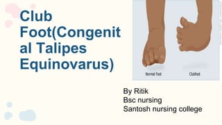
- 1. Club Foot(Congenit al Talipes Equinovarus) By Ritik Bsc nursing Santosh nursing college
- 3. Club foot is a congenital abnormally of the foot and lower leg involving abnormality of bone are structural and soft tissue The foot may be divided into forefoot, midfoot and the hind foot. INTRODUCTION
- 5. DEFINITION The term clubfoot is used to describe a common deformity in which the foot is twisted out of its normal shape or position. Any foot deformity involving the ankle is known as”tallipes” derived from “talus” meaning ankle and “pes” meaning foot.
- 8. INCIDENCE. ● Occurs approx. in 1-2of every 1000 live birth. ● In affected families, clubfeet are about 30 times more frequent in offspring. ● Male are affected in about 65% of case
- 9. RISK FACTORS ● Risk factors include: ● Sex. Clubfoot is more common in males. ● Family history. If either one of the parents or their other children have had clubfoot, the baby is more likely to have it as well. It’s also more common if the baby has another birth defect. ● Smoking during pregnancy. If a woman with a family history of clubfoot smokes during pregnancy, her baby’s risk of the condition may be 20 times greater than average.
- 10. RISK FACTORS ● Not enough amniotic fluid during pregnancy. Too little of the fluid that surrounds the baby in the womb may increase the risk of clubfoot. ● Getting an infection or using illicit drugs during pregnancy. These can increase the risk of clubfoot as well.
- 11. Causes ● The cause of clubfoot is unknown (idiopathic). But scientists do know that clubfoot is not caused by the position of the baby in the womb (fetus). ● In some cases, clubfoot can be associated with other abnormalities of the skeleton that are present at birth (congenital), such as spina bifida. ● Clubfoot can also be the result of problems that affect the nerve, muscle, and bone systems, such as stroke or brain injury. ● Extrinsic associations include teratogenic agents (eg, sodium aminopterin), oligohydramnios, and congenital constriction rings
- 12. TYPES CLUB FOOT Talipes varus Talipes calcaneu s Talipes equinus Talipes equinovarus Talipes valus
- 13. 1. Talipes varus : there is an inversion or bending inward of foot 2. Talipes valgus : there is an eversion or bending outward of foot 3. Talipes equinue : there is planter flexion and toe is lower than heel 4. Talipes calcaneus : there is dorsiflexion and toe higher than heel 5. Talipes equinovarus : the foot is in plantar flexion(cavus) and adductus and hindfoot demonstrates. Type
- 14. CLINICAL FEATURES 1. Malposition of the Foot 2. Equinus Deformity 3. Hindfoot Varus 4. Forefoot Adduction 5. Midfoot Cavus 6. Tight Achilles 7. Muscular Imbalance 8. Smaller Foot Size 9. Limited Range of Motion
- 15. ASSOCIATED ABNORMALY 1. Other Musculoskeletal Abnormalities: such as hip dysplasia, limb length discrepancy, or spinal abnormalities like scoliosis. 2. Congenital Conditions: spina bifida, a neural tube defect affecting the spine; and Larsen syndrome 3. Neuromuscular Disorders: such as cerebral palsy, spinal muscular atrophy, or myelomeningocele 4. Metabolic Disorders: such as mucopolysaccharidosis and diastrophic dysplasia, have been associated with the development of clubfoot. 5. Genetic Factors: Having a family history of clubfoot increases the risk of a child developing the condition.
- 16. 1. Medical History: The healthcare provider will begin by gathering information about the patient’s medical history, including any known risk factors or family history of clubfoot. 2. Physical Examination: A thorough physical examination of the affected foot is essential to assess the severity and characteristics of the clubfoot. The healthcare provider will look for the following signs: ● Abnormal foot position: The foot may be turned inward and downward, with the sole facing inward. Diagnostic evaluation
- 17. ● Muscle tightness or atrophy: The calf muscles may appear smaller or tighter on the affected side. ● Skin creases: Abnormal or absent skin creases may be observed around the foot and ankle. ● Deformities: The bones and joints of the foot will be examined for any bony abnormalities or joint contractures. ● Limited range of motion: The foot may have a restricted range of motion, particularly in the ankle and subtalar joints. Diagnostic evaluation
- 18. 3.Imaging Studies: In certain cases, imaging studies may be ordered to further evaluate the clubfoot. These can include: ● X-rays: X-ray images can help assess the alignment and bony structures of the foot, including the bones of the ankle, foot arch, and metatarsals. ● Ultrasound: In infants, an ultrasound may be performed to evaluate the soft tissues, ligaments, and joint structures of the foot. ● MRI (Magnetic Resonance Imaging): MRI may be used in complex cases or to assess associated abnormalities of the bones, tendons, or nerves. Diagnostic evaluation
- 19. X-ray of clubfoot MRI of club foot
- 21. Management 1. Non-surgical treatment includes manipulating the foot inta a corrected position and then holding it in position with a cast or splint with tapes 2. Physiotherapy is always an important part of the treatment. It may begin once the child is 3 months old. The therapist manipulates the affected foot and may also tape it. 3. Strething and casting: it is also known as the ponseti method. The foot is manipulated into a correct position and a cast is placed to maintain that position ● Repositioning and recasting is repeated for every 1 to 2 week for 2 to 4 months, each time bringing the foot towards the normal position
- 22. 4. The ponseti technique has become the most widely practiced method for initial treatment of infants born with clubfoot. The corrective process utilising the ponseti technique can be divided into two phases: a. The treatment phase : During which the deformity is corrected completely. ● Gentle manipulation and casting is performed on a weekly basis ● Each cast holds the foot in the correct position, allowing it to gradually re shape b. The maintenance phase: During which a brace is utilized to prevent recurrence ● The final cast remain in place for three weeks, after which the infant foot is placed into a removable orthotic device. ● The orthosis is worn 23 hours per day for three months and then during the night time for several years Management
- 24. SURGERY 1. Achilles tenotomy : It is a small surgical procedure in which the Achilles tendon (heel cord) is cut. It allows the ankle to flex upwards (dorsiflex). 2. Osteotomy: It is a surgical procedure that involves cutting and reshaping a bone near a damaged joint to minimize stress on the affected area 3. Fusion and arthrodesis : the orthopedic surgeon manually straightens out the damaged joint, removes the cartilage, and then stabilizes the bone so that they heal together. 4. Club foot repair : it is a surgical repair of the birth defect which involves lengthening or shortening the tendons of the foot
- 26. Nursing diagnosis: They are as follows: 1. Impaired physical mobility related to abnormal foot 2. Disturbed body image related to permanent alternation in structure and /or function 3. Deficient knowledge related to the condition prognosis treatment self care and discharge needs NURSING MANAGEMENT
- 27. 4. Risk for peripheral neurovascular dysfunction related to mechanical compression 5. Risk for impaired skin integrity related to cast application traction or surgery 6. Risk for impaired parenting related to maladaptive coping strategies secondary to diagnosis of talipes deformity. Nursing diagnosis
- 28. ● Nursing object: Impaired physical mobility related to abnormal foot Nursing interventions 1. Assessment and monitoring 2. Educate the patient and family 3. Assistive devices and equipment 4. Pain management 5. Skin care Nursing management
- 29. Nursing objective: Disturbed body image related to permanent alternation in structure and /or function Nursing interventions 1. Education and counseling 2. Encourage self-expression 3. Body positivity and acceptance 4. Peer support and social integration 5. Collaboration with other healthcare professionals 6. Family involvement NURSING MANAGEMENT
- 30. Nursing objective: Deficient knowledge related to the condition prognosis treatment self care and discharge needs Nursing interventions: 1. Assess learning needs 2. Provide information about clubfoot 3. Teach self-care techniques 4. Facilitate discussions with other families 5. Plan for discharge and follow-up care NURSING MANAGEMENT
- 31. Nursing objective: Risk for peripheral neurovascular dysfunction related to mechanical compression Risk for peripheral neurovascular dysfunction related to mechanical compression Nursing interventions: 1. Assess neurovascular status 2. Encourage regular repositioning 3. Monitor footwear and orthotic devices 4. Implement appropriate padding and positioning 5. Monitor for complications NURSING MANAGEMENT
- 32. Nursing objective: Risk for impaired skin integrity related to cast application traction or surgery Nursing interventions: 1. Regular skin assessments 2. Ensure proper cast/traction application 3. Monitor for pressure points: 4. Maintain skin hygiene: 5. Manage pain and discomfort 6. Regular repositioning and movement Nursing management
- 33. 1. Mobility limitations 2. Muscle imbalances and weakness: 3. Joint stiffness 4. Psychosocial impacts 5. Skin breakdown or pressure sores 6. Delayed milestones Complications
- 34. 1. Prenatal care 2. Genetic counseling 3. Avoiding risk factors 4. Maintaining a healthy pregnancy 5. Early detection and treatment Prevention
- 35. 1. Definition and causes 2. Prevalence and risk factors: 3. Treatment options 4. Rehabilitation and long-term care 5. Emotional support 6. Success stories Health education
- 36. ● Sharma R., “Essentials of Pediatric Nursing”, Jaypee publishers, 3rd edition Pg No. 343-545 Bibliography
- 37. THANK YOU
Editor's Notes
- Varus bending inward Valus bending outward Equinue:toe lower the heel