SlideShare uma empresa Scribd logo
1 de 16
By
Willie Francis
BOH/BDS III
SMHS, UPNG
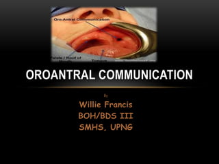
OROANTRAL COMMUNICATION
OUTLINE
• What is Oroantral communication?
• What are the causes of oroantral communication?
• How is oroantral communication diagnosed in patients?
• How do we treat Oroantral communication?
• Conclusion – recommendations!
WHAT IS OROANTRAL COMMUNICATION?
Oroantral communication is simply described as the unnatural communication between the
maxillary sinus and the oral cavity (Doran 2008:2). It is one of the complications which can
occur when doing extraction of the upper molars because sometimes their roots are close to
the maxillary sinus.
When this Oroantral communications occur it can cause problems for both the patient and the
operator. Problems such as:
• Patient not impressed with the operator
• For the operator it is not a practice builder
• Possible medico-legal action
• Removal of bone that may needed for implants (sinus repair and lift/augmentation may be
needed).
• Removal of bony support for dentures
WHY IS OROANTRAL COMMUNICATION A
PROBLEM?
When Oroantral communication is created, it allows food, fluids or smoke from the mouth to flow via the maxillary
sinus into the nasal cavity
Not only that but also it gives access for bacteria and other microorganisms to travel from the oral cavity into the
nose and vice versa.
This can set up a maxillary sinusitis, which mainly depends on how long the communication between the oral
cavity and the maxillary sinus lasts for, may yield either an acute/chronic maxillary sinusitis (Doran 2008:2).
Maxillary sinusitis is a condition that affects the area in which Oroantral communication has occurred.
• Oroantral communication has occurred. The following are some of the factors that are caused by maxillary
sinusitis:
• Sinusitis pain may occur in the cheek, around the eye or in the forward
• Pain felt on upper teeth which can be mistaken for a tooth ache
• Person to feel malaise with a headache and perhaps a stuffy nose
• Discharge of pus into the nose (not noticed until beginning to recover)
• Swelling of the face over the sinus
• Nasal discharge from back of the nose down to the throat
WHAT ARE THE CAUSES OF OROANTRAL
COMMUNICATION?
Some of the underlying factors which may contribute to the aetiology of Oroantral
communications are:
• Exodontia
• Tumors
• Osteomyelitis
• Trauma
• Dentigerous cysts
• Correlation of septal perforations
CONT….
However, Oroantral communication is mainly caused by tooth extractions. Studies have shown that out of the
2,038 teeth that were extracted perforation occurred in 77 of it (of these 38 teeth were from males & 39 were
from females) (Doran 2008:2). These perforations occur mostly when an extraction of the upper first molar is
done. Some of the posterior teeth have roots which are long and may grow into the sinus therefor, when these
teeth are being extracted (Doran 2008:2).
In the dental clinic there are some surgical procedures that can trigger Oroantral communications. These
procedures do not purposely create Oroantral communications but because of the operators mistakes and also
accidents that leads to its formation. Some of these are (Doran 2008:2):
• Apicectomies of maxillary premolars & molars (perforations occurred in 10.4% of teeth).
• Plunging an elevator through the bony floor during root tip removal.
• Forcing root tips or tooth into sinus.
• Penetration while exposing impacted teeth.
• Perforation during incorrect curettage.
• Fracture of segment of the alveolar process containing several teeth with tearing of floor of antrum
• Luxating an impacted 3rdmolar into the antrum whilst attempting to remove it.
HOW IS OROANTRAL COMMUNICATION
DIAGNOSED?
• Diagnosis of acute Oroantral communication – if there is a possibility of Oroantral
communication then the operator should check the extracted tooth for adherent bone; an
adherent bone may stick onto the root of the extracted. Also a nose blowing test must be
carried out, if air from the sinus is felt entering the oral cavity then Oroantral cavity is
suspected. The size of the defect is determined in order for treatment to begin; if the
defect has a diameter of less than 2mm no treatment is required because it
spontaneously heal up (Doran 2008:2).
• Diagnosis of chronic – the Oroantral communication is likely to become chronic if the
diameter of the connection between the oral cavity and the maxillary sinus is more than
5mm, wound dehiscence and enucleation of a cyst. Chronic Oroantral communication
develops four to six weeks after an extraction is made. The patient may have problems
with eating, drinking or smoking, will be subject to chronic maxillary sinusitis and also
experience purulent discharge from the nose(Doran 2008:2).
HOW DO WE TREAT OROANTRAL
COMMUNICATION?
Oroantral communication is treated according to its severity. Chronic cases of Oroantral communication are
treated by surgical procedures while acute cases do not need surgery.
Treatment of acute Oroantral communication: there is no specific treatment of acute Oroantral communication,
however the following can be done;
• The defect should not be probed with needles or any other instruments
• Promote good blood cloth
• The gingival margin around the socket should be approximated as close as possible
• Thare has to be some physical agents placed in the socket to stop excessive bleeding, e.g: surgicel,
spongostan or haemocollagene
• Antibiotics should be prescribed (amoxicillin, doxycycline)
• Nasal decongestants can be used (ephedrine nasal drops, oxymetazoline)
• Steam inhalation can also be used (menthol and eucalyptus)
• Antiseptic mouth-wash can also be used (corsodyl)
• The patient is asked not to blow his/her nose and also not to smoke
CONT..
Treatment of chronic Oroantral communication: there are two ways which involves surgical
procedures that can be used for this treatment (Doran 2008:2).
1. Buccal advancement flap (most common)
• Broad base providing good blood supply.
• Periosteum scored parallel to base of flap to allow greater mobilisation of flap.
• OAC / OAF mucosa excised.
• Alveolus reduced in height.
• Palatal mucosa incised & mobilized.
• Flap brought across defect & secured with sutures.
• There must be no / minimal tension on the flap.
• Disadvantage of reduction of buccal vestibular depth; reshapes in 4 -8 weeks as flap adapts to
underlying bone.
CONT…
2. Palatal Rotational Advancement Flap (Doran 2008:2).
• Advantages of insured vascularity (greater palatine vessels)& thickness of tissue more
like crest of ridge.
• OAC / OAF mucosa excised.
• Buccal mucosa incised & mobilised.
• Flap brought across defect & secured with sutures.
• There must be no / minimal tension on the flap.
• Allows for the maintenance of the vestibularsulcus depth.
• Indicated in cases of unsuccessful buccal flap closure.
• Disadvantage of raw surface left behind; can be covered with a plate or Coe-pack.
CONCLUSION - RECOMENDATION
• It can be concluded that Oroantral communication is mainly caused by the extraction of
posterior teeth on the maxilla. If not treated the defect may become infected leading to
other health problems. Therefore, it is recommended that before any extraction of the
upper teeth is done the patient should be assessed carefully. A proper radiograph should
be taken to give a proper picture of the position of the teeth and if it is a possible OAC
then proper precautions should be taken.
REFERENCE
• Doran, J. MD, 2008, Oro-Antral Communication: Aetiology, Diagnosis, Avoidaance &
Repair, 2nd edn, East Greenstead, Victoria.

Mais conteúdo relacionado

Mais procurados

Arthrocentesis of the temporomandibular joint
Arthrocentesis of the temporomandibular jointArthrocentesis of the temporomandibular joint
Arthrocentesis of the temporomandibular jointAhmed Adawy
 
Local Anesthesia in Oral and Maxillofacial Surgery
Local Anesthesia in Oral and Maxillofacial SurgeryLocal Anesthesia in Oral and Maxillofacial Surgery
Local Anesthesia in Oral and Maxillofacial SurgerySapna Vadera
 
Complication of extraction
Complication of extractionComplication of extraction
Complication of extractionPushp Shah
 
Oro – antral communication
Oro – antral  communicationOro – antral  communication
Oro – antral communicationCFFP
 
Mandibular osteotomies in orthognathic surgery of Face
Mandibular osteotomies in orthognathic surgery of FaceMandibular osteotomies in orthognathic surgery of Face
Mandibular osteotomies in orthognathic surgery of FaceSapna Vadera
 
Maxillary Local Anesthesia
Maxillary Local AnesthesiaMaxillary Local Anesthesia
Maxillary Local AnesthesiaIAU Dent
 
Hypermobility and ankylosis
Hypermobility and ankylosisHypermobility and ankylosis
Hypermobility and ankylosisHanan Shanab
 
Odontogeniccysts OKC
Odontogeniccysts OKCOdontogeniccysts OKC
Odontogeniccysts OKCMaryam Arbab
 
Temporomandibular joint ankylosis
Temporomandibular   joint ankylosisTemporomandibular   joint ankylosis
Temporomandibular joint ankylosisJamil Kifayatullah
 
Management of Impacted third molars
Management of Impacted third molarsManagement of Impacted third molars
Management of Impacted third molarsDr Rayan Malick
 

Mais procurados (20)

Arthrocentesis of the temporomandibular joint
Arthrocentesis of the temporomandibular jointArthrocentesis of the temporomandibular joint
Arthrocentesis of the temporomandibular joint
 
Oroantral Communication and Fistula
Oroantral Communication and FistulaOroantral Communication and Fistula
Oroantral Communication and Fistula
 
Oroantral fistula
Oroantral fistulaOroantral fistula
Oroantral fistula
 
Local Anesthesia in Oral and Maxillofacial Surgery
Local Anesthesia in Oral and Maxillofacial SurgeryLocal Anesthesia in Oral and Maxillofacial Surgery
Local Anesthesia in Oral and Maxillofacial Surgery
 
Complication of extraction
Complication of extractionComplication of extraction
Complication of extraction
 
Oroantral Fistula
Oroantral FistulaOroantral Fistula
Oroantral Fistula
 
Oro – antral communication
Oro – antral  communicationOro – antral  communication
Oro – antral communication
 
Dry socket
Dry socketDry socket
Dry socket
 
Mandibular nerve blocks techniques
Mandibular nerve blocks techniques Mandibular nerve blocks techniques
Mandibular nerve blocks techniques
 
Tmj ankylosis
Tmj ankylosisTmj ankylosis
Tmj ankylosis
 
Mandibular osteotomies in orthognathic surgery of Face
Mandibular osteotomies in orthognathic surgery of FaceMandibular osteotomies in orthognathic surgery of Face
Mandibular osteotomies in orthognathic surgery of Face
 
Maxillary Local Anesthesia
Maxillary Local AnesthesiaMaxillary Local Anesthesia
Maxillary Local Anesthesia
 
Hypermobility and ankylosis
Hypermobility and ankylosisHypermobility and ankylosis
Hypermobility and ankylosis
 
5. Vestibuloplasty.pptx
5. Vestibuloplasty.pptx5. Vestibuloplasty.pptx
5. Vestibuloplasty.pptx
 
Odontogeniccysts OKC
Odontogeniccysts OKCOdontogeniccysts OKC
Odontogeniccysts OKC
 
Temporomandibular joint ankylosis
Temporomandibular   joint ankylosisTemporomandibular   joint ankylosis
Temporomandibular joint ankylosis
 
Instrument in oral surgery
Instrument in oral surgeryInstrument in oral surgery
Instrument in oral surgery
 
Osteomyelitis of jaws dikiohs
Osteomyelitis of jaws dikiohsOsteomyelitis of jaws dikiohs
Osteomyelitis of jaws dikiohs
 
Flaps in oral surgery
Flaps in oral surgeryFlaps in oral surgery
Flaps in oral surgery
 
Management of Impacted third molars
Management of Impacted third molarsManagement of Impacted third molars
Management of Impacted third molars
 

Semelhante a Oroantral communication

Management of-extraction-complications
Management of-extraction-complicationsManagement of-extraction-complications
Management of-extraction-complicationsWeam Faroun
 
PULP AND PERIRADICULAR DISEASES.pptx
PULP AND PERIRADICULAR DISEASES.pptxPULP AND PERIRADICULAR DISEASES.pptx
PULP AND PERIRADICULAR DISEASES.pptxDr. Saurav Paul
 
Complication and management of tooth extraction albayati
Complication and management of tooth extraction albayatiComplication and management of tooth extraction albayati
Complication and management of tooth extraction albayatiAHMED ALBAYATI
 
Endodontic surgery ppt dr. ahmed elfatory
Endodontic surgery ppt  dr. ahmed elfatoryEndodontic surgery ppt  dr. ahmed elfatory
Endodontic surgery ppt dr. ahmed elfatoryaabdesalam
 
Complications occur During Dental Extraction and their Management
Complications occur During Dental Extraction and their ManagementComplications occur During Dental Extraction and their Management
Complications occur During Dental Extraction and their ManagementIraqi Dental Academy
 
Complication of Exodontia.pptx
Complication of Exodontia.pptxComplication of Exodontia.pptx
Complication of Exodontia.pptxStarrkate
 
Complications of exodontia
Complications of exodontiaComplications of exodontia
Complications of exodontiaYanimo
 
Management of Open Bite
Management of Open Bite Management of Open Bite
Management of Open Bite MAZVINNIHAL
 
OROANTRAL FISTULA mohammad abusad.pptx
OROANTRAL FISTULA mohammad abusad.pptxOROANTRAL FISTULA mohammad abusad.pptx
OROANTRAL FISTULA mohammad abusad.pptxMohamadAbusaad
 
Orthodontic & endodontics correlation.pptx
Orthodontic & endodontics correlation.pptxOrthodontic & endodontics correlation.pptx
Orthodontic & endodontics correlation.pptxHazimrizk1
 
ORTHODONTIC TREATMENT PLANING.pdf
ORTHODONTIC TREATMENT PLANING.pdfORTHODONTIC TREATMENT PLANING.pdf
ORTHODONTIC TREATMENT PLANING.pdfamarnath956154
 
Acute apical abscess dr anirudh singh chauhan
Acute apical abscess   dr anirudh singh chauhanAcute apical abscess   dr anirudh singh chauhan
Acute apical abscess dr anirudh singh chauhanAnirudh Singh Chauhan
 
Treatment of deep caries and pulp exposure
Treatment of deep caries and pulp exposureTreatment of deep caries and pulp exposure
Treatment of deep caries and pulp exposureMohammed_Yazdi
 

Semelhante a Oroantral communication (20)

Management of-extraction-complications
Management of-extraction-complicationsManagement of-extraction-complications
Management of-extraction-complications
 
PULP AND PERIRADICULAR DISEASES.pptx
PULP AND PERIRADICULAR DISEASES.pptxPULP AND PERIRADICULAR DISEASES.pptx
PULP AND PERIRADICULAR DISEASES.pptx
 
Complication and management of tooth extraction albayati
Complication and management of tooth extraction albayatiComplication and management of tooth extraction albayati
Complication and management of tooth extraction albayati
 
Periotome
PeriotomePeriotome
Periotome
 
Pre prosthetic surgery
Pre prosthetic surgeryPre prosthetic surgery
Pre prosthetic surgery
 
Exodontia
ExodontiaExodontia
Exodontia
 
Endodontic surgery ppt dr. ahmed elfatory
Endodontic surgery ppt  dr. ahmed elfatoryEndodontic surgery ppt  dr. ahmed elfatory
Endodontic surgery ppt dr. ahmed elfatory
 
serial extraction
 serial extraction  serial extraction
serial extraction
 
Teeth Extraction.ppt
Teeth Extraction.pptTeeth Extraction.ppt
Teeth Extraction.ppt
 
Complications occur During Dental Extraction and their Management
Complications occur During Dental Extraction and their ManagementComplications occur During Dental Extraction and their Management
Complications occur During Dental Extraction and their Management
 
Complication of Exodontia.pptx
Complication of Exodontia.pptxComplication of Exodontia.pptx
Complication of Exodontia.pptx
 
INTERCEPTIVE ORTHODONTICS
INTERCEPTIVE ORTHODONTICSINTERCEPTIVE ORTHODONTICS
INTERCEPTIVE ORTHODONTICS
 
Oro-antral fistula
Oro-antral fistulaOro-antral fistula
Oro-antral fistula
 
Complications of exodontia
Complications of exodontiaComplications of exodontia
Complications of exodontia
 
Management of Open Bite
Management of Open Bite Management of Open Bite
Management of Open Bite
 
OROANTRAL FISTULA mohammad abusad.pptx
OROANTRAL FISTULA mohammad abusad.pptxOROANTRAL FISTULA mohammad abusad.pptx
OROANTRAL FISTULA mohammad abusad.pptx
 
Orthodontic & endodontics correlation.pptx
Orthodontic & endodontics correlation.pptxOrthodontic & endodontics correlation.pptx
Orthodontic & endodontics correlation.pptx
 
ORTHODONTIC TREATMENT PLANING.pdf
ORTHODONTIC TREATMENT PLANING.pdfORTHODONTIC TREATMENT PLANING.pdf
ORTHODONTIC TREATMENT PLANING.pdf
 
Acute apical abscess dr anirudh singh chauhan
Acute apical abscess   dr anirudh singh chauhanAcute apical abscess   dr anirudh singh chauhan
Acute apical abscess dr anirudh singh chauhan
 
Treatment of deep caries and pulp exposure
Treatment of deep caries and pulp exposureTreatment of deep caries and pulp exposure
Treatment of deep caries and pulp exposure
 

Oroantral communication

  • 1. By Willie Francis BOH/BDS III SMHS, UPNG OROANTRAL COMMUNICATION
  • 2. OUTLINE • What is Oroantral communication? • What are the causes of oroantral communication? • How is oroantral communication diagnosed in patients? • How do we treat Oroantral communication? • Conclusion – recommendations!
  • 3. WHAT IS OROANTRAL COMMUNICATION? Oroantral communication is simply described as the unnatural communication between the maxillary sinus and the oral cavity (Doran 2008:2). It is one of the complications which can occur when doing extraction of the upper molars because sometimes their roots are close to the maxillary sinus. When this Oroantral communications occur it can cause problems for both the patient and the operator. Problems such as: • Patient not impressed with the operator • For the operator it is not a practice builder • Possible medico-legal action • Removal of bone that may needed for implants (sinus repair and lift/augmentation may be needed). • Removal of bony support for dentures
  • 4.
  • 5. WHY IS OROANTRAL COMMUNICATION A PROBLEM? When Oroantral communication is created, it allows food, fluids or smoke from the mouth to flow via the maxillary sinus into the nasal cavity Not only that but also it gives access for bacteria and other microorganisms to travel from the oral cavity into the nose and vice versa. This can set up a maxillary sinusitis, which mainly depends on how long the communication between the oral cavity and the maxillary sinus lasts for, may yield either an acute/chronic maxillary sinusitis (Doran 2008:2). Maxillary sinusitis is a condition that affects the area in which Oroantral communication has occurred. • Oroantral communication has occurred. The following are some of the factors that are caused by maxillary sinusitis: • Sinusitis pain may occur in the cheek, around the eye or in the forward • Pain felt on upper teeth which can be mistaken for a tooth ache • Person to feel malaise with a headache and perhaps a stuffy nose • Discharge of pus into the nose (not noticed until beginning to recover) • Swelling of the face over the sinus • Nasal discharge from back of the nose down to the throat
  • 6. WHAT ARE THE CAUSES OF OROANTRAL COMMUNICATION? Some of the underlying factors which may contribute to the aetiology of Oroantral communications are: • Exodontia • Tumors • Osteomyelitis • Trauma • Dentigerous cysts • Correlation of septal perforations
  • 7. CONT…. However, Oroantral communication is mainly caused by tooth extractions. Studies have shown that out of the 2,038 teeth that were extracted perforation occurred in 77 of it (of these 38 teeth were from males & 39 were from females) (Doran 2008:2). These perforations occur mostly when an extraction of the upper first molar is done. Some of the posterior teeth have roots which are long and may grow into the sinus therefor, when these teeth are being extracted (Doran 2008:2). In the dental clinic there are some surgical procedures that can trigger Oroantral communications. These procedures do not purposely create Oroantral communications but because of the operators mistakes and also accidents that leads to its formation. Some of these are (Doran 2008:2): • Apicectomies of maxillary premolars & molars (perforations occurred in 10.4% of teeth). • Plunging an elevator through the bony floor during root tip removal. • Forcing root tips or tooth into sinus. • Penetration while exposing impacted teeth. • Perforation during incorrect curettage. • Fracture of segment of the alveolar process containing several teeth with tearing of floor of antrum • Luxating an impacted 3rdmolar into the antrum whilst attempting to remove it.
  • 8. HOW IS OROANTRAL COMMUNICATION DIAGNOSED? • Diagnosis of acute Oroantral communication – if there is a possibility of Oroantral communication then the operator should check the extracted tooth for adherent bone; an adherent bone may stick onto the root of the extracted. Also a nose blowing test must be carried out, if air from the sinus is felt entering the oral cavity then Oroantral cavity is suspected. The size of the defect is determined in order for treatment to begin; if the defect has a diameter of less than 2mm no treatment is required because it spontaneously heal up (Doran 2008:2). • Diagnosis of chronic – the Oroantral communication is likely to become chronic if the diameter of the connection between the oral cavity and the maxillary sinus is more than 5mm, wound dehiscence and enucleation of a cyst. Chronic Oroantral communication develops four to six weeks after an extraction is made. The patient may have problems with eating, drinking or smoking, will be subject to chronic maxillary sinusitis and also experience purulent discharge from the nose(Doran 2008:2).
  • 9. HOW DO WE TREAT OROANTRAL COMMUNICATION? Oroantral communication is treated according to its severity. Chronic cases of Oroantral communication are treated by surgical procedures while acute cases do not need surgery. Treatment of acute Oroantral communication: there is no specific treatment of acute Oroantral communication, however the following can be done; • The defect should not be probed with needles or any other instruments • Promote good blood cloth • The gingival margin around the socket should be approximated as close as possible • Thare has to be some physical agents placed in the socket to stop excessive bleeding, e.g: surgicel, spongostan or haemocollagene • Antibiotics should be prescribed (amoxicillin, doxycycline) • Nasal decongestants can be used (ephedrine nasal drops, oxymetazoline) • Steam inhalation can also be used (menthol and eucalyptus) • Antiseptic mouth-wash can also be used (corsodyl) • The patient is asked not to blow his/her nose and also not to smoke
  • 10. CONT.. Treatment of chronic Oroantral communication: there are two ways which involves surgical procedures that can be used for this treatment (Doran 2008:2). 1. Buccal advancement flap (most common) • Broad base providing good blood supply. • Periosteum scored parallel to base of flap to allow greater mobilisation of flap. • OAC / OAF mucosa excised. • Alveolus reduced in height. • Palatal mucosa incised & mobilized. • Flap brought across defect & secured with sutures. • There must be no / minimal tension on the flap. • Disadvantage of reduction of buccal vestibular depth; reshapes in 4 -8 weeks as flap adapts to underlying bone.
  • 11.
  • 12.
  • 13. CONT… 2. Palatal Rotational Advancement Flap (Doran 2008:2). • Advantages of insured vascularity (greater palatine vessels)& thickness of tissue more like crest of ridge. • OAC / OAF mucosa excised. • Buccal mucosa incised & mobilised. • Flap brought across defect & secured with sutures. • There must be no / minimal tension on the flap. • Allows for the maintenance of the vestibularsulcus depth. • Indicated in cases of unsuccessful buccal flap closure. • Disadvantage of raw surface left behind; can be covered with a plate or Coe-pack.
  • 14.
  • 15. CONCLUSION - RECOMENDATION • It can be concluded that Oroantral communication is mainly caused by the extraction of posterior teeth on the maxilla. If not treated the defect may become infected leading to other health problems. Therefore, it is recommended that before any extraction of the upper teeth is done the patient should be assessed carefully. A proper radiograph should be taken to give a proper picture of the position of the teeth and if it is a possible OAC then proper precautions should be taken.
  • 16. REFERENCE • Doran, J. MD, 2008, Oro-Antral Communication: Aetiology, Diagnosis, Avoidaance & Repair, 2nd edn, East Greenstead, Victoria.