SlideShare uma empresa Scribd logo
1 de 9
Baixar para ler offline
Edited March02,2016
Updated on Septembre 30, 2016
Medical Imaging of PneumoThorax (PNO4)
Dr WALIF CHBEIR
V- Diagnosis
* The diagnosis of PNO is suspected in stable patients with dyspnea or pleuritic chest pain and
is confirmed with upright inspiratory chest x-ray. Radiolucent air and the absence of lung
markings juxtaposed between a shrunken lobe or lung and the parietal pleura are diagnostic of
pneumothorax.
- Tracheal deviation and mediastinal shift occur with large pneumothoraces.
* Small pneumothoraces (eg, < 10%) are sometimes overlooked on chest x-ray. In patients
with possible pneumothorax, lung markings should be traced to the edge of the pleura on chest
x-ray. Conditions that mimic pneumothorax radiographically include emphysematous bullae,
skinfolds, folded bed sheets, and overlap of stomach or bowel markings on lung fields. -
Ultrasonography (done at the bedside during initial resuscitation) and CT are more sensitive for
small pneumothoraces than chest x-ray.
* Traumatic PNO: Diagnosis is usually made by chest x-ray. Ultrasonography (done at the
bedside during initial resuscitation) and CT are more sensitive for small pneumothoraces than
chest x-ray.
* The size of the pneumothorax, stated as percent of the hemithorax that is vacant, can be
estimated by x-ray findings. The numerical size is valuable mainly for quantifying progression
and resolution rather than for determining prognosis.
* Open PNO: The diagnosis is made clinically and requires inspecting the entire chest wall
surface.
* In critical care and in patients with severe ARDS (ARDS & PNO) , Patients with
pneumothorax did not have the traditional clinical and radiologic signs and the most repeatable
finding may be a subtle drop in oxygenation measurements without another obvious cause
(Loculated Pneumothorax: A Special Challenge In Critical Care) .
The diagnosis of pneumothorax in critical illness is made from the history and examination of
the patient and confirmed, where possible, by radiological investigation. The factors that are
important in the history relate to the underlying disease process and any potential for iatrogenic
pneumothorax .
The early and accurate diagnosis of pneumothorax in ARDS patients is mandatory since this
complication carries an increased mortality. Furthermore, small pneumothoraces in these
patients can cause severe hemodynamic or pulmonary compromise. This is the reason why
pneumothorax must always be suspected in any patient with ARDS who experiences an acute
worsening in respiratory function, accompanied with dyspnea and hypoxemia, which is usually
unresponded to oxygen therapy.
- Portable chest X-ray is the first diagnostic evaluation imaging being used and the procedure
of choice for the documentation of lung underlying pathology or the presents of lines, tubes or
devices. Nevertheless, often exhibits diagnostic disadvantages, taking into account that
pneumothoraces in ARDS patients may have unusual, as well as subtle features and small sized
pneumothoraces or loculated pneumothoraces, can be missed on chest X-ray. Furthermore,
other types of air leaks, such as pneumomediastinum and interstitial pulmonary emphysema,
may be more difficulty observed by chest radiographs .
- Cases have been described in medical literature, referring to patients presenting clinical
deterioration but unchanged chest X-ray and functioning chest drains (Acute respiratory
distress syndrome and pneumothorax; ref 14). This is the reason why, especially in patients under
mechanical ventilation, serial and daily chest radiographs are necessary in the evaluation of
underlying lung pathology.
- There for, if a pneumothorax is suspected and is unrevealed on chest X-ray, a more
specific diagnostic imaging like chest-computed tomography (CT) is necessary. CT scan in
patients with ARDS, as explained above , can reveal a variety of abnormalities , is helpful in
understanding the extent of the underlying lung parenchyma distraction and is quite more
sensitive in identifying pneumomediastinum andpneumothorax, which are frequently observed
in patients with ARDS.
- Nevertheless, chest-CT evaluation is seldom employed in patients with ARDS, especially
patients with severe respiratory failure under mechanical ventilation, mostly due to problems
concerning the transfer and monitoring of these critically ill patients.
- Due to these technical difficulties of chest-CT, an essential diagnostic method in critically ill
patient gaining respect is lung-ultrasonography, a relatively easy to perform, portable and
inexpensive diagnostic imaging. Lung-ultrasonography can prove an alternative diagnostic
procedure in the difficult diagnosis of pneumothorax in critically ill patients with severe
ARDS, which not only permits bedside assessment of lung pathology but also assists in the
evaluation of mechanical ventilation parameters, as well as the evaluation of lung
overdistension and PEEP-induced lung recruitment .
- In the same setting, Loculated pneumothorax provides only subtle clinical clues. The only
clinical evidence may be deteriorating oxygenation without another obvious cause. US findings
may be equivocal . The abscence of lung sliding may be caused by pno but it has others causes
as well . The presence of Lung Sliding indicates that there is no PNO but alone doen't exclude
the diagnostic, while the abscence of lung sliding only indicates that there may be one ( because
it has others causes as well). Scan the entire chest for B lines. (Clinical chest US: from the ICU to the bronchoscopic
suite).
PNO that is not in immediate contact with the chest wall will not be identified on US ( e.g.
Loculated PNO against medistinum).
Be prepared for chest CT scans if ever Lung US is inconclusive for this hard-to-catch
complication of mechanical ventilation in patients with ARDS.
* Tension pneumothorax is a clinical diagnosis, not a radiographic diagnosis, because the
respiratory and hemodynamic consequences of tension pneumothorax do not have radiographic
equivalents in many circumstances. Radiographic signs of tension (mediastinal shift, inversion
of diaphragm, enlargement of affected hemithorax) can occur in the absence of adverse
physiologic effects, and the physiologic effects of pleural tension may be present without
radiographic signs of tension ( critically ill p.) .
- In ARDS, the diseased noncompliant lung may not collapse in the presence of a
pneumothorax, and the controralateral lung may be too stiff to allow mediastinal shift. Thus,
tension pneumothorax in ARDS can present as a loculated paracardiac or subpulmonic air
collection with little or no mediastinal shift and only slight changes of the cardiac contour.
- Treatment should not be delayed pending radiographic confirmation. Although cardiac
tamponade also can cause hypotension, neck vein distention, and sometimes respiratory
distress, tension pneumothorax can be differentiated clinically by its unilateral absence of breath
sounds and hyperresonance to percussion.
- Although non-specific, the association of respiratory and haemodynamic signs found with a
tension pneumothorax are a medical emergency. Severe haemodynamic compromise will
require urgent needle decompression of the pneumothorax before its diagnosis being confirmed
radiologically. Fortunately this situation is uncommon and there is frequently time for
radiological investigations to help establish the diagnosis of a simple pneumothorax.
VI- Significant Points
* A large pneumothorax is radiographically defined as one with > 2 cm from pleural surface to
lung edge; this is an objective indication for drainage
* Don’t wait for a radiograph if there are clinical signs of a tension pneumothorax.
Tension pneumothorax is a medical emergency and may require immediate needle
decompression before radiological investigation.
- Treat the patient not the radiograph. Don’t act on a radiographic appearance if it does not fit
the clinical picture. Get an expert opinion on the radiograph first.
* Skin folds, companion shadows, the scapula, and previous lung surgery or chest drain
placement may all mimic pneumothoraxes on XRay Chest.
* In the supine patient, pneumothoraxes are best seen at the lung bases and adjacent to the
heart.
The “deep sulcus sign” describes a costophrenic angle that extends more inferiorly than
usual as a result of air lying in the costophrenic angle. The liver appears more radiolucent than
usual due to air lying anteriorly in the costophrenic angle, and on the left side, air will
outline the medial aspect of the hemidiaphragm under the heart.
* What other radiological investigations may be used to confirm the diagnosis of PNO in acute
traumatic setting and in critically ill patients?
- A radiograph with the patient in a lateral decubitus position, with the affected side uppermost,
can be helpful in demonstrating a lung edge.
- In patients well enough to be transported, thoracic computed tomography can be helpful in
locating the position of a pneumothorax and accurately siting a chest drain.
- US and CT are more sensitive than Chest X-Ray Radiography in detection PNO. CT is the
gold standard. US is done at the bedside during initial resuscitation of trauma patient and in
critically ill patient in ICU.
- The early and accurate diagnosis of pneumothorax in ARDS patients is mandatory since this
complication carries an increased mortality. Portable chest X-ray is the first diagnostic
evaluation imaging. If a pneumothorax is suspected and is unrevealed on chest X-ray, Lung
USG is now an alternative method to Chest CT mostly due to the cumbersome nature of this
technique in critically ill patients.
* The negative predictive value of Chest US for lung sliding is reported as 99.2–100%,
indicating that the presence of sliding effectively rules out a pneumothorax. For some authors,
lung sliding ALONE does not exclude PNO and scanning of the entire anterior chest for B-
Lines is mandatory (Ultrasonography in the ICU: Practical Applications).
- However, the absence of lung sliding does not necessarily indicate that a pneumothorax is
present. In fact, Lung sliding is abolished in a variety of conditions other than pneumothorax.
- Lung Sliding and B lines are not present on a patient with PNO. M Mode can help
differenciate between a seashore sign or Stratosphere or bar code signs ( SeaShore = no PNO).
* Blind chest drain placement into a loculated pneumothorax may lead to an iatrogenic air leak
from direct trauma to the pleura and worsening the patient’s clinical condition.
* An immediate post-treatment radiograph is essential to detect complications and ensure a
satisfactory drain position
* A chest drain apparently well positioned on frontal radiograph may be lying in the soft
tissues, in a lung fissure, or within the substance of the lung.
VII- References
1- Fahad M AlHameed, MD, AmBIM, FCCP, FRCPC; Chief Editor: Eugene C Lin, MD.
Pneumothorax Imaging: Overview, Radiography, Computed Tomography, in emedecine /
Medscape, Updated: Oct 04, 2015, (Page consulted February 12, 2016).
[http://emedicine.medscape.com/article/360796-overview#showall]
2-Patricia Carroll, RN,C, CEN, RRT, MS Loculated Pneumothorax: A Special Challenge In
Critical Care., in Clinical Update for the Professional Nurse, September 2000. Clinical Update
is an educational newsletter provided by Atrium Medical Corporation.
[http://www.atriummed.com/en/chest_drainage/Clinical%20Updates/ClinicalUpdateSept00.pdf]
3- Chon KS, vanSonnenberg E, D'Agostino HB, O'Laoide RM, Colt HG, Hart E: CT- guided
catheter drainage of loculated thoracic air collections in mechanically ventilated patients
with acute respiratory distress syndrome. American Journal of Roentgenology
1999;173(5):1345-1350, in PubMed - indexed for MEDLINE , PMID: 10541116.
[http://www.ajronline.org/doi/abs/10.2214/ajr.173.5.10541116]
4- Emergency, Emergency! in General Quiz 001, Copyright © 2003 LearningRadiology.com.
(Page consulted February 12, 2016).
[http://learningradiology.com/new/quizzes/quiz0004/index0004.htm].
5- Dr Ayush Goel et al. Pneumothorax: ultrasound, in © 2005–2016 Radiopaedia.org
[http://radiopaedia.org/articles/pneumothorax-ultrasound. Page consulted February 12, 2016. ]
6- Terrance Healey, MD- Pneumothorax, Primary Spontaneous, in
STATdx Copyrigh © 2016 Elsevier, Inc. All rights reserved.
Chest > Diagnosis > Pleural Diseases > Pneumothorax. (Page consulted February 12, 2016).
[https://my.statdx.com/document/pneumothorax-primary-spontaneous/eb1b4e8a-d718-45c8-9935-
34da254c9af3?searchTerm=Pneumothorax,%20Primary%20Spontaneous]
7- Lubna F Husain, Laura Hagopian, Derek Wayman, William E Baker, and Kristin A
Carmody: Sonographic diagnosis of pneumothorax, in J Emerg Trauma Shock. 2012 Jan-
Mar; 5(1): 76–81, in PMC free articles, PMCID: PMC3299161, Received 2011 Jun 1;
Accepted 2011 Jun 6, ( Page consulted February 12, 2016).
[http://www.ncbi.nlm.nih.gov/pmc/articles/PMC3299161]
8- Jain, Arpana (et al.). Thoracic Ultrasonography in the Critically Ill, in Paula Ferrada
(Ed.), Ultrasonography in the ICU: Practical Applications Pages 37-52, © 2015 Springer
International Publishing, eBook ISBN 978-3-319-11876-5, in google Books, (Page consulted
February 12, 2016).
[https://books.google.com.lb/books?id=9Fx1CQAAQBAJ&pg=PA51&lpg=PA51&dq=loculated+pneumothora
x+and+ultrasonography&source=bl&ots=re2jhh0
lu&sig=lOXixQ68hsRD1ZegQCQSpQ9YTs&hl=fr&sa=X&ved=0ahUKEwj566y21uzKAhWDthoKHauLDO0
Q6AEIVjAI#v=onepage&q=loculated%20pneumothorax%20and%20ultrasonography&f=false].
9- Arun Nagdev, MD, and Michael Murphy, MD: Focus On: Ultrasound Detection of
Traumatic Anterior Pneumothorax, in Copyright © 2014 American College of Emergency
Physicians American website, December 2008. (Page consulted February 12, 2016).
[http://www.acep.org/Clinical---Practice-Management/Focus-On--Ultrasound-Detection-of-
Traumatic-Anterior-Pneumothorax ]
10- A R O’Connor, W E Morgan. Radiological review of pneumothorax, in BMJ
2005;330:1493–7, in PMC free articles, PMCID: PMC558461
[http://www.ncbi.nlm.nih.gov/pmc/articles/PMC558461]
11- Pneumothorax From Wikipedia, the free encyclopedia. Page was last modified on 16
January 2016, at 22:13, (page consulted February 12, 2016).
[https://en.wikipedia.org/wiki/Pneumothorax]
12- Pneumothorax in Collins, Jannette; Stern, Eric J. Chest Radiology: The Essentials, 2nd
Edition. Copyright ©2008 Lippincott Williams & Wilkins. P.148-152.
13- Richard W. Light, MD. Pneumothorax in Merck Manual, Professionnal Version,
[http://www.merckmanuals.com/professional/pulmonary-disorders/mediastinal-and-pleural-
disorders/pneumothorax], Last full review/revision September 2014, ( page consulted February
12, 2016).
14- Ashley Miller : Lung Ultrasound - Pneumothorax, in ICMteaching.com. © 2011.
(Page consulted February 12, 2016).
[http://www.icmteaching.com/ultrasound/lung%20ultrasound/styled-125/]
15- Mitlehner W, Friedrich M, Dissmann W. Value of computer tomography in the
detection of bullae and blebs in patients with primary spontaneous pneumothorax, in
Respiration. 1992. 59(4):2217, in PubMed - indexed for MEDLINE ( PMID: 1485007), (Page
consulted February 12, 2016).
[http://www.ncbi.nlm.nih.gov/pubmed/?term=PMID%3A+1485007]
16- James J Rankine, Antony N Thomas, Dorothee Fluechter. Diagnosis of pneumothorax in
critically ill adults Postgrad Med J 2000;76:399–404, in in PMC free articles, PMCID:
PMC1741653
[http://www.ncbi.nlm.nih.gov/pmc/articles/PMC1741653/]
17- Skaarup SH, Folkersen BH. Ultrasound-assisted aspiration of loculated pneumothorax:
A new technique, in J Clin Ultrasound. 2015 Dec 16. doi: 10.1002/jcu.22326, in PubMed,
PMID: 26676092, (Page consulted February 12, 2016).
[http://www.ncbi.nlm.nih.gov/pubmed/26676092]
18- Paul Stark, MD. Imaging of pneumothorax, in UpToDate, Literature review current
through: Jan 2016, This topic last updated: Feb 3, 2015, (Page consulted February 12, 2016).
[www.uptodate.com/contents/imaging-of-pneumothorax].
19- Eirini Terzi, & al: Acute respiratory distress syndrome and pneumothorax, in J
Thorac Dis 2014;6(S4):S435-S442, in www.jthoracdis.com, Submitted Aug 15, 2014.
Accepted for publication Aug 19, 2014, (Page consulted February 12, 2016).
[http://www.jthoracdis.com/article/view/3101/3676].
20- Grace M. Thomas, MD - Stephen Jones, MD - Clint M. Gerdes, MD: Supine
pneumothorax:
in ACR- Case in point, Wednesday, August 6, 2014, ( Page consulted February 12, 2016).
[https://3s.acr.org/CIP/SearchCaseView.aspx?CaseId=KsGsjCvXCLg%253d]
21- IM Tocino, MH Miller and WR Fairfax: Distribution of pneumothorax in the supine and
semirecumbent critically ill adult, in AJR 144:901-905, May 1985, in PubMed (PMID:
3872573), (Page consulted February 12, 2016).
[http://www.ajronline.org/doi/abs/10.2214/ajr.144.5.901]
22- Wang J.S. ·Doelken P. Pleural Ultrasonography in the Intensive Care Unit, in Bolliger CT,
Herth FJF, Mayo PH, Miyazawa T, Beamis JF (eds): Clinical Chest Ultrasound: From the ICU
to the Bronchoscopy Suite, in Prog Respir Res. Basel, Karger, 2009, vol 37, pp 82–88,
Published online: 3/25/2009, eISBN: 978-3-8055-8643-6 (Online), in Google Books.
https://books.google.com.lb/books?id=OF4SS1mgZRkC&pg=PA78&lpg=PA78&dq=loculated+pneumothorax
+and+ultrasonography&source=bl&ots=JMu4GtHHIW&sig=0lUnTRfU9_x0k5kG80akbcFvC3I&hl=fr&sa=X
&ved=0ahUKEwj566y21uzKAhWDthoKHauLDO0Q6AEIPTAE#v=onepage&q=loculated%20pneumothorax
%20and%20ultrasonography&f=false
23- Thomas G. Weiser, MD, MPH. Pneumothorax (Traumatic) in Merck Manual,
Professionnal Version, Last full review/revision August 2014, (page consulted February 12,
2016).
[http://www.merckmanuals.com/professional/injuries-poisoning/thoracic
trauma/pneumothorax-(traumatic) ],
24-Thomas G. Weiser, MD, MPH. Pneumothorax (Tension) in Merck Manual, Professionnal
Version, Last full review/revision August 2014, ( Page consulted February 12, 2016).
[http://www.merckmanuals.com/professional/injuries-poisoning/thoracic-
trauma/pneumothorax-(tension)]
25- Thomas G. Weiser, MD, MPH. Pneumothorax (Open) (Sucking Chest Wound) in Merck
Manual, Professionnal Version, Last full review/revision August 2014, ( Page consulted
February 12, 2016).
[http://www.merckmanuals.com/professional/injuries-poisoning/thoracic-
trauma/pneumothorax-(open) ],

Mais conteúdo relacionado

Mais de Walif Chbeir

Pachygyria, Polymicrogyria
Pachygyria, Polymicrogyria Pachygyria, Polymicrogyria
Pachygyria, Polymicrogyria Walif Chbeir
 
Carotid Artery Stenosis | Walif Chbeir
Carotid Artery Stenosis  |  Walif ChbeirCarotid Artery Stenosis  |  Walif Chbeir
Carotid Artery Stenosis | Walif ChbeirWalif Chbeir
 
Posterolateral Corner(PLC) Injury of Knee.
 Posterolateral Corner(PLC) Injury of Knee. Posterolateral Corner(PLC) Injury of Knee.
Posterolateral Corner(PLC) Injury of Knee.Walif Chbeir
 
Walif Chbeir: Cerebral Cavernous Malformation
Walif Chbeir: Cerebral Cavernous MalformationWalif Chbeir: Cerebral Cavernous Malformation
Walif Chbeir: Cerebral Cavernous MalformationWalif Chbeir
 
Medical Imaging of Pneumothorax (PNO)–FIGURES 2-Walif Chbeir
Medical Imaging of Pneumothorax (PNO)–FIGURES 2-Walif ChbeirMedical Imaging of Pneumothorax (PNO)–FIGURES 2-Walif Chbeir
Medical Imaging of Pneumothorax (PNO)–FIGURES 2-Walif ChbeirWalif Chbeir
 
Medical Imaging of Pneumothorax (PNO)–FIGURES-Walif Chbeir
Medical Imaging of Pneumothorax (PNO)–FIGURES-Walif ChbeirMedical Imaging of Pneumothorax (PNO)–FIGURES-Walif Chbeir
Medical Imaging of Pneumothorax (PNO)–FIGURES-Walif ChbeirWalif Chbeir
 
Normal Labral Variant Figures II - Walif Chbeir
Normal Labral Variant Figures II - Walif ChbeirNormal Labral Variant Figures II - Walif Chbeir
Normal Labral Variant Figures II - Walif ChbeirWalif Chbeir
 
Doppler of Lower Limb Arteries. Technical Aspects.
Doppler of Lower Limb Arteries. Technical Aspects.Doppler of Lower Limb Arteries. Technical Aspects.
Doppler of Lower Limb Arteries. Technical Aspects.Walif Chbeir
 
Ankle-Brachial Index (ABI) --Walif Chbeir
Ankle-Brachial Index (ABI) --Walif ChbeirAnkle-Brachial Index (ABI) --Walif Chbeir
Ankle-Brachial Index (ABI) --Walif ChbeirWalif Chbeir
 
Normal Labral Variants - Walif Chbeir
Normal Labral Variants - Walif ChbeirNormal Labral Variants - Walif Chbeir
Normal Labral Variants - Walif ChbeirWalif Chbeir
 
Medical Imaging of Pneumothorax (PNO)-Walif Chbeir
Medical Imaging of Pneumothorax (PNO)-Walif ChbeirMedical Imaging of Pneumothorax (PNO)-Walif Chbeir
Medical Imaging of Pneumothorax (PNO)-Walif ChbeirWalif Chbeir
 

Mais de Walif Chbeir (11)

Pachygyria, Polymicrogyria
Pachygyria, Polymicrogyria Pachygyria, Polymicrogyria
Pachygyria, Polymicrogyria
 
Carotid Artery Stenosis | Walif Chbeir
Carotid Artery Stenosis  |  Walif ChbeirCarotid Artery Stenosis  |  Walif Chbeir
Carotid Artery Stenosis | Walif Chbeir
 
Posterolateral Corner(PLC) Injury of Knee.
 Posterolateral Corner(PLC) Injury of Knee. Posterolateral Corner(PLC) Injury of Knee.
Posterolateral Corner(PLC) Injury of Knee.
 
Walif Chbeir: Cerebral Cavernous Malformation
Walif Chbeir: Cerebral Cavernous MalformationWalif Chbeir: Cerebral Cavernous Malformation
Walif Chbeir: Cerebral Cavernous Malformation
 
Medical Imaging of Pneumothorax (PNO)–FIGURES 2-Walif Chbeir
Medical Imaging of Pneumothorax (PNO)–FIGURES 2-Walif ChbeirMedical Imaging of Pneumothorax (PNO)–FIGURES 2-Walif Chbeir
Medical Imaging of Pneumothorax (PNO)–FIGURES 2-Walif Chbeir
 
Medical Imaging of Pneumothorax (PNO)–FIGURES-Walif Chbeir
Medical Imaging of Pneumothorax (PNO)–FIGURES-Walif ChbeirMedical Imaging of Pneumothorax (PNO)–FIGURES-Walif Chbeir
Medical Imaging of Pneumothorax (PNO)–FIGURES-Walif Chbeir
 
Normal Labral Variant Figures II - Walif Chbeir
Normal Labral Variant Figures II - Walif ChbeirNormal Labral Variant Figures II - Walif Chbeir
Normal Labral Variant Figures II - Walif Chbeir
 
Doppler of Lower Limb Arteries. Technical Aspects.
Doppler of Lower Limb Arteries. Technical Aspects.Doppler of Lower Limb Arteries. Technical Aspects.
Doppler of Lower Limb Arteries. Technical Aspects.
 
Ankle-Brachial Index (ABI) --Walif Chbeir
Ankle-Brachial Index (ABI) --Walif ChbeirAnkle-Brachial Index (ABI) --Walif Chbeir
Ankle-Brachial Index (ABI) --Walif Chbeir
 
Normal Labral Variants - Walif Chbeir
Normal Labral Variants - Walif ChbeirNormal Labral Variants - Walif Chbeir
Normal Labral Variants - Walif Chbeir
 
Medical Imaging of Pneumothorax (PNO)-Walif Chbeir
Medical Imaging of Pneumothorax (PNO)-Walif ChbeirMedical Imaging of Pneumothorax (PNO)-Walif Chbeir
Medical Imaging of Pneumothorax (PNO)-Walif Chbeir
 

Último

Call Girls Hosur Just Call 7001305949 Top Class Call Girl Service Available
Call Girls Hosur Just Call 7001305949 Top Class Call Girl Service AvailableCall Girls Hosur Just Call 7001305949 Top Class Call Girl Service Available
Call Girls Hosur Just Call 7001305949 Top Class Call Girl Service Availablenarwatsonia7
 
Kolkata Call Girls Services 9907093804 @24x7 High Class Babes Here Call Now
Kolkata Call Girls Services 9907093804 @24x7 High Class Babes Here Call NowKolkata Call Girls Services 9907093804 @24x7 High Class Babes Here Call Now
Kolkata Call Girls Services 9907093804 @24x7 High Class Babes Here Call NowNehru place Escorts
 
call girls in munirka DELHI 🔝 >༒9540349809 🔝 genuine Escort Service 🔝✔️✔️
call girls in munirka  DELHI 🔝 >༒9540349809 🔝 genuine Escort Service 🔝✔️✔️call girls in munirka  DELHI 🔝 >༒9540349809 🔝 genuine Escort Service 🔝✔️✔️
call girls in munirka DELHI 🔝 >༒9540349809 🔝 genuine Escort Service 🔝✔️✔️saminamagar
 
Russian Call Girl Brookfield - 7001305949 Escorts Service 50% Off with Cash O...
Russian Call Girl Brookfield - 7001305949 Escorts Service 50% Off with Cash O...Russian Call Girl Brookfield - 7001305949 Escorts Service 50% Off with Cash O...
Russian Call Girl Brookfield - 7001305949 Escorts Service 50% Off with Cash O...narwatsonia7
 
Call Girls Frazer Town Just Call 7001305949 Top Class Call Girl Service Avail...
Call Girls Frazer Town Just Call 7001305949 Top Class Call Girl Service Avail...Call Girls Frazer Town Just Call 7001305949 Top Class Call Girl Service Avail...
Call Girls Frazer Town Just Call 7001305949 Top Class Call Girl Service Avail...narwatsonia7
 
Book Call Girls in Yelahanka - For 7001305949 Cheap & Best with original Photos
Book Call Girls in Yelahanka - For 7001305949 Cheap & Best with original PhotosBook Call Girls in Yelahanka - For 7001305949 Cheap & Best with original Photos
Book Call Girls in Yelahanka - For 7001305949 Cheap & Best with original Photosnarwatsonia7
 
Russian Call Girls Gunjur Mugalur Road : 7001305949 High Profile Model Escort...
Russian Call Girls Gunjur Mugalur Road : 7001305949 High Profile Model Escort...Russian Call Girls Gunjur Mugalur Road : 7001305949 High Profile Model Escort...
Russian Call Girls Gunjur Mugalur Road : 7001305949 High Profile Model Escort...narwatsonia7
 
call girls in Connaught Place DELHI 🔝 >༒9540349809 🔝 genuine Escort Service ...
call girls in Connaught Place  DELHI 🔝 >༒9540349809 🔝 genuine Escort Service ...call girls in Connaught Place  DELHI 🔝 >༒9540349809 🔝 genuine Escort Service ...
call girls in Connaught Place DELHI 🔝 >༒9540349809 🔝 genuine Escort Service ...saminamagar
 
Call Girls Service Noida Maya 9711199012 Independent Escort Service Noida
Call Girls Service Noida Maya 9711199012 Independent Escort Service NoidaCall Girls Service Noida Maya 9711199012 Independent Escort Service Noida
Call Girls Service Noida Maya 9711199012 Independent Escort Service NoidaPooja Gupta
 
Call Girls Thane Just Call 9910780858 Get High Class Call Girls Service
Call Girls Thane Just Call 9910780858 Get High Class Call Girls ServiceCall Girls Thane Just Call 9910780858 Get High Class Call Girls Service
Call Girls Thane Just Call 9910780858 Get High Class Call Girls Servicesonalikaur4
 
Call Girls Whitefield Just Call 7001305949 Top Class Call Girl Service Available
Call Girls Whitefield Just Call 7001305949 Top Class Call Girl Service AvailableCall Girls Whitefield Just Call 7001305949 Top Class Call Girl Service Available
Call Girls Whitefield Just Call 7001305949 Top Class Call Girl Service Availablenarwatsonia7
 
Call Girls Jayanagar Just Call 7001305949 Top Class Call Girl Service Available
Call Girls Jayanagar Just Call 7001305949 Top Class Call Girl Service AvailableCall Girls Jayanagar Just Call 7001305949 Top Class Call Girl Service Available
Call Girls Jayanagar Just Call 7001305949 Top Class Call Girl Service Availablenarwatsonia7
 
High Profile Call Girls Jaipur Vani 8445551418 Independent Escort Service Jaipur
High Profile Call Girls Jaipur Vani 8445551418 Independent Escort Service JaipurHigh Profile Call Girls Jaipur Vani 8445551418 Independent Escort Service Jaipur
High Profile Call Girls Jaipur Vani 8445551418 Independent Escort Service Jaipurparulsinha
 
Glomerular Filtration and determinants of glomerular filtration .pptx
Glomerular Filtration and  determinants of glomerular filtration .pptxGlomerular Filtration and  determinants of glomerular filtration .pptx
Glomerular Filtration and determinants of glomerular filtration .pptxDr.Nusrat Tariq
 
Call Girls Service Chennai Jiya 7001305949 Independent Escort Service Chennai
Call Girls Service Chennai Jiya 7001305949 Independent Escort Service ChennaiCall Girls Service Chennai Jiya 7001305949 Independent Escort Service Chennai
Call Girls Service Chennai Jiya 7001305949 Independent Escort Service ChennaiNehru place Escorts
 
VIP Call Girls Mumbai Arpita 9910780858 Independent Escort Service Mumbai
VIP Call Girls Mumbai Arpita 9910780858 Independent Escort Service MumbaiVIP Call Girls Mumbai Arpita 9910780858 Independent Escort Service Mumbai
VIP Call Girls Mumbai Arpita 9910780858 Independent Escort Service Mumbaisonalikaur4
 
Call Girls ITPL Just Call 7001305949 Top Class Call Girl Service Available
Call Girls ITPL Just Call 7001305949 Top Class Call Girl Service AvailableCall Girls ITPL Just Call 7001305949 Top Class Call Girl Service Available
Call Girls ITPL Just Call 7001305949 Top Class Call Girl Service Availablenarwatsonia7
 
College Call Girls Vyasarpadi Whatsapp 7001305949 Independent Escort Service
College Call Girls Vyasarpadi Whatsapp 7001305949 Independent Escort ServiceCollege Call Girls Vyasarpadi Whatsapp 7001305949 Independent Escort Service
College Call Girls Vyasarpadi Whatsapp 7001305949 Independent Escort ServiceNehru place Escorts
 
Call Girls Kanakapura Road Just Call 7001305949 Top Class Call Girl Service A...
Call Girls Kanakapura Road Just Call 7001305949 Top Class Call Girl Service A...Call Girls Kanakapura Road Just Call 7001305949 Top Class Call Girl Service A...
Call Girls Kanakapura Road Just Call 7001305949 Top Class Call Girl Service A...narwatsonia7
 
Call Girl Service Bidadi - For 7001305949 Cheap & Best with original Photos
Call Girl Service Bidadi - For 7001305949 Cheap & Best with original PhotosCall Girl Service Bidadi - For 7001305949 Cheap & Best with original Photos
Call Girl Service Bidadi - For 7001305949 Cheap & Best with original Photosnarwatsonia7
 

Último (20)

Call Girls Hosur Just Call 7001305949 Top Class Call Girl Service Available
Call Girls Hosur Just Call 7001305949 Top Class Call Girl Service AvailableCall Girls Hosur Just Call 7001305949 Top Class Call Girl Service Available
Call Girls Hosur Just Call 7001305949 Top Class Call Girl Service Available
 
Kolkata Call Girls Services 9907093804 @24x7 High Class Babes Here Call Now
Kolkata Call Girls Services 9907093804 @24x7 High Class Babes Here Call NowKolkata Call Girls Services 9907093804 @24x7 High Class Babes Here Call Now
Kolkata Call Girls Services 9907093804 @24x7 High Class Babes Here Call Now
 
call girls in munirka DELHI 🔝 >༒9540349809 🔝 genuine Escort Service 🔝✔️✔️
call girls in munirka  DELHI 🔝 >༒9540349809 🔝 genuine Escort Service 🔝✔️✔️call girls in munirka  DELHI 🔝 >༒9540349809 🔝 genuine Escort Service 🔝✔️✔️
call girls in munirka DELHI 🔝 >༒9540349809 🔝 genuine Escort Service 🔝✔️✔️
 
Russian Call Girl Brookfield - 7001305949 Escorts Service 50% Off with Cash O...
Russian Call Girl Brookfield - 7001305949 Escorts Service 50% Off with Cash O...Russian Call Girl Brookfield - 7001305949 Escorts Service 50% Off with Cash O...
Russian Call Girl Brookfield - 7001305949 Escorts Service 50% Off with Cash O...
 
Call Girls Frazer Town Just Call 7001305949 Top Class Call Girl Service Avail...
Call Girls Frazer Town Just Call 7001305949 Top Class Call Girl Service Avail...Call Girls Frazer Town Just Call 7001305949 Top Class Call Girl Service Avail...
Call Girls Frazer Town Just Call 7001305949 Top Class Call Girl Service Avail...
 
Book Call Girls in Yelahanka - For 7001305949 Cheap & Best with original Photos
Book Call Girls in Yelahanka - For 7001305949 Cheap & Best with original PhotosBook Call Girls in Yelahanka - For 7001305949 Cheap & Best with original Photos
Book Call Girls in Yelahanka - For 7001305949 Cheap & Best with original Photos
 
Russian Call Girls Gunjur Mugalur Road : 7001305949 High Profile Model Escort...
Russian Call Girls Gunjur Mugalur Road : 7001305949 High Profile Model Escort...Russian Call Girls Gunjur Mugalur Road : 7001305949 High Profile Model Escort...
Russian Call Girls Gunjur Mugalur Road : 7001305949 High Profile Model Escort...
 
call girls in Connaught Place DELHI 🔝 >༒9540349809 🔝 genuine Escort Service ...
call girls in Connaught Place  DELHI 🔝 >༒9540349809 🔝 genuine Escort Service ...call girls in Connaught Place  DELHI 🔝 >༒9540349809 🔝 genuine Escort Service ...
call girls in Connaught Place DELHI 🔝 >༒9540349809 🔝 genuine Escort Service ...
 
Call Girls Service Noida Maya 9711199012 Independent Escort Service Noida
Call Girls Service Noida Maya 9711199012 Independent Escort Service NoidaCall Girls Service Noida Maya 9711199012 Independent Escort Service Noida
Call Girls Service Noida Maya 9711199012 Independent Escort Service Noida
 
Call Girls Thane Just Call 9910780858 Get High Class Call Girls Service
Call Girls Thane Just Call 9910780858 Get High Class Call Girls ServiceCall Girls Thane Just Call 9910780858 Get High Class Call Girls Service
Call Girls Thane Just Call 9910780858 Get High Class Call Girls Service
 
Call Girls Whitefield Just Call 7001305949 Top Class Call Girl Service Available
Call Girls Whitefield Just Call 7001305949 Top Class Call Girl Service AvailableCall Girls Whitefield Just Call 7001305949 Top Class Call Girl Service Available
Call Girls Whitefield Just Call 7001305949 Top Class Call Girl Service Available
 
Call Girls Jayanagar Just Call 7001305949 Top Class Call Girl Service Available
Call Girls Jayanagar Just Call 7001305949 Top Class Call Girl Service AvailableCall Girls Jayanagar Just Call 7001305949 Top Class Call Girl Service Available
Call Girls Jayanagar Just Call 7001305949 Top Class Call Girl Service Available
 
High Profile Call Girls Jaipur Vani 8445551418 Independent Escort Service Jaipur
High Profile Call Girls Jaipur Vani 8445551418 Independent Escort Service JaipurHigh Profile Call Girls Jaipur Vani 8445551418 Independent Escort Service Jaipur
High Profile Call Girls Jaipur Vani 8445551418 Independent Escort Service Jaipur
 
Glomerular Filtration and determinants of glomerular filtration .pptx
Glomerular Filtration and  determinants of glomerular filtration .pptxGlomerular Filtration and  determinants of glomerular filtration .pptx
Glomerular Filtration and determinants of glomerular filtration .pptx
 
Call Girls Service Chennai Jiya 7001305949 Independent Escort Service Chennai
Call Girls Service Chennai Jiya 7001305949 Independent Escort Service ChennaiCall Girls Service Chennai Jiya 7001305949 Independent Escort Service Chennai
Call Girls Service Chennai Jiya 7001305949 Independent Escort Service Chennai
 
VIP Call Girls Mumbai Arpita 9910780858 Independent Escort Service Mumbai
VIP Call Girls Mumbai Arpita 9910780858 Independent Escort Service MumbaiVIP Call Girls Mumbai Arpita 9910780858 Independent Escort Service Mumbai
VIP Call Girls Mumbai Arpita 9910780858 Independent Escort Service Mumbai
 
Call Girls ITPL Just Call 7001305949 Top Class Call Girl Service Available
Call Girls ITPL Just Call 7001305949 Top Class Call Girl Service AvailableCall Girls ITPL Just Call 7001305949 Top Class Call Girl Service Available
Call Girls ITPL Just Call 7001305949 Top Class Call Girl Service Available
 
College Call Girls Vyasarpadi Whatsapp 7001305949 Independent Escort Service
College Call Girls Vyasarpadi Whatsapp 7001305949 Independent Escort ServiceCollege Call Girls Vyasarpadi Whatsapp 7001305949 Independent Escort Service
College Call Girls Vyasarpadi Whatsapp 7001305949 Independent Escort Service
 
Call Girls Kanakapura Road Just Call 7001305949 Top Class Call Girl Service A...
Call Girls Kanakapura Road Just Call 7001305949 Top Class Call Girl Service A...Call Girls Kanakapura Road Just Call 7001305949 Top Class Call Girl Service A...
Call Girls Kanakapura Road Just Call 7001305949 Top Class Call Girl Service A...
 
Call Girl Service Bidadi - For 7001305949 Cheap & Best with original Photos
Call Girl Service Bidadi - For 7001305949 Cheap & Best with original PhotosCall Girl Service Bidadi - For 7001305949 Cheap & Best with original Photos
Call Girl Service Bidadi - For 7001305949 Cheap & Best with original Photos
 

Walif Chbeir: Medical Imaging of PneumoThorax (PNO)–4

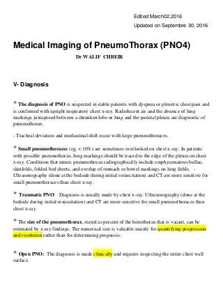
  • 1. Edited March02,2016 Updated on Septembre 30, 2016 Medical Imaging of PneumoThorax (PNO4) Dr WALIF CHBEIR V- Diagnosis * The diagnosis of PNO is suspected in stable patients with dyspnea or pleuritic chest pain and is confirmed with upright inspiratory chest x-ray. Radiolucent air and the absence of lung markings juxtaposed between a shrunken lobe or lung and the parietal pleura are diagnostic of pneumothorax. - Tracheal deviation and mediastinal shift occur with large pneumothoraces. * Small pneumothoraces (eg, < 10%) are sometimes overlooked on chest x-ray. In patients with possible pneumothorax, lung markings should be traced to the edge of the pleura on chest x-ray. Conditions that mimic pneumothorax radiographically include emphysematous bullae, skinfolds, folded bed sheets, and overlap of stomach or bowel markings on lung fields. - Ultrasonography (done at the bedside during initial resuscitation) and CT are more sensitive for small pneumothoraces than chest x-ray. * Traumatic PNO: Diagnosis is usually made by chest x-ray. Ultrasonography (done at the bedside during initial resuscitation) and CT are more sensitive for small pneumothoraces than chest x-ray. * The size of the pneumothorax, stated as percent of the hemithorax that is vacant, can be estimated by x-ray findings. The numerical size is valuable mainly for quantifying progression and resolution rather than for determining prognosis. * Open PNO: The diagnosis is made clinically and requires inspecting the entire chest wall surface.
  • 2. * In critical care and in patients with severe ARDS (ARDS & PNO) , Patients with pneumothorax did not have the traditional clinical and radiologic signs and the most repeatable finding may be a subtle drop in oxygenation measurements without another obvious cause (Loculated Pneumothorax: A Special Challenge In Critical Care) . The diagnosis of pneumothorax in critical illness is made from the history and examination of the patient and confirmed, where possible, by radiological investigation. The factors that are important in the history relate to the underlying disease process and any potential for iatrogenic pneumothorax . The early and accurate diagnosis of pneumothorax in ARDS patients is mandatory since this complication carries an increased mortality. Furthermore, small pneumothoraces in these patients can cause severe hemodynamic or pulmonary compromise. This is the reason why pneumothorax must always be suspected in any patient with ARDS who experiences an acute worsening in respiratory function, accompanied with dyspnea and hypoxemia, which is usually unresponded to oxygen therapy. - Portable chest X-ray is the first diagnostic evaluation imaging being used and the procedure of choice for the documentation of lung underlying pathology or the presents of lines, tubes or devices. Nevertheless, often exhibits diagnostic disadvantages, taking into account that pneumothoraces in ARDS patients may have unusual, as well as subtle features and small sized pneumothoraces or loculated pneumothoraces, can be missed on chest X-ray. Furthermore, other types of air leaks, such as pneumomediastinum and interstitial pulmonary emphysema, may be more difficulty observed by chest radiographs . - Cases have been described in medical literature, referring to patients presenting clinical deterioration but unchanged chest X-ray and functioning chest drains (Acute respiratory distress syndrome and pneumothorax; ref 14). This is the reason why, especially in patients under mechanical ventilation, serial and daily chest radiographs are necessary in the evaluation of underlying lung pathology. - There for, if a pneumothorax is suspected and is unrevealed on chest X-ray, a more specific diagnostic imaging like chest-computed tomography (CT) is necessary. CT scan in patients with ARDS, as explained above , can reveal a variety of abnormalities , is helpful in understanding the extent of the underlying lung parenchyma distraction and is quite more sensitive in identifying pneumomediastinum andpneumothorax, which are frequently observed in patients with ARDS. - Nevertheless, chest-CT evaluation is seldom employed in patients with ARDS, especially patients with severe respiratory failure under mechanical ventilation, mostly due to problems concerning the transfer and monitoring of these critically ill patients. - Due to these technical difficulties of chest-CT, an essential diagnostic method in critically ill patient gaining respect is lung-ultrasonography, a relatively easy to perform, portable and
  • 3. inexpensive diagnostic imaging. Lung-ultrasonography can prove an alternative diagnostic procedure in the difficult diagnosis of pneumothorax in critically ill patients with severe ARDS, which not only permits bedside assessment of lung pathology but also assists in the evaluation of mechanical ventilation parameters, as well as the evaluation of lung overdistension and PEEP-induced lung recruitment . - In the same setting, Loculated pneumothorax provides only subtle clinical clues. The only clinical evidence may be deteriorating oxygenation without another obvious cause. US findings may be equivocal . The abscence of lung sliding may be caused by pno but it has others causes as well . The presence of Lung Sliding indicates that there is no PNO but alone doen't exclude the diagnostic, while the abscence of lung sliding only indicates that there may be one ( because it has others causes as well). Scan the entire chest for B lines. (Clinical chest US: from the ICU to the bronchoscopic suite). PNO that is not in immediate contact with the chest wall will not be identified on US ( e.g. Loculated PNO against medistinum). Be prepared for chest CT scans if ever Lung US is inconclusive for this hard-to-catch complication of mechanical ventilation in patients with ARDS. * Tension pneumothorax is a clinical diagnosis, not a radiographic diagnosis, because the respiratory and hemodynamic consequences of tension pneumothorax do not have radiographic equivalents in many circumstances. Radiographic signs of tension (mediastinal shift, inversion of diaphragm, enlargement of affected hemithorax) can occur in the absence of adverse physiologic effects, and the physiologic effects of pleural tension may be present without radiographic signs of tension ( critically ill p.) . - In ARDS, the diseased noncompliant lung may not collapse in the presence of a pneumothorax, and the controralateral lung may be too stiff to allow mediastinal shift. Thus, tension pneumothorax in ARDS can present as a loculated paracardiac or subpulmonic air collection with little or no mediastinal shift and only slight changes of the cardiac contour. - Treatment should not be delayed pending radiographic confirmation. Although cardiac tamponade also can cause hypotension, neck vein distention, and sometimes respiratory distress, tension pneumothorax can be differentiated clinically by its unilateral absence of breath sounds and hyperresonance to percussion. - Although non-specific, the association of respiratory and haemodynamic signs found with a tension pneumothorax are a medical emergency. Severe haemodynamic compromise will require urgent needle decompression of the pneumothorax before its diagnosis being confirmed radiologically. Fortunately this situation is uncommon and there is frequently time for radiological investigations to help establish the diagnosis of a simple pneumothorax.
  • 4. VI- Significant Points * A large pneumothorax is radiographically defined as one with > 2 cm from pleural surface to lung edge; this is an objective indication for drainage * Don’t wait for a radiograph if there are clinical signs of a tension pneumothorax. Tension pneumothorax is a medical emergency and may require immediate needle decompression before radiological investigation. - Treat the patient not the radiograph. Don’t act on a radiographic appearance if it does not fit the clinical picture. Get an expert opinion on the radiograph first. * Skin folds, companion shadows, the scapula, and previous lung surgery or chest drain placement may all mimic pneumothoraxes on XRay Chest. * In the supine patient, pneumothoraxes are best seen at the lung bases and adjacent to the heart. The “deep sulcus sign” describes a costophrenic angle that extends more inferiorly than usual as a result of air lying in the costophrenic angle. The liver appears more radiolucent than usual due to air lying anteriorly in the costophrenic angle, and on the left side, air will outline the medial aspect of the hemidiaphragm under the heart. * What other radiological investigations may be used to confirm the diagnosis of PNO in acute traumatic setting and in critically ill patients? - A radiograph with the patient in a lateral decubitus position, with the affected side uppermost, can be helpful in demonstrating a lung edge. - In patients well enough to be transported, thoracic computed tomography can be helpful in locating the position of a pneumothorax and accurately siting a chest drain. - US and CT are more sensitive than Chest X-Ray Radiography in detection PNO. CT is the gold standard. US is done at the bedside during initial resuscitation of trauma patient and in critically ill patient in ICU. - The early and accurate diagnosis of pneumothorax in ARDS patients is mandatory since this complication carries an increased mortality. Portable chest X-ray is the first diagnostic
  • 5. evaluation imaging. If a pneumothorax is suspected and is unrevealed on chest X-ray, Lung USG is now an alternative method to Chest CT mostly due to the cumbersome nature of this technique in critically ill patients. * The negative predictive value of Chest US for lung sliding is reported as 99.2–100%, indicating that the presence of sliding effectively rules out a pneumothorax. For some authors, lung sliding ALONE does not exclude PNO and scanning of the entire anterior chest for B- Lines is mandatory (Ultrasonography in the ICU: Practical Applications). - However, the absence of lung sliding does not necessarily indicate that a pneumothorax is present. In fact, Lung sliding is abolished in a variety of conditions other than pneumothorax. - Lung Sliding and B lines are not present on a patient with PNO. M Mode can help differenciate between a seashore sign or Stratosphere or bar code signs ( SeaShore = no PNO). * Blind chest drain placement into a loculated pneumothorax may lead to an iatrogenic air leak from direct trauma to the pleura and worsening the patient’s clinical condition. * An immediate post-treatment radiograph is essential to detect complications and ensure a satisfactory drain position * A chest drain apparently well positioned on frontal radiograph may be lying in the soft tissues, in a lung fissure, or within the substance of the lung. VII- References 1- Fahad M AlHameed, MD, AmBIM, FCCP, FRCPC; Chief Editor: Eugene C Lin, MD. Pneumothorax Imaging: Overview, Radiography, Computed Tomography, in emedecine / Medscape, Updated: Oct 04, 2015, (Page consulted February 12, 2016). [http://emedicine.medscape.com/article/360796-overview#showall] 2-Patricia Carroll, RN,C, CEN, RRT, MS Loculated Pneumothorax: A Special Challenge In Critical Care., in Clinical Update for the Professional Nurse, September 2000. Clinical Update is an educational newsletter provided by Atrium Medical Corporation. [http://www.atriummed.com/en/chest_drainage/Clinical%20Updates/ClinicalUpdateSept00.pdf]
  • 6. 3- Chon KS, vanSonnenberg E, D'Agostino HB, O'Laoide RM, Colt HG, Hart E: CT- guided catheter drainage of loculated thoracic air collections in mechanically ventilated patients with acute respiratory distress syndrome. American Journal of Roentgenology 1999;173(5):1345-1350, in PubMed - indexed for MEDLINE , PMID: 10541116. [http://www.ajronline.org/doi/abs/10.2214/ajr.173.5.10541116] 4- Emergency, Emergency! in General Quiz 001, Copyright © 2003 LearningRadiology.com. (Page consulted February 12, 2016). [http://learningradiology.com/new/quizzes/quiz0004/index0004.htm]. 5- Dr Ayush Goel et al. Pneumothorax: ultrasound, in © 2005–2016 Radiopaedia.org [http://radiopaedia.org/articles/pneumothorax-ultrasound. Page consulted February 12, 2016. ] 6- Terrance Healey, MD- Pneumothorax, Primary Spontaneous, in STATdx Copyrigh © 2016 Elsevier, Inc. All rights reserved. Chest > Diagnosis > Pleural Diseases > Pneumothorax. (Page consulted February 12, 2016). [https://my.statdx.com/document/pneumothorax-primary-spontaneous/eb1b4e8a-d718-45c8-9935- 34da254c9af3?searchTerm=Pneumothorax,%20Primary%20Spontaneous] 7- Lubna F Husain, Laura Hagopian, Derek Wayman, William E Baker, and Kristin A Carmody: Sonographic diagnosis of pneumothorax, in J Emerg Trauma Shock. 2012 Jan- Mar; 5(1): 76–81, in PMC free articles, PMCID: PMC3299161, Received 2011 Jun 1; Accepted 2011 Jun 6, ( Page consulted February 12, 2016). [http://www.ncbi.nlm.nih.gov/pmc/articles/PMC3299161] 8- Jain, Arpana (et al.). Thoracic Ultrasonography in the Critically Ill, in Paula Ferrada (Ed.), Ultrasonography in the ICU: Practical Applications Pages 37-52, © 2015 Springer International Publishing, eBook ISBN 978-3-319-11876-5, in google Books, (Page consulted February 12, 2016). [https://books.google.com.lb/books?id=9Fx1CQAAQBAJ&pg=PA51&lpg=PA51&dq=loculated+pneumothora x+and+ultrasonography&source=bl&ots=re2jhh0 lu&sig=lOXixQ68hsRD1ZegQCQSpQ9YTs&hl=fr&sa=X&ved=0ahUKEwj566y21uzKAhWDthoKHauLDO0 Q6AEIVjAI#v=onepage&q=loculated%20pneumothorax%20and%20ultrasonography&f=false]. 9- Arun Nagdev, MD, and Michael Murphy, MD: Focus On: Ultrasound Detection of Traumatic Anterior Pneumothorax, in Copyright © 2014 American College of Emergency Physicians American website, December 2008. (Page consulted February 12, 2016). [http://www.acep.org/Clinical---Practice-Management/Focus-On--Ultrasound-Detection-of- Traumatic-Anterior-Pneumothorax ]
  • 7. 10- A R O’Connor, W E Morgan. Radiological review of pneumothorax, in BMJ 2005;330:1493–7, in PMC free articles, PMCID: PMC558461 [http://www.ncbi.nlm.nih.gov/pmc/articles/PMC558461] 11- Pneumothorax From Wikipedia, the free encyclopedia. Page was last modified on 16 January 2016, at 22:13, (page consulted February 12, 2016). [https://en.wikipedia.org/wiki/Pneumothorax] 12- Pneumothorax in Collins, Jannette; Stern, Eric J. Chest Radiology: The Essentials, 2nd Edition. Copyright ©2008 Lippincott Williams & Wilkins. P.148-152. 13- Richard W. Light, MD. Pneumothorax in Merck Manual, Professionnal Version, [http://www.merckmanuals.com/professional/pulmonary-disorders/mediastinal-and-pleural- disorders/pneumothorax], Last full review/revision September 2014, ( page consulted February 12, 2016). 14- Ashley Miller : Lung Ultrasound - Pneumothorax, in ICMteaching.com. © 2011. (Page consulted February 12, 2016). [http://www.icmteaching.com/ultrasound/lung%20ultrasound/styled-125/] 15- Mitlehner W, Friedrich M, Dissmann W. Value of computer tomography in the detection of bullae and blebs in patients with primary spontaneous pneumothorax, in Respiration. 1992. 59(4):2217, in PubMed - indexed for MEDLINE ( PMID: 1485007), (Page consulted February 12, 2016). [http://www.ncbi.nlm.nih.gov/pubmed/?term=PMID%3A+1485007] 16- James J Rankine, Antony N Thomas, Dorothee Fluechter. Diagnosis of pneumothorax in critically ill adults Postgrad Med J 2000;76:399–404, in in PMC free articles, PMCID: PMC1741653 [http://www.ncbi.nlm.nih.gov/pmc/articles/PMC1741653/] 17- Skaarup SH, Folkersen BH. Ultrasound-assisted aspiration of loculated pneumothorax: A new technique, in J Clin Ultrasound. 2015 Dec 16. doi: 10.1002/jcu.22326, in PubMed, PMID: 26676092, (Page consulted February 12, 2016). [http://www.ncbi.nlm.nih.gov/pubmed/26676092] 18- Paul Stark, MD. Imaging of pneumothorax, in UpToDate, Literature review current through: Jan 2016, This topic last updated: Feb 3, 2015, (Page consulted February 12, 2016).
  • 8. [www.uptodate.com/contents/imaging-of-pneumothorax]. 19- Eirini Terzi, & al: Acute respiratory distress syndrome and pneumothorax, in J Thorac Dis 2014;6(S4):S435-S442, in www.jthoracdis.com, Submitted Aug 15, 2014. Accepted for publication Aug 19, 2014, (Page consulted February 12, 2016). [http://www.jthoracdis.com/article/view/3101/3676]. 20- Grace M. Thomas, MD - Stephen Jones, MD - Clint M. Gerdes, MD: Supine pneumothorax: in ACR- Case in point, Wednesday, August 6, 2014, ( Page consulted February 12, 2016). [https://3s.acr.org/CIP/SearchCaseView.aspx?CaseId=KsGsjCvXCLg%253d] 21- IM Tocino, MH Miller and WR Fairfax: Distribution of pneumothorax in the supine and semirecumbent critically ill adult, in AJR 144:901-905, May 1985, in PubMed (PMID: 3872573), (Page consulted February 12, 2016). [http://www.ajronline.org/doi/abs/10.2214/ajr.144.5.901] 22- Wang J.S. ·Doelken P. Pleural Ultrasonography in the Intensive Care Unit, in Bolliger CT, Herth FJF, Mayo PH, Miyazawa T, Beamis JF (eds): Clinical Chest Ultrasound: From the ICU to the Bronchoscopy Suite, in Prog Respir Res. Basel, Karger, 2009, vol 37, pp 82–88, Published online: 3/25/2009, eISBN: 978-3-8055-8643-6 (Online), in Google Books. https://books.google.com.lb/books?id=OF4SS1mgZRkC&pg=PA78&lpg=PA78&dq=loculated+pneumothorax +and+ultrasonography&source=bl&ots=JMu4GtHHIW&sig=0lUnTRfU9_x0k5kG80akbcFvC3I&hl=fr&sa=X &ved=0ahUKEwj566y21uzKAhWDthoKHauLDO0Q6AEIPTAE#v=onepage&q=loculated%20pneumothorax %20and%20ultrasonography&f=false 23- Thomas G. Weiser, MD, MPH. Pneumothorax (Traumatic) in Merck Manual, Professionnal Version, Last full review/revision August 2014, (page consulted February 12, 2016). [http://www.merckmanuals.com/professional/injuries-poisoning/thoracic trauma/pneumothorax-(traumatic) ], 24-Thomas G. Weiser, MD, MPH. Pneumothorax (Tension) in Merck Manual, Professionnal Version, Last full review/revision August 2014, ( Page consulted February 12, 2016). [http://www.merckmanuals.com/professional/injuries-poisoning/thoracic- trauma/pneumothorax-(tension)]
  • 9. 25- Thomas G. Weiser, MD, MPH. Pneumothorax (Open) (Sucking Chest Wound) in Merck Manual, Professionnal Version, Last full review/revision August 2014, ( Page consulted February 12, 2016). [http://www.merckmanuals.com/professional/injuries-poisoning/thoracic- trauma/pneumothorax-(open) ],