SlideShare uma empresa Scribd logo
1 de 15
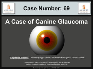
Case Number: 69
A Case of Canine Glaucoma
Permission granted only for viewing on SEVPAC website
1Stephanie Shrader, 1Jennifer (Jey) Koehler, 2Roxanne Rodriguez, 2Phillip Moore
1Department of Pathobiology and 2Department of Clinical Sciences
Auburn University, College of Veterinary Medicine (AUCVM)
 4-year-old, castrated male, Labrador retriever mix
 One-week history of blepharospasm (OD)
 Intraocular pressure (IOP): 50-mmHg
 Normal: 12-mmHg to 25-mmHg
 Placed on triple antibiotic ophthalmic ointment and
referred to the AUCVM
SIGNALMENT AND HISTORY
Permission granted only for viewing on SEVPAC website
 Right eye: blepharospasm, buphthalmia, aqueous
flare, corneal edema, posterior synechiae, vascular
attenuation, cupped optic disc, IOP of 47-mmHg
 Left eye: reportedly unremarkable
 CBC and chemistry values: WNL
 Treated with latanoprost and dorzolamide
(decrease IOP) and tramadol (pain relief)
Owners opted for enucleation of the right globe
OPHTHALMIC EXAMINATION
Permission granted only for viewing on SEVPAC website
CORNEAL EDEMA
100-um
Permission granted only for viewing on SEVPAC website
20-um
H&E
H&E
CLOSED IRIDOCORNEAL ANGLE
Permission granted only for viewing on SEVPAC website
100-um
500-um
H&E
H&E
POSTERIOR SYNECHIAE
Permission granted only for viewing on SEVPAC website
100-um
20-um
H&E
H&E
Permission granted only for viewing on SEVPAC website
20-um
RETINAL DEGENERATION
Nerve fiber layer
Inner plexiform layer
Inner nuclear layer
Outer plexiform layer
Outer nuclear layer
Rods and conesH&E
H&E H&E
20-um
20-um
Right globe: Lymphohistiocytic endophthalmitis,
multifocal, scant, subacute with iridocorneal
angle closure, posterior synechiae, retinal
degeneration and atrophy, and corneal stromal
edema
MORPHOLOGIC DIAGNOSIS
Permission granted only for viewing on SEVPAC website
http://blogs.edweek.org/edweek/the_startup_blog/2014/02/but_wait_theres_more_building_a_solution_to_users_actual_problems_edthena.html
Permission granted only for viewing on SEVPAC website
DESCEMET’S MEMBRANE
Permission granted only for viewing on SEVPAC website
20-um
H&E PAS
Right globe: Goniodysgenesis with minimal
lymphohistiocytic endophthalmitis, iridocorneal
angle closure, posterior synechiae, retinal
degeneration and atrophy, and corneal stromal
edema
FINAL
MORPHOLOGIC DIAGNOSIS
Permission granted only for viewing on SEVPAC website
 Congenital anomaly of the iridocorneal angle
 Abnormal intraocular fluid egress1
 Risk factor for development of primary glaucoma
 Associated with abnormal development of:
 Pectinate ligament
 Trabecular meshwork
 Ciliary cleft
 Absence of concurrent ocular or systemic disease2
GONIODYSGENESIS
Permission granted only for viewing on SEVPAC website
 Hallmark histologic feature:
 Solid sheet of iridal tissue extending to the distorted
terminus of Descemet’s membrane3
 Although congenital, clinical signs in dogs manifest
at 4-8 years of age4,5
 Following diagnosis in one eye, the contralateral
eye may also develop glaucoma
 Clinical course can range from days to years
GONIODYSGENESIS
Permission granted only for viewing on SEVPAC website
1. R. Sampaolesi, et al. Goniodysgenesis or Late Congenital Glaucoma.
Pigmentary Glaucoma. In: The Glaucomas. Vol. 1, Pediatric
Glaucomas. Berlin: Springer. 311-66. 2009.
2. "Read-Only Case Details Reviewed: May 2012." The Joint Pathology
Center, n.d. Web. 14 Apr. 2015.
<http://www.askjpc.org/vspo/show_page.php?id=713>.
3. R. Dubielzig, et al. Veterinary Ocular Pathology: A Comparative
Review. Edinburgh: Saunders/Elsevier. 45-46. 2010.
4. K Gelatt. The canine glaucomas. In: Veterinary Ophthalmology, 3rd
Ed. (ed. Gelatt KN) Lea & Febiger, Philadelphia, 701–754. 1999.
5. C Reilly, et al. Canine goniodysgenesis-related glaucoma: a
morphologic review of 100 cases looking at inflammation and pigment
dispersion. Veterinary Ophthalmology. 9(6):253-254. 2006.
REFERENCES
Permission granted only for viewing on SEVPAC website
QUESTIONS??
http://galleryhip.com/tumblr-brown-eyes-collage.html
Permission granted only for viewing on SEVPAC website

Mais conteúdo relacionado

Mais procurados

Acute Congestive Glaucoma / Optic Neuritis / Painful Loss Of Vision by Dr. Mu...
Acute Congestive Glaucoma / Optic Neuritis / Painful Loss Of Vision by Dr. Mu...Acute Congestive Glaucoma / Optic Neuritis / Painful Loss Of Vision by Dr. Mu...
Acute Congestive Glaucoma / Optic Neuritis / Painful Loss Of Vision by Dr. Mu...Zeeshan Hameed
 
Glaucoma suspect,ocular hypertension, steroid induced glaucoma
Glaucoma suspect,ocular hypertension, steroid induced glaucomaGlaucoma suspect,ocular hypertension, steroid induced glaucoma
Glaucoma suspect,ocular hypertension, steroid induced glaucomaBipin Bista
 
Glaucoma slides
Glaucoma slidesGlaucoma slides
Glaucoma slidesBapi Das
 
Glaucoma treatment
Glaucoma treatmentGlaucoma treatment
Glaucoma treatmentLOVEFUCK
 
Approach to monocular blindness
Approach to monocular blindnessApproach to monocular blindness
Approach to monocular blindnessNeurologyKota
 
Glaucoma word document
Glaucoma word document Glaucoma word document
Glaucoma word document ayesharuqsar
 
Glaucoma
GlaucomaGlaucoma
Glaucomanazaika
 
Glaucoma 3 primary open angle glaucoma,dr.k.n.jha, 03.11.16
Glaucoma 3 primary open angle glaucoma,dr.k.n.jha, 03.11.16Glaucoma 3 primary open angle glaucoma,dr.k.n.jha, 03.11.16
Glaucoma 3 primary open angle glaucoma,dr.k.n.jha, 03.11.16ophthalmgmcri
 
Normal Tension Glaucoma
Normal Tension GlaucomaNormal Tension Glaucoma
Normal Tension Glaucomaeknath87
 
Steroid Induced Glaucoma - Dr Shylesh B Dabke
Steroid Induced Glaucoma - Dr Shylesh B DabkeSteroid Induced Glaucoma - Dr Shylesh B Dabke
Steroid Induced Glaucoma - Dr Shylesh B DabkeShylesh Dabke
 

Mais procurados (20)

Acute Congestive Glaucoma / Optic Neuritis / Painful Loss Of Vision by Dr. Mu...
Acute Congestive Glaucoma / Optic Neuritis / Painful Loss Of Vision by Dr. Mu...Acute Congestive Glaucoma / Optic Neuritis / Painful Loss Of Vision by Dr. Mu...
Acute Congestive Glaucoma / Optic Neuritis / Painful Loss Of Vision by Dr. Mu...
 
Glaucoma suspect,ocular hypertension, steroid induced glaucoma
Glaucoma suspect,ocular hypertension, steroid induced glaucomaGlaucoma suspect,ocular hypertension, steroid induced glaucoma
Glaucoma suspect,ocular hypertension, steroid induced glaucoma
 
POAG AND PACG
POAG AND PACGPOAG AND PACG
POAG AND PACG
 
Clinical round ophthalmic revision
Clinical round ophthalmic revisionClinical round ophthalmic revision
Clinical round ophthalmic revision
 
Glaucoma slides
Glaucoma slidesGlaucoma slides
Glaucoma slides
 
Glaucoma treatment
Glaucoma treatmentGlaucoma treatment
Glaucoma treatment
 
Opthalmology Quiz
Opthalmology QuizOpthalmology Quiz
Opthalmology Quiz
 
Primary Open Angle Glaucoma
Primary Open Angle GlaucomaPrimary Open Angle Glaucoma
Primary Open Angle Glaucoma
 
Glaucoma
GlaucomaGlaucoma
Glaucoma
 
Glaucoma
GlaucomaGlaucoma
Glaucoma
 
5th year Course Book/Ophthalmology
5th year Course Book/Ophthalmology5th year Course Book/Ophthalmology
5th year Course Book/Ophthalmology
 
Approach to monocular blindness
Approach to monocular blindnessApproach to monocular blindness
Approach to monocular blindness
 
Glaucoma word document
Glaucoma word document Glaucoma word document
Glaucoma word document
 
Glaucoma
GlaucomaGlaucoma
Glaucoma
 
Glaucoma
GlaucomaGlaucoma
Glaucoma
 
steroid glaucoma
steroid glaucomasteroid glaucoma
steroid glaucoma
 
Glaucoma 3 primary open angle glaucoma,dr.k.n.jha, 03.11.16
Glaucoma 3 primary open angle glaucoma,dr.k.n.jha, 03.11.16Glaucoma 3 primary open angle glaucoma,dr.k.n.jha, 03.11.16
Glaucoma 3 primary open angle glaucoma,dr.k.n.jha, 03.11.16
 
Normal Tension Glaucoma
Normal Tension GlaucomaNormal Tension Glaucoma
Normal Tension Glaucoma
 
History taking in ophthalmology
History taking in ophthalmologyHistory taking in ophthalmology
History taking in ophthalmology
 
Steroid Induced Glaucoma - Dr Shylesh B Dabke
Steroid Induced Glaucoma - Dr Shylesh B DabkeSteroid Induced Glaucoma - Dr Shylesh B Dabke
Steroid Induced Glaucoma - Dr Shylesh B Dabke
 

Destaque

Case Report : Chorodial Melanoma With Secondary Glaucoma In Asian Race.
Case Report : Chorodial Melanoma With Secondary Glaucoma In Asian Race.Case Report : Chorodial Melanoma With Secondary Glaucoma In Asian Race.
Case Report : Chorodial Melanoma With Secondary Glaucoma In Asian Race.Dr. Jagannath Boramani
 
Strep Throat
Strep ThroatStrep Throat
Strep Throatfitango
 
CASE STUDY: OB/GYN - UTI & Domestic Violence
CASE STUDY:  OB/GYN - UTI & Domestic ViolenceCASE STUDY:  OB/GYN - UTI & Domestic Violence
CASE STUDY: OB/GYN - UTI & Domestic ViolenceAnn Hinnen Sparks
 
An Unusual case of Glaucoma
An Unusual case of GlaucomaAn Unusual case of Glaucoma
An Unusual case of GlaucomaAayush Tandon
 
Posterior polar cataract
Posterior polar cataractPosterior polar cataract
Posterior polar cataractSumeet Agrawal
 
Glaucoma 4 therapy of glaucomas, dr.k.n.jha,09.11.16
Glaucoma 4 therapy of glaucomas, dr.k.n.jha,09.11.16Glaucoma 4 therapy of glaucomas, dr.k.n.jha,09.11.16
Glaucoma 4 therapy of glaucomas, dr.k.n.jha,09.11.16ophthalmgmcri
 
Otitis externa and media
Otitis externa and mediaOtitis externa and media
Otitis externa and mediaKapil Dhital
 

Destaque (12)

Case Report : Chorodial Melanoma With Secondary Glaucoma In Asian Race.
Case Report : Chorodial Melanoma With Secondary Glaucoma In Asian Race.Case Report : Chorodial Melanoma With Secondary Glaucoma In Asian Race.
Case Report : Chorodial Melanoma With Secondary Glaucoma In Asian Race.
 
optic neuritis
optic neuritisoptic neuritis
optic neuritis
 
Strep Throat
Strep ThroatStrep Throat
Strep Throat
 
CASE STUDY: OB/GYN - UTI & Domestic Violence
CASE STUDY:  OB/GYN - UTI & Domestic ViolenceCASE STUDY:  OB/GYN - UTI & Domestic Violence
CASE STUDY: OB/GYN - UTI & Domestic Violence
 
An Unusual case of Glaucoma
An Unusual case of GlaucomaAn Unusual case of Glaucoma
An Unusual case of Glaucoma
 
Acute Otitis Media
Acute Otitis Media Acute Otitis Media
Acute Otitis Media
 
Posterior polar cataract
Posterior polar cataractPosterior polar cataract
Posterior polar cataract
 
Glaucoma 4 therapy of glaucomas, dr.k.n.jha,09.11.16
Glaucoma 4 therapy of glaucomas, dr.k.n.jha,09.11.16Glaucoma 4 therapy of glaucomas, dr.k.n.jha,09.11.16
Glaucoma 4 therapy of glaucomas, dr.k.n.jha,09.11.16
 
Otitis externa and media
Otitis externa and mediaOtitis externa and media
Otitis externa and media
 
UTI Case Presentation
UTI Case PresentationUTI Case Presentation
UTI Case Presentation
 
Optic nerve 1
Optic nerve 1Optic nerve 1
Optic nerve 1
 
Glaucoma
GlaucomaGlaucoma
Glaucoma
 

Semelhante a 69-Shrader

Semelhante a 69-Shrader (20)

Glaucomatous Optic Atrophy
Glaucomatous Optic AtrophyGlaucomatous Optic Atrophy
Glaucomatous Optic Atrophy
 
Pseudoexfoliation glaucoma
Pseudoexfoliation glaucomaPseudoexfoliation glaucoma
Pseudoexfoliation glaucoma
 
Heterochromia
HeterochromiaHeterochromia
Heterochromia
 
Lecture pspos
Lecture   psposLecture   pspos
Lecture pspos
 
Glaucoma
GlaucomaGlaucoma
Glaucoma
 
Primary open angle glaucoma (poag)kalpy
Primary open angle glaucoma (poag)kalpyPrimary open angle glaucoma (poag)kalpy
Primary open angle glaucoma (poag)kalpy
 
Bilateral lens capsule rupture in a patient with previously undiagnosed alpor...
Bilateral lens capsule rupture in a patient with previously undiagnosed alpor...Bilateral lens capsule rupture in a patient with previously undiagnosed alpor...
Bilateral lens capsule rupture in a patient with previously undiagnosed alpor...
 
Presentation 1 exfoliation and uveitis
Presentation 1 exfoliation and uveitisPresentation 1 exfoliation and uveitis
Presentation 1 exfoliation and uveitis
 
Cataracts in paediatric patients
Cataracts in paediatric patients Cataracts in paediatric patients
Cataracts in paediatric patients
 
Glaucoma and cataract include treatment
Glaucoma and cataract include treatmentGlaucoma and cataract include treatment
Glaucoma and cataract include treatment
 
Glaucoma HNS.pptx
Glaucoma HNS.pptxGlaucoma HNS.pptx
Glaucoma HNS.pptx
 
Congenital glaucoma -Evaluation
Congenital glaucoma  -EvaluationCongenital glaucoma  -Evaluation
Congenital glaucoma -Evaluation
 
Central retinal artery occlusion
Central retinal artery occlusionCentral retinal artery occlusion
Central retinal artery occlusion
 
Iridocorneal endothelial (ICE) syndrome
Iridocorneal endothelial (ICE) syndrome Iridocorneal endothelial (ICE) syndrome
Iridocorneal endothelial (ICE) syndrome
 
Ocular sarcoidosis
Ocular sarcoidosisOcular sarcoidosis
Ocular sarcoidosis
 
Primary open angle glaucoma(POAG)
Primary open angle glaucoma(POAG)Primary open angle glaucoma(POAG)
Primary open angle glaucoma(POAG)
 
Glaucoma 2nd class
Glaucoma 2nd classGlaucoma 2nd class
Glaucoma 2nd class
 
Ocular sarcoidosis
Ocular sarcoidosisOcular sarcoidosis
Ocular sarcoidosis
 
Ang.Closure Glaucoma
Ang.Closure GlaucomaAng.Closure Glaucoma
Ang.Closure Glaucoma
 
Glaucoma
GlaucomaGlaucoma
Glaucoma
 

69-Shrader

  • 1. Case Number: 69 A Case of Canine Glaucoma Permission granted only for viewing on SEVPAC website 1Stephanie Shrader, 1Jennifer (Jey) Koehler, 2Roxanne Rodriguez, 2Phillip Moore 1Department of Pathobiology and 2Department of Clinical Sciences Auburn University, College of Veterinary Medicine (AUCVM)
  • 2.  4-year-old, castrated male, Labrador retriever mix  One-week history of blepharospasm (OD)  Intraocular pressure (IOP): 50-mmHg  Normal: 12-mmHg to 25-mmHg  Placed on triple antibiotic ophthalmic ointment and referred to the AUCVM SIGNALMENT AND HISTORY Permission granted only for viewing on SEVPAC website
  • 3.  Right eye: blepharospasm, buphthalmia, aqueous flare, corneal edema, posterior synechiae, vascular attenuation, cupped optic disc, IOP of 47-mmHg  Left eye: reportedly unremarkable  CBC and chemistry values: WNL  Treated with latanoprost and dorzolamide (decrease IOP) and tramadol (pain relief) Owners opted for enucleation of the right globe OPHTHALMIC EXAMINATION Permission granted only for viewing on SEVPAC website
  • 4. CORNEAL EDEMA 100-um Permission granted only for viewing on SEVPAC website 20-um H&E H&E
  • 5. CLOSED IRIDOCORNEAL ANGLE Permission granted only for viewing on SEVPAC website 100-um 500-um H&E H&E
  • 6. POSTERIOR SYNECHIAE Permission granted only for viewing on SEVPAC website 100-um 20-um H&E H&E
  • 7. Permission granted only for viewing on SEVPAC website 20-um RETINAL DEGENERATION Nerve fiber layer Inner plexiform layer Inner nuclear layer Outer plexiform layer Outer nuclear layer Rods and conesH&E H&E H&E 20-um 20-um
  • 8. Right globe: Lymphohistiocytic endophthalmitis, multifocal, scant, subacute with iridocorneal angle closure, posterior synechiae, retinal degeneration and atrophy, and corneal stromal edema MORPHOLOGIC DIAGNOSIS Permission granted only for viewing on SEVPAC website
  • 10. DESCEMET’S MEMBRANE Permission granted only for viewing on SEVPAC website 20-um H&E PAS
  • 11. Right globe: Goniodysgenesis with minimal lymphohistiocytic endophthalmitis, iridocorneal angle closure, posterior synechiae, retinal degeneration and atrophy, and corneal stromal edema FINAL MORPHOLOGIC DIAGNOSIS Permission granted only for viewing on SEVPAC website
  • 12.  Congenital anomaly of the iridocorneal angle  Abnormal intraocular fluid egress1  Risk factor for development of primary glaucoma  Associated with abnormal development of:  Pectinate ligament  Trabecular meshwork  Ciliary cleft  Absence of concurrent ocular or systemic disease2 GONIODYSGENESIS Permission granted only for viewing on SEVPAC website
  • 13.  Hallmark histologic feature:  Solid sheet of iridal tissue extending to the distorted terminus of Descemet’s membrane3  Although congenital, clinical signs in dogs manifest at 4-8 years of age4,5  Following diagnosis in one eye, the contralateral eye may also develop glaucoma  Clinical course can range from days to years GONIODYSGENESIS Permission granted only for viewing on SEVPAC website
  • 14. 1. R. Sampaolesi, et al. Goniodysgenesis or Late Congenital Glaucoma. Pigmentary Glaucoma. In: The Glaucomas. Vol. 1, Pediatric Glaucomas. Berlin: Springer. 311-66. 2009. 2. "Read-Only Case Details Reviewed: May 2012." The Joint Pathology Center, n.d. Web. 14 Apr. 2015. <http://www.askjpc.org/vspo/show_page.php?id=713>. 3. R. Dubielzig, et al. Veterinary Ocular Pathology: A Comparative Review. Edinburgh: Saunders/Elsevier. 45-46. 2010. 4. K Gelatt. The canine glaucomas. In: Veterinary Ophthalmology, 3rd Ed. (ed. Gelatt KN) Lea & Febiger, Philadelphia, 701–754. 1999. 5. C Reilly, et al. Canine goniodysgenesis-related glaucoma: a morphologic review of 100 cases looking at inflammation and pigment dispersion. Veterinary Ophthalmology. 9(6):253-254. 2006. REFERENCES Permission granted only for viewing on SEVPAC website