
Recommended
More Related Content
What's hot
What's hot (20)
Similar to Esophageal Cancer Anatomy, Risk Factors, Staging and Treatment
Similar to Esophageal Cancer Anatomy, Risk Factors, Staging and Treatment (20)
More from Silah Aysha
More from Silah Aysha (20)
Recently uploaded
Recently uploaded (20)
Esophageal Cancer Anatomy, Risk Factors, Staging and Treatment
- 2. Clinical Anatomy❚ Hollow muscular tube 25 cm in length which spans from the cricopharyngeus at the cricoid cartilage to gastroesophageal junction (Extends from C7-T10). ❚ Has 4 constrictions- ❙ At starting(cricophyrangeal junction) ❙ crossed by aortic arch(9’inch) ❙ crossed by left bronchus(11’inch) ❙ Pierces the diaphragm(15’inch) ❚ Histologically 4 layers: mucosa, submucosa, muscular & fibrous layer. FIGURE Anatomy of the esophagus
- 3. Contd… Four regions of the esophagus: ❚ ❚ ❚ ❚ Cervical = cricoid cartilage to thoracic inlet (15–18 cm from the incisor). Upper thoracic = thoracic inlet to arch of aorta (18–24 cm). Midthoracic = arch of aorta to inferior pulmonary vein (24–32 cm). Lower thoracic =inferior pulmonary vein to GE junction (32–40 cm). Figure Anatomy landmarks and recorded distance from incisors used to divide the esophagus compartments. of the esophagus with the into GE,topographic gastroesophageal.
- 4. Lymphatic Drainage ❚ Rich mucosal and submucosal lymphatic system. ❚ The submucosal lymphatics may extend long distances (proximal and distal margins used for RTP have traditionally been a minimum of 5 cm). ❚ The submucosal plexus drains into the regional lymph nodes in the cervical, mediastinal, paraesophageal, left gastric, and celiac axis regions Figure Lymphatic drainage of the esophagus with anatomically defined lymph node basins
- 5. Epidemiology ❚ Esophageal cancer is the 6th leading cause of cancer deaths. ❚ accounts for 1% of all malignancy & 6% of all GI malignancy. ❚ Most common in China, Iran, South Africa, India and the former Soviet Union. ❚ The incidence rises steadily with age, reaching a peak in the 6th to 7th decade of life. ❚ Male : Female = 3.5 : 1 ❚ African-American males : White males = 5:1
- 6. Contd… ❚ Worldwide SCC responsible for most of the cases. ❚ Adenocarcinoma now accounts for over 50% of esophageal cancer in the USA, due to association with GERD , Barretts’s esophagus & obesity. ❚ SCC usually occurs in the middle 3rd of the esophagus (the ratio of upper : middle : lower is 15 : 50 : 35). ❚ Adenocarcinoma is most common in the lower 3rd of the esophagus, accounting for over 65% of cases.
- 7. Risk Factors : Squamous Cell Carcinoma ❚ Smoking and alcohol (80% - 90%) ❚ Dietary factors ❙ N-nitroso compounds (animal carcinogens) ❙ Pickled vegetables and other food-products ❙ Toxin-producing fungi(Fusarium, aspergillus, penicillium) ❙ Betel nut chewing ❙ Ingestion of very hot foods and beverages (such as tea) ❚ Underlying esophageal diseases (such as achalasia, plummer vinson syndrome, celiac disease, Tylosis) ❚ Geneticabnormalities: ❙ p53 mutation, loss of 3p and 9q alleli, amp. Cyclin D1 &. EGFR
- 8. Risk Factors: Adenocarcinoma ❚ Associated with Barretts’s esophagus, GERD & hiatal hernia. ❚ Obesity (3 to 4 fold risk) ❚ Smoking (2 to 3 fold risk) ❚ Increased esophageal acid exposure such as Zollinger-Ellison syndrome. Fig. Barretts’s esophagus Barrett’s esophagus is a metaplasia of the esophageal epithelial lining. The squamous epithelium is replaced by columnar epithelium,with 0.5% annual rate of neoplastic transformation.
- 9. Pattern of spread ❚ No serosal covering, direct invasion of contiguous structures occurs early. ❚ Commonly spread by lymphatics (70%) ❚ Lymph node involvement increases with T stage. ❙ ❙ T1 – 14 to 21% T2 – 38 to 60% ❚ 25% - 30% hematogenous metastases at time of presentation. ❚ ❚ Most common site of metastases are ❙ lung, liver, pleura, bone, kidney & adrenal gland Median survival with distant metastases – 6 to 12 months
- 11. Clinical Features ❚ It is commonly associated with the symptoms of dysphagia, wt. loss, pain, anorexia, and vomiting ❚ Symptoms often start 3 to 4 months before diagnosis ❚ Dysphagia - in more than 90% pt. Odynophagia - in 50% of pt. ❚ Wt. loss – more than 5 % of total body wt. in 40 – 70% pt. associated with worst prognosis.
- 12. Contd… Complications: ❚ Cachexia, Malnutrition, dehydration, anaemia,. ❚ Aspiration pneumonia. ❚ Distant metastasis. ❚ Invasion of near by structures: e.g. ❙ Recurrent laryngeal nerve →Hoarseness of voice ❙ Trachea →Stridor & TOF→ cough, choking & cyanosis ❙ Perforation into the pleural cavity →Empyema ❙ back pain in celiac axis node involvement
- 13. AJCC TNM classification ❚ a: Includes nodes previously labeled as “M1a” ❚ b : “M1a” designation is no longer recognized in the 7th edn. of the AJCC system
- 14. Staging : Squamous cell carcinoma
- 15. Staging : Adenocarcinoma Group T N M Grade 0 Tis (HGD) N0 M0 1, X IA T1 1-2, X IB T1 3 T2 1-2, X IIA T2 3 IIB T3 T1-2 N1 IIIA T1-2 N2 T3 N1 T4a N0 Any IIIB T3 N2 IIIC T4a N1-2 T4b Any Any N3 IV Any Any M1
- 16. Diagnostic Workup ❚ Detailed history & Physical examination: Dysphagia, odynophagia, hoarseness, wt. loss, use of tobacco, nitrosamines, history of GERD. Examine for cervical or supraclavicular adenopathy. ❚ Confirmation of diagnosis: ❙ EGD: allow direct visualization and biopsy, measure proximal & distal distance of tumor from incisor, presence of Barrett’s esophagus. Early, superficial cancer Circumferential ulceration esophageal cancer Malignant stricture of esophagus
- 17. ❚Staging: ❙ CT chest and abdomen: Essential for staging because it can identify extension beyond the esophageal wall, enlarged lymph nodes and visceral metastases. Figure Esophageal cancer with tracheal invasion. CT scan shows circumferential wall thickening of the proximal esophagus (arrowheads), which shows irregular interface with the posterior wall of the trachea (arrows), indicating direct extension into the lumen Figure Esophageal cancer with aortic invasion. An arc (bent arrow) of the contact between esophageal cancer (arrows) and the the aorta (arrowheads) is more than 90 degrees, indicating aortic invasion.
- 18. Endoscopic Ultrasonography ❚ EUS: ❙ assess the depth of penetration and LN involvement. Limited by the degree of obstruction. ❙ Compared with EUS, CT is not a reliable tool for evaluation of the extent of tumor in the esophageal wall. Fig. —55-year-old man with T2 esophageal tumor (m) shown on endoscopic sonogram. Note alternating hyperechoic and hypoechoic layers (arrowheads) of normal esophageal wall as seen on sonography. Innermost layer is hyperechoic and corresponds to superficial mucosa. Second layer is hypoechoic and corresponds to deep mucosa and muscularis mucosae. Third layer is again hyperechoic and corresponds to submucosa and its interface with muscularis propria. Fourth layer is hypoechoic and corresponds to muscularis propria, and outer fifth layer is hyperechoic and corresponds to adventitia.
- 19. PET Scan ❚ most recently, proven to be valuable staging tool ❚ can detect up to 15–20% of metastases not seen on CT and EUS ❚ low accuracy in detecting local nodal disease compared to CT / EUS ❚ Value in evaluating response to Chemo Therapy & Radio Therapy ❚ addition of PET to CT can improve specificity and accuracy of non- invasive staging Figure Distant lymph node metastases of esophageal cancer detected by integrated CT PET. A, Integrated CT PET demonstrates para-aortic lymph node metastases showing increased FDG uptake (arrowheads). B, Corresponding CT image shows lymph nodes (arrowheads) measuring 5 to 8 mm in diameter. Based on size criteria, these lymph nodes may be considered benign on CT scan
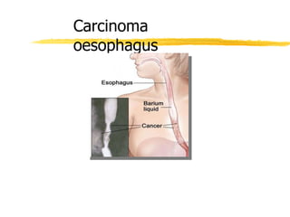
- 20. ❚ Barium swallow: ❙ can delineate proximal and distal margins as well as TEF ❙ Helpful for correlation with simulation film. ❚ Bronchoscopy: rule-out fistula in midesophageal lesions. ❚Routine Investigations: CBC, chemistries, LFTs. Cancer lower 1/3 Filling defect (ulcerative type)Rat tail appearance Apple core appearance
- 21. Treatment
- 22. Management depends upon: ❚ Site of disease ❚ Extent of disease involvement ❚ Co-morbid conditions ❚ Patientpreference.
- 23. Surgery ❚ Prerequisite for surgery ❙ disease should be 5 cm beyond cricophyrangeus. ❚ Surgery indications ❙ Lower 1/3 rd oesophageal ds involving GE junction. ❙ Tumor size <5 cm . ❙ palliative surgery ❚ 5-Year OS for surgery alone is 20–25% (no significant difference between surgical techniques according to results of 2 meta-analyses) ❚ Local failure rate around 19–57% when used alone ❚ surgical morbidity/mortality related to experience of the surgeons.
- 24. Types of Surgery ❚ Transhiatal esophagectomy) ORRINGER`S APPROACH(: for tumors anywhere in esophagus or gastric cardia. No thoracotomy. Blunt dissection of the thoracic esophagus. Left with cervical anastomosis. Limitations are lack of exposure of midesophagus and direct visualization and dissection of the subcarinal LN cannot be performed. ❚ Right thoracotomy (Ivor-Lewis procedure): good for exposure of mid to upper esophageal lesions. Left with thoracic or cervical anastomosis. • Mc keown 3 phase oesophagectomy ❚ Left thoracotomy: appropriate for lower third of esophagus and gastric cardia. Left with low-to-midthoracic anastomosis. ❚ Radical (en block) resection: for tumor anywhere in esophagus or gastric cardia. Left with cervical or thoracic anastomosis. Benefit is more extensive lymphadenectomy and potentially better survival, but increased operative risk.
- 25. Chemotherapy ❚ No data proving that chemotherapy alone provides improved survival or palliation. Partial response, not long-term remission, is the rule ❚Indication ❙ Used in combination with radiation for locally advancedcancers ❙ Used as single treatment modality in stage IVdisease ❙ Combination chemoradiotherapy has been used preoperatively in a combined modality approach to esophageal Ca in hopes of controlling occult metastatic disease and improving the resectability rate.
- 26. ❚ Platinum doublet is preferred over single agents ❚ Cisplatin plus 5-FU or docetaxel are commonly used combinations Regimens: ❚ Paclitaxel and carboplatin ❚ Cisplatin and 5-FU or capecitabine ❚ Oxaliplatin and 5-FU or capecitabine ❚ Paclitaxel or docetaxel and cisplatin ❚ Carboplatin and 5-FU ❚ Irinotecan and cisplatin ❚ Oxaliplatin, docetaxel and capecitabine ❚ Epirubicin, cisplatin and 5-FU (Only for adenocarcinoma)
- 27. PALLIATIVE TREATMENT If patient is not fit for major surgery If there is blood spread If there is adjacent organ spread If there is peritoneal / liver spread
- 28. PALLIATIVE PROCEDURES Radiotherapy ( External or intraluminal brachytherapy) Traction tubes like Celastin or MBtubes through open surgery Pulsion tubes like self expandable metal tube Endoscopic laser Photodynamic therapy
- 29. Conclusion ❚ Esophageal cancer is the 6th leading cause of cancer deaths. ❚ Adenocarcinoma now accounts for over 50% of esophageal cancer in the USA, due to association with GERD & obesity. ❚ Dysphagia and weight loss are the two most common presentations in patients with esophageal cancer. ❚ Endoscopic ultrasound (EUS) is necessary to accompany a complete workup for proper staging and diagnosis of esophageal cancer. ❚ Surgery is the standard of care for early-stage esophageal cancer. ❚ Preoperative chemotherapy and radiation is the standard option for locally advanced esophageal cancer in surgically eligible patients.
- 30. Thank You