
Recomendados
Mais conteúdo relacionado
Semelhante a UPPER RESPIRATORY TRACT INFECTIONS_015624.pptx
Semelhante a UPPER RESPIRATORY TRACT INFECTIONS_015624.pptx (20)
Mais de ShubhrimaKhan
Mais de ShubhrimaKhan (20)
Último
Último (20)
UPPER RESPIRATORY TRACT INFECTIONS_015624.pptx
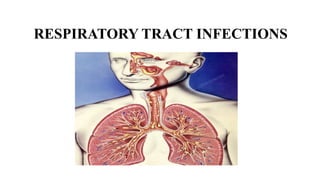
- 2. RELATED ANATOMY & PHYSIOLOGY • The respiratory system consists of the nose, pharynx (throat), larynx (voice box), trachea (windpipe), bronchi and lungs. • Structurally, the respiratory system consists of two parts: 1. The upper respiratory system includes the nose, pharynx, larynx and associated structures. 2. The lower respiratory system includes the trachea, bronchi and lungs • The lower respiratory tract fills most of the Thoracic Cavity
- 4. CONT.. • Respiratory system functions mainly as gas exchange system for O2 and CO2 cellular respiration (energy production) • It closely tied to circulatory system • Functionally the organ of the respiratory system can also be subdivided into: 1. Conducting division: passages that serve only for airflow 2. Respiratory division: alveoli and gas exchange areas
- 6. CONT.. 1. The conducting zone consists of a series of interconnecting cavities and tubes both outside and within the lungs. • These include the nose, nasal cavity, pharynx, larynx, trachea, bronchi and bronchioles. • Their function is to filter, warm, and moisten air and conduct it into the lungs
- 7. CONT.. 2. The respiratory zone consists of tubes and tissues within the lungs where gas exchange occurs. • These include the alveolar ducts, alveolar sacs and alveoli. (Gas exchange occur in these zone)
- 9. Respiratory mucosa • It covers most of the nose, pharynx, larynx, trachea, bronchi and bronchioles • It contains mucus producing goblet cells and ciliated epithelium. • Mucus can trap contaminants. • Cilia move them up towards mouth
- 11. NOSE • It provides an entrance for air in which air is filtered by hairs inside the nostrils. • It has two portions : the external and internal. • The external portion is supported by a framework of bone and cartilage covered with skin and lined with mucous membrane. • The internal portion is a large cavity beneath the skull, merging with the external nose anteriorly and communicating with the throat posteriorly.
- 13. CONT.. • The anterior portion of the nasal cavity just inside the nostrils, called the nasal vestibule is surrounded by cartilage. • The superior part of the nasal cavity is surrounded by bone. • A vertical partition, the nasal septum divides the nasal cavity into right and left sides. • The anterior portion of the nasal septum consists primarily of hyaline cartilage.
- 15. PARANASAL SINUSES • Four bones of the skull contain paired air spaces called the paranasal sinuses - frontal, ethmoidal, sphenoidal, maxillary. • Add resonance to voice. • Communicate with the nasal cavity by ducts. • Lined by pseudostratified ciliated columnar epithelium.
- 17. PHARYNX (THROAT) • The pharynx is a funnel-shaped tube about 13 cm or 5” long that starts at the internal nares and extends to the junction with esophagus and trachea • It is a common passage for air and food. • divided into three regions: a. Nasopharynx: includes uvula, tonsils (adenoids), auditory tube b. Oropharynx: extended from soft palate to level of hyoid bone c. Laryngopharynx: from hyoid bone to esophagus/larynx
- 19. LARYNX • Larynx or Voice box is a short, cylindrical airway ends in the trachea. It is about 5 cm long. • Opening into larynx = glottis • Boundaries: – Superiorly it attaches to hyoid bone and opens into laryngopharynx – Inferiorly trachea. – Posteriorly esophagus • Functions: - Prevent food from entering lower respiratory system - Sound, speech, etc.
- 21. TRACHEA • Flexible cylindrical tube, Size is 4 – 5 inch long & 1 inch diameter, also called windpipe. • Extends from larynx to bronchi • Surrounded by “C” – shaped bands of cartilage, ends joined by bands of muscle tissue • Cartilage provide rigidity to the tracheal wall, holds walls open, prevents collapse.
- 22. BRONCHI • Trachea divides into two branches called bronchi. • Each bronchus enters lung and continues to divide into smaller and smaller branches = bronchi, then = bronchioles • 2 primary bronchi branches into 5 secondary bronchi (1 for each lobe of lung) • Each of these branches into tertiary bronchi • Bronchioles - smallest branches of “respiratory tree” - <1mm diameter - no supportive cartilage
- 23. LUNGS • Lung occupies most of the space within the thoracic cavity. • Toward the midline, the lungs are separated from each other by the mediastinum and this is called the mediastinal surface. • The relatively broad, rounded surface in contact with the thoracic wall is called the costal surface of the lung. • The superior end of the lungs narrows to a rounded tip known as the apex. • The inferior end of the lungs, known as the base. • Right lung has 3 lobes and left lung has 2 lobes
- 25. Pleura and Pleural Cavity • The outer surface of each lung and the adjacent internal thoracic wall are lined by two layered serous membrane called pleura. • The outer surface of each lung is tightly covered by the visceral pleura. • while the internal thoracic walls, the lateral surfaces of the mediastinum, and the superior surface of the diaphragm are lined by the parietal pleura. • The potential space between the serous membrane layers is a pleural cavity. • The fluid inside the pleural cavity pleural fluid which acts as a lubricant, ensuring minimal friction during breathing.
- 28. ALVEOLI • Smallest bronchioles (respiratory bronchioles) have clusters (grapelike clusters) of tiny sacs branching off = alveoli • 300-500 Million alveoli/lung • Enveloped by capillaries • These are functional unit of respiratory system • Actual site of gas exchange with blood
- 29. ALVEOLI
- 31. • Infections of airway above glottis or vocal cords. It includes – – Rhinitis – Sinusitis – Tonsillitis – Pharyngitis – Laryngitis – Otitis media URTIs
- 32. Rhinitis • Rhinitis is inflammation and swelling of the mucous membrane of the nose, characterized by a runny nose and stuffiness and usually caused by the common cold or a seasonal allergy. • Symptoms of rhinitis include a runny nose, sneezing, and stuffiness. • Rhinitis is classified as allergic or nonallergic. The cause of nonallergic rhinitis is usually a viral infection, although irritants can cause it.
- 34. Cont.. • Rhinitis may be acute or chronic. Acute rhinitis commonly results from viral infections but may also be a result of allergies, bacteria, or other causes. Chronic rhinitis usually occurs with chronic sinusitis (chronic rhinosinusitis). • Typically, the diagnosis is based on the symptoms. • The various forms of rhinitis are treated in various ways, such as with antibiotics, antihistamines, surgery, desensitization injections (sometimes called allergy shots), and avoidance of irritants.
- 35. Sinusitis • Sinusitis is an inflammation, or swelling, of the tissue lining the sinuses. • Sinuses are structures inside face that are normally filled with air. Bacterial infections, viral infections and allergies can irritate them, causing them to get blocked and filled with fluid. • This can cause pressure and pain in face, nasal congestion (a stuffy nose) and other symptoms.
- 37. Cont.. • Types of sinusitis • According to duration: acute, subacute, chronic • According to cause: bacterial, viral or fungal. • Acute sinusitis symptoms last less than 4 weeks. It’s usually caused by viruses like the common cold. • Subacute sinusitis symptoms last 4 to 12 weeks. • Chronic sinusitis symptoms last at least 12 weeks. Bacteria are usually the cause.
- 38. Cont.. • Viruses, like the ones that cause the common cold (Rhinovirus, Influenza virus, Para influenza virus), cause most cases of sinusitis. • Bacteria (Streptococcus pneumoniae, Haemophiles influenzae) can cause sinusitis, or they can infect after a case of viral sinusitis. • Sinus infections caused by fungus are usually more serious than other forms of sinusitis. They’re more likely to happen in weakened immune system.
- 39. Cont.. • There are many treatment options for sinusitis, depending on the symptoms and underlying causes. It includes – • Intranasal steroid sprays. • Antibiotics • Topical antihistamine sprays or oral pills. • Leukotriene antagonists, like montelukast. • Surgery to treat structural issues, polyps or fungal infections.
- 40. Tonsillitis • Tonsillitis is inflammation of the tonsils, two oval-shaped pads of tissue at the back of the throat — one tonsil on each side. • Tonsillitis is most often caused by common viruses, but bacterial infections also can be the cause. The most common bacterium causing tonsillitis is Streptococcus pyogenes (group A streptococcus) • Signs and symptoms of tonsillitis include swollen tonsils, sore throat, difficulty swallowing, fever and tender lymph nodes on the sides of the neck.
- 42. Cont.. • Management • The patient is advised to bed rest and consume high amounts of fluids • Analgesics such as paracetamol are given to relieve the pain • Antibiotic therapy • Tonsillectomy: usually performed only when tonsillitis occurs frequently, doesn't respond to other treatments or causes serious complications.
- 43. Pharyngitis • The inflammation of pharynx is known as pharyngitis. Etiology: Viruses are the commonest cause of pharyngitis although bacteria and sometimes even fungi inflame the pharynx. Clinical Features 1. Mild inflammation: low-grade fever, malaise and some discomfort in the throat 2. Moderate to severe infections: headache, dysphagia, odynophagia, malaise and high fever. 3. In very severe cases there can be edema of the soft palate and cervical lymph node enlargement.
- 45. Cont.. Diagnosis • The culture of throat swabs is helpful in identifying the bacterial causative agents of pharyngitis. Management • Bed rest, increased fluid intake, gargling the throat with salt water and analgesics are the main components in the management of pharyngitis. • When the symptoms do not disappear spontaneously antibiotics such as penicillin can be given. If the patient is allergic to penicillin, erythromycin can be prescribed.
- 46. Laryngitis • Laryngitis is an inflammation of voice box (larynx) from overuse, irritation or infection. Etiology • Having a respiratory infection, such as a cold, bronchitis or sinusitis • Exposure to irritating substances, such as cigarette smoke, excessive alcohol intake, workplace chemicals • Overusing your voice, by speaking too much, speaking too loudly, shouting or singing Symptoms: Hoarseness, Weak voice or voice loss, Tickling sensation, Sore throat, Dry throat, Dry cough
- 48. Cont.. Types: • Acute laryngitis: Most cases of laryngitis are temporary and improve after the underlying cause gets better. • Chronic laryngitis: Laryngitis that lasts longer than three weeks. Diagnosis: • Throat swab culture • Laryngoscopy • Biopsy
- 49. Cont.. Management • Corticosteroids • Antibiotics • Pain medications: acetaminophen or ibuprofen. • Voice therapy. Prevention • Avoid smoking and stay away from secondhand smoke • Limit alcohol and caffeine • Drink plenty of water • Keep spicy foods out of your diet. • Include a variety of healthy foods in your diet • Avoid clearing your throat. • Avoid upper respiratory infections
- 50. Otitis media • Otitis media is a group of inflammatory diseases of the middle ear. Types 1. Acute otitis media (AOM): an infection of rapid onset that usually presents with ear pain. Most commonly found in young children. 2. Otitis media with effusion (OME): typically not associated with symptoms, although occasionally a feeling of fullness is described. it is defined as the presence of non-infectious fluid in the middle ear which may persist for weeks or months often after an episode of acute otitis media. 3. Chronic suppurative otitis media (CSOM): it is middle ear inflammation that results in a perforated tympanic membrane with discharge from the ear for more than six weeks.
- 52. Cont.. Etiology • Either bacteria or viruses may be involved. • Risk factors include exposure to smoke, use of pacifiers, and attending daycare, having cleft lip and palate or Down syndrome. Clinical manifestation • Ear pain, fever, hearing loss, tenderness on touch of the skin above the ear, purulent discharge from the ears, irritability, ear blocking sensation. Diagnosis • Audiometry • Tympanogram • Temporal bone CT and MRI
- 53. Cont.. Management • Oral and topical pain killers: Paracetamol (acetaminophen), ibuprofen, benzocaine ear drops • Antibiotics Prevention • Vaccination: PCV, pentavalent • Exclusive Breastfeeding