

Recommended
More Related Content
What's hot
What's hot (20)
Similar to MDCT (2)
Similar to MDCT (2) (20)
Recently uploaded
Recently uploaded (20)
MDCT (2)
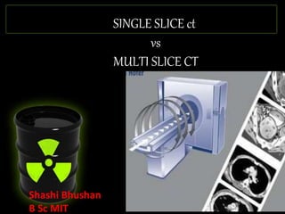
- 1. SINGLE SLICE ct vs MULTI SLICE CT Shashi Bhushan B Sc MIT
- 2. BEFORE CT •Entire areas of body inaccessible to radiography (brain, retroperitoneum,etc.) •Some useful diagnostic procedures were either potentially harmful or considerably uncomfortable (exploratory laparotomy, peumoencephalography)
- 3. WHY CT? •Better contrast resolution •No superimposition of tissues. •3D imaging
- 4. PRINCIPLES OF CT • Radiographic tube emits x-rays while rotating axially around patient. • Array of detectors on opposite side of patient detects x-rays transmitted through patient. • Computer algorithms use digitized data from detectors to create axial tomographic images of body. • CT = tomography + algorithms + high speed digital computer
- 6. Development of Workable CT Scanner • 1963- Cormack in S. Africa develops algorithm for accurate reconstruction of images from radiographic projections. • 1971- Hounsfield, a computer engineer in England produces first working CT scanner used clinically on patients. Cormack and Hounsfield awarded Nobel Prize in medicine and physiology in 1979.
- 7. Sir Godfrey Hounsfield with a prototype CT scanner in 1974
- 9. Helical (spiral) CT • Simultaneous patient translation and x-ray scanning generates volume of data. • X-ray beam traces a helix of raw data from which axial images must be generated • Each rotation generates data specific to an angled plane of section. • To create true axial image, data points above and below desired section must be interpolated to estimate value in axial plane. • Thus, interval between reconstructed transaxial images can be chosen retrospectively.
- 10. • Transverse images can be reconstructed at any z-axis position.
- 11. • Movement of x-ray tube is not a spiral. • Appears so because of translation movement of the patient.
- 12. INTERPOLATION
- 13. Technological considerations of helical CT • Slip-ring technology (no electrical cables connecting gantry to ground) allows source detector assembly to rotate continuously. • Previously, frequent, abrupt changes between scans were necessary to permit winding and unwinding of cables.
- 14. SLIP RING Introduced in 1990 allowed continuous rotation of the tube. Power and signals transmitted to gantry using Brushes on Static Rings. No need to start and stop the rotation. Scan time as fast as 0.3 sec.Brushes Static Rings.
- 15. Type of spiral CT Single slice spiral CT Multi slice Spiral CT (MSCT)
- 16. Single Slice CT Scan 1.) Acquires one slice at a time. The table moves to the start the acquisition of next slice. 2.) Long acquisition time 3.) Creates artifacts in images (partial volume artifact). A) Single Slice CT Scan
- 17. Single Slice Helical CT Scan 1.) Tube rotates and Couch moves continuously 2.) X-ray tube is always on and rotating 3.) Instead of one slice it scans a volume 4.) Covers larger area of the patient in one single breath hold 5.) Less artifact compared to single slice CT Scan Note: Development of Slip ring Technology helped the development of Helical scan Systems. Single Slice Helical CT Scan
- 18. B) Multi Detector CT • Introduced in 1998 • It had the ability to acquire four or more images with each rotation of the gantry, which has made the significant impact in patient throughput. • They are combined with high heat capacity x- ray tubes.
- 20. Multi Detector System Multi Slice Helical CT Scan
- 21. Multi slice Ct Scan 1.) Uses the principle of Helical scanner 2.)Acquires Multiple slices per tube rotation 3.) Covers more volume of the patient in one rotation and less time.
- 23. MSCT: Concepts and Issues • Detector Thickness: the Z-axis size of each basic detector element • Detector Collimation: total combined size of elements “linked” to acquire individual slices. • Beam Collimation: total Z-axis x-ray beam width. • Slice Thickness: determined by detector collimation and not by x-ray beam collimation.
- 24. What Helped the development of Multi Slice CT Scan 1.) High heat capacity X-ray tubes (upto 8 MHU). 2.) Multi Detector Technology. 3.) Dual focal spot Technology. 4.) Fast Reconstruction Algorithm maintaining good image quality.
- 25. Tubes with High Heat capacity were developed
- 26. Development of Dual focal Spot Technology
- 29. COMPARISON OF SINGLE SLICE ct AND MULTI-SLICE CT Detector configuration Reconstructions Pitch image quality
- 30. 1. DETECTORS CONFIGURATION MS- detector array segmented in z axis, a mosaic SS- long, narrow array with length of single detector aligned in z axis.
- 31. Mosaic Detector (for 16 slice CT) • 16 cells in Z direction --each cell 1.25 mm (in Z axis). • 16 cells (Z) x 912 cells (transverse) = 14592 total cells.
- 32. 2. RECONSTRUCTIONS SS- reconstruct images of SAME thickness with different image indexing (table increment intervals) MS- acquire 3D raw data that are contiguous in space. Therefore can reconstruct images at various thicknesses AND at different intervals. –If image index < image thickness, results in overlapping slices –Must have raw data available for any type of reconstruction
- 33. RECONSTRUCTIONS algorithms SS and MS are similar First step done by machine, z axis interpolator works on raw data to weight projections nearest the slice location most heavily. Second step selected by user: for soft tissue images, want to suppress noise and increase low contrast sensitivity. For bone want higher contrast.
- 34. 3D RECONSTRUCTIONS • Multiplanar reconstruction (MPR) • Volume rendering (VR) • Maximum intensity projection(MIP) • Surface Shaded Display (SSD) .
- 35. • A volume is built by stacking the axial slices. The software then cuts slices through the volume in a different plane 1. Multiplanar reconstruction
- 36. • Volume rendering is a set of techniques used to display a 2D projection of a 3D discretely sampled dat a set 2. Volume rendering
- 37. • voxels with maximum intensity traced from the viewpoint to the plane of projection. 3. Maximum intensity projection
- 38. 4. Surface Shaded Display(SSD)
- 39. 3. PITCH • Beam pitch: relates patient translation per 360 degree revolution to the width of the x-ray beam • Slice pitch: has the same form as beam pitch except the denominator is the slice thickness rather than the beam width.
- 40. Pitch • Pitch= ratio of the table increment during a 360 degree rotation to the collimated beam thickness. • Pitch = Table Movement per Rotation Slice Collimation 40
- 41. For single slice CT, pitch is defined as the patient couch movement per 360 rotation divided by slice thickness . 41
- 42. For MSCT, there are two definitions for pitch a) Beam pitch b) Slice pitch 42
- 43. 43
- 44. Data Acquisition Pitch = Table Movement per Rotation Slice Collimation • Contiguous Spiral Pitch = 1 (10 mm / 10 mm) • Extended (Non-Contiguous) Spiral Pitch = 2 (20 mm/ 10 mm) • Overlapping Spiral Pitch = 1/2 ( 5 mm / 10 mm)
- 46. Pitch and Noise • To reconstruct image, projections must be collected over 180degree gantry rotation and fan angle of x-ray beam (45 degree), about 2/3 of spiral. • Since reconstruction algorithms need fixed number of projections to make image and since pitch only affects how these projections are distributed in spiral, not the number of projections, pitch does not affect noise • No difference between SS and MS
- 47. 4. image quality Comparison b/n ssct & msct SSCT • Resolution. • Noise. • Motion blur. • Partial volume artifact. MSCT • Resolution. • Noise. • Motion blur. • Partial volume artifact.
- 49. Advantages of multi-slice over single-slice •Same acquisition in shorter time •Thin slices give better z-axis resolution •Scan larger volumes in the same time
- 50. 50
- 51. 51
- 52. 52
- 53. applications Of Msct •significant reduction in scan time. • four to eight times faster than with single slice spiral CT. •The speed of acquisition allows greater coverage with thinner slice thickness- enabling high quality reconstructions and CT angiograms. •allows better image quality for other post-processing techniques like virtual endoscopy.
- 54. IsotropicImaging
- 55. IsotropicImaging
- 56. Cardiac Imaging 1.) Noninvasive CT angiography can be used to rule out significant disease in major vessels and their branches. 2.) CT Angiography can pinpoint blockages,stenoses,aneurysms, or other kinds of lesions 56
- 57. 1.) Multiplanar Visualization is highly helpful for examination of musculoskeletal system 2.) Fastly replacing X-ray myelogram for spine and nervous system 57
- 58. Virtual endoscopy This is useful in acute stroke to assess cerebral ischaemia and areas at risk and results from this can be used to prevent stroke progression.
- 59. Multi-slice scanning has done a lot to transform the way we use CT in the last four years • Improvements in existing techniques • New techniques Power and speed of machines increasing all the time • 16 slice scanners now available • Reconstruction and data handling / viewing have to keep pace Dose from CT becoming increasingly important • With power comes responsibility to use it carefully Conclusions
Editor's Notes
- Computation of an unknown value using known values on either side. Performed by program – INTERPOLATION ALGORITHM
- More robust x-ray tubes and generators were developed to allow high tube current for prolonged duration. Also needed to be lightweight enough to be mounted in slip ring gantry.
- 64 slice is introduced in 2004
- Interpolation algorithms.
- Image reconstruction in CT is a mathematical process that generates images from X-ray projection data acquired at many different angles around the patient. Image reconstruction has a fundamental impact on image quality and therefore on radiation dose.
- new CT scanners offer isotropic or near isotropic, resolution, display of images does not need to be restricted to the conventional axial images. Instead, it is possible for a software program to build a volume by "stacking" the individual slices one on top of the other. The program may then display the volume in an alternative manner
- Multiplanar reconstruction (MPR) is the simplest method of reconstruction.
- maximum intensity projection (MIP) is a volume rendering method for 3D data that projects in the visualization plane the voxels with maximum intensity that fall in the way of parallel rays traced from the viewpoint to the plane of projection.
- Slice pitch is not as important as beam pitch in CT.
- Isotropy is uniformity in all orientations
- The other major advantage of MSCT is in evaluation of sick patients such as poly-trauma victims, pediatric and unco-operative patients.