2. investigations and control of disease progression in out-
break situations. Despite these manifold advantages,
detection of DNA does not yield information about the
presence of viable organisms.
Multiplexing qPCR detection offers several advantages,
including reduction of sample volume and handling
time (reducing the analysis time, cost and opportunities
for lab contamination). Also, false-negative results can
be reduced through co-amplification of internal controls
in each sample, and using multiple redundant genetic
markers for each organism reduces the chance that
strain variants are missed. Amplification of multiple sig-
nature sequences per organism will also reduce false-
positive results in complex samples. False positives can
be an issue if detection relies on single targets when
analyzing environmental samples, due to the presence of
homologous sequences in related organisms or unknown
sources [1,2]. Therefore, it is essential to validate the
qPCR using multiple strains, including of closely related
organisms.
The selection of suitable signature sequences is an
essential requirement for reliable PCR assays. The suit-
ability of signature sequences may be based on their
function, e.g. detection of virulence factors supplies
important information. But also the stability of their
association with the pathogen is of importance. For
instance, virulent B. anthracis can be recognized by its
virulence plasmids pXO1 and pXO2 [3] which contain
genes that confer toxin production and capsule synthesis
activities, respectively. However, there are also chromo-
somally encoded factors that are important for the full
virulence of B. anthracis [4]. Also, recent studies have
shown the occurrence of a plasmid homologous to
pXO1 in a pathogenic B. cereus strain [5] as well as
genes homologous to genes on pXO2 in environmental
Bacillus isolates [2]. This underscores the importance of
inclusion of a chromosomal signature for B. anthracis in
addition to the detection of plasmid genes. Similarly,
virulent Y. pestis possesses 3 plasmids involved in viru-
lence, but these plasmids are not stable and pathogenic
Y. pestis lacking any of these plasmids exists [6].
Several reports have described real-time PCR assays
for the detection of B. anthracis [7-10], Y. pestis
[6,11,12] and F. tularensis [13-15]. Some assays were
designed in multiplex format, but only few of these
included internal controls for DNA amplification [10,16]
and none included an internal control for successful
DNA extraction. Here, we report the highly reliable and
sensitive detection of these three pathogens that we
achieved by developing multiplex qPCRs for 3 organ-
ism-specific markers and 1 internal control. By using a
B. thuringiensis gene as internal control, it is possible to
use the highly refractory spores of this near relative of
B. anthracis as a control for both DNA extraction and
qPCR amplification. The assays were extensively vali-
dated and were used on different real-time PCR plat-
forms. The multiplex qPCRs are being applied in
screening protocols and our setup allows straightforward
expansion of the detection capabilities by inclusion of
additional pathogens.
Results
Design of multiplex hydrolysis probe assays
A selection of signature sequences for the specific detec-
tion and partial characterization of B. anthracis, F.
tularensis and Y. pestis was based on previous reports
[4-6,8,11-14,17], and sequence data accessible via public
databases (NCBI/EMBL). Additional sequences were
obtained from sspE genes from a number of strains
from the Bacillus cereus group in our culture collection
and from the cry1 gene from B. thuringiensis strain
ATCC 29730. Based on signature sequence alignments,
regions were identified that were shared by all strain
variants and sufficiently different from homologous
sequences to design selective oligonucleotides for multi-
plex real-time qPCR assays (see Table 1).
In order to achieve a reliable as well as rapid method
for the detection of B. anthracis, Y. pestis and F. tular-
ensis, the cry1 gene from B. thuringiensis was included
in the multiplex qPCR assays. Inclusion of this gene per-
mitted the development of B. thuringiensis spores as
internal control for DNA extraction as well as amplifica-
tion. The amount of spores that must be added to the
samples before DNA extraction to obtain the desired Cq
value was determined from serial dilutions of the spores.
Specificity and coverage of strain diversity
A DNA panel from the Bacterial and Eukaryal species
listed in Additional file 1 Table S1 was used to validate
the specificity of the developed real-time qPCR assays.
The pathogen-specific targets showed no cross-reactivity
and very near relatives could be differentiated as evi-
denced by the absence of amplification from various
members of the Bacillus cereus group, Yersinia pseudo-
tuberculosis, Y. enterocolitica and Francisella philomira-
gia. From the latter species, one out of the four strains
that were tested showed very weak amplification of the
multicopy sequence ISFtu2, but none of the strains
showed amplification of the F. tularensis signature
sequence fopA. The assays detected all available strains
from the targeted organisms. Nevertheless, the genomic
marker ypo393 was amplified from only one strain
(NCTC 10329) out of four from a Y. pestis cluster from
Nairobi. Additional information about the pathogens
could be derived from the detection of particular plas-
mid combinations in the B. anthracis and Y. pestis
assays, and from the detection of the pdpD gene [14] in
the F. tularensis assay. This was confirmed by the
Janse et al. BMC Microbiology 2010, 10:314
http://www.biomedcentral.com/1471-2180/10/314
Page 2 of 12
3. anticipated absence of the pdpD gene in the 16 F. tular-
ensis holarctica strains we tested. However, the probe
designed for pdpD detection could not discriminate
between subspecies tularensis and novicida. Based on
the available sequences from F. tularensis mediasiatica,
amplification of pdpD from this subspecies will occur as
well, however, we did not have genomic materials to
verify this. Amplification of the pla target from Rattus
rattus DNA was unexpected and seemed to indicate
cross-reactivity. To confirm pla amplification we investi-
gated DNA from 10 rats, including 3 from the related
species Rattus norvegicus (Additional file 1 Table S1).
Sequencing of the amplification product from these
samples revealed the presence of a pla gene highly simi-
lar to that of Y. pestis (99% identity), while no sequences
with any homology to these sequences were encoun-
tered in the published rat genome. Therefore, the ampli-
fication does not invalidate our assay but highlights the
fact that the pla gene alone is not a sufficient diagnostic
marker for the presence of Y. pestis. The internal con-
trol gene cry1 was amplified from several Bacillus cul-
tures in addition to B. thuringiensis.
Efficiency, dynamic range, precision and detection limit
Ten-fold independent serial dilutions from purified target
amplicons (PCR products containing target sequences)
were used to generate calibration curves and calculate
PCR amplification efficiencies. As shown in Table 2 effi-
ciencies for the different targets ranged between 94.5%
and 94.8% for B. anthracis, between 95.9% and 98.2% for
F. tularensis and between 93.1% and 93.2% for Y. pestis.
The efficiency for amplification of the internal control
target cry1 varied slightly between the assays and was
near that of the organism-specific targets. The reaction
was linear over 6 orders of magnitude, from 1.5·102
to
1.5·107
target copies per reaction (data not shown).
Table 1 Primers and probes for multiplex qPCR
Organism Target Oligo function Oligo name Sequence 5’-3’a
B. anthracis sspE Forward primer spEpri_f CGACTGAAACAAATGTACAAGCAGTA
Reverse primer spEpri_r CGTCTGTTTCAGTTGCAAATTCTG
Probe Tqpro_spE FAM-TGCTAGCATTCAAAGCACAAATGCTAGTT-BHQ1
cya Forward primer cyapri_f AGGTAGATTTATAGAAAAAAACATTACGGG
Reverse primer cyapri_r GCTGACGTAGGGATGGTATT
Probe Tqpro_cya JOE-CCACTCAATATAAGCTTTATTACCAGGAGC-BHQ1
capB Forward primer caBpri2_f AGCAAATGTTGGAGTGATTGTAAATG
Reverse primer caBpri2_r AAAGTAATCCAAGTATTCACTTTCAATAG
Probe Tqpro_caB CFR590-AGGTCCCATAACATCCATATGATCTTCTAA-BHQ2
F. tularensis fopA Forward primer foApri_f GCGCTTTGACTAACAAGGACA
Reverse primer foApri_r CCAGCACCTGATGGAGAGTT
Probe Tqpro_foA FAM-TGGCCAGTGGTACTTAGGTGTAGATGCTA-BHQ1
ISFtu2 Forward primer isfpri2_f CAAGCAATTGGTAGATCAGTTGG
Reverse primer isfpri2_r GACAACAATATTTCTATTGGATTACCTAAA
Probe Tqpro_isf JOE-ACCACTAAAATCCATGCTATGACTGATG-BHQ1
pdpD Forward primer pdDpri_f TCAATGGCTCAGAGACATCAATTAAAAGAA
Reverse primer pdDpri_r CACAGCTCCAAGAGTACTATTTCC
Probe Tqpro_pdD CFR590-ACCAAATCAAAATCCTGCTGAGCAGA-BHQ2
Y. pestis ypo393 Forward primer yp93pri_f AGATAGTGTGACTGGTCTTGTTTCA
Reverse primer yp93pri_r AGATGCAGATTGTATTGTAAACAATGAC
Probe Tqpro_yp93 FAM-ACTTCCTGATATATTGGAAATCTTCTTCTC-BHQ1
caf1 Forward primer cafpri_f CCAGCCCGCATCACT
Reverse primer cafpri_r ATCTGTAAAGTTAACAGATGTGCTAGT
Probe Tqpro_caf JOE-AGCGTACCAACAAGTAATTCTGTATCGATG-BHQ1
pla Forward primer plapri_f ATGAGAGATCTTACTTTCCGTGAGAA
Reverse primer plapri_r GACTTTGGCATTAGGTGTGACATA
Probe Tqpro_pla CFR590-TCCGGCTCACGTTATTATGGTACCG-BHQ2
B. thuringiensis cry1 Forward primer crypri_f GCAACTATGAGTAGTGGGAGTAATTTAC
Reverse primer crypri_r TTCATTGCCTGAATTGAAGACATGAG
Probe Tqpro_cry Cy5-ACGTAAATACACT-BHQ2-TGATCCATTTGAAAAG-P
a
CFR590 = CALFluor Red 590, BHQ = Black Hole quencher, P = phosporylation
Janse et al. BMC Microbiology 2010, 10:314
http://www.biomedcentral.com/1471-2180/10/314
Page 3 of 12
4. The precision of the different qPCR assays was calcu-
lated from 4 replicates of 5 independent dilutions. Mean
Cq values and standard deviations (SD) were calculated
from each dilution. As shown in Table 2 there is a high
repeatability for the different targets, with SDs around
0.05 Cq. Only at very low concentrations (high Cq
values) near the limit of detection, the SD exceeded 1
Cq (data not shown).
To determine the analytical sensitivity for each single
target, dilutions of target amplicons near the detection
limit were measured by using the developed assays. The
analytical sensitivity for genomic DNA was calculated
from dilutions of purified genomic DNA from selected
pathogens. The fraction of positive reactions in replicate
dilutions were scored and a probit analysis was used to
calculate the limit of detection (LOD), which is the low-
est concentration at which 95% of positive samples are
detected. The LOD for single targets could be expressed
as copy numbers as the target amplicons were of known
size. Table 2 shows LODs of below 10 copies for the
various targets. For genomic DNA, LODs based on the
most sensitive target were for B. anthracis15.7 fg, for F.
tularensis 0.6 fg and for Y. pestis 29.6 fg.
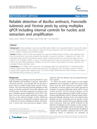
Co-amplification targets in multiplex assay
Large concentration differences between DNA templates
in a multiplex PCR may lead to competition for reaction
components and impaired amplification of the rarer
templates. Divergence of target concentrations could
originate from different copy numbers of the targets
within the pathogen genome, or from differences
between the numbers of organisms that are detected
simultaneously. Although there is limited copy number
variation for the selected targets, multicopy sequences
such as insertion sequences and plasmid genes could
outnumber single-copy targets by a factor of more than
200 [3,18]. To exclude an inhibitory effect of the domi-
nant amplification product in the multiplex reaction,
dilution series of the high copy number targets (cya, pla
and ISFtu2) were made in the presence of a constant
and low concentration of the other targets from that
organism, and measured by the multiplex qPCRs (Figure
1A-C). No inhibitory effect (increasing Cq) was
observed, even if the excess target considerably exceeded
the maximum ratio that could be anticipated.
Significant concentration differences are possible
between the pathogen specific targets and the internal
control target, as these organisms could be mixed in
very different quantities. Inhibition of the internal con-
trol (IC) by excess pathogen DNA is not a problem as
the function of the IC is to exclude false negative results
(a positive pathogen signal makes an additional IC signal
irrelevant). In contrast, it is essential that inhibition of
pathogen targets by the internal control is prevented.
To determine the boundaries within which IC B. thurin-
giensis DNA could be added to pathogen DNA without
interfering with the detection of low pathogen concen-
trations, a dilution series of the IC target amplicon (cry1
gene) was made in the presence of a constant and low
concentration of pathogen targets and measured by the
multiplex qPCRs. As shown in Figure 1D-F, the amplifi-
cation of 20 copies of pathogen targets was inhibited
(increasing Cq) if more than 200 copies of the internal
control target were present for B. anthracis or more
than 2000 copies for Y. pestis and F. tularensis. More-
over, rare targets were still detectable at much higher
excess ratios of internal control, even though at higher
Cq values.
Table 2 Precision and detection limits of the multiplex PCRs
organism Target Efficiency (%) Repeatability
(SD of Cq)a
LOD target amplicons (copies/reaction)b
LOD gDNA (fg/reaction)b
B. anthracis sspE 94.5 0.045 2.6 (1.6-7.5) 22.6 (9.9-148.5)
cya 94.7 0.057 6.5 (3.7-19.6) 50.5 (19.1-408.3)
capB 94.8 0.051 3.6 (2.0-10.7) 15.7 (9.9-78.9)
F. tularensis fopA 98.2 0.042 7.2 (3.5-24.7) 11.8 (5.5-66.4)
ISFtu2 98.1 0.075 4.1 (2.2-12.8) 0.6 (0.2-3.4)
pdpD 95.9 0.067 6.1 (3.1-20) 4.2 (2.5-25.6)
Y. pestis ypo0393 93.1 0.057 1.7 (1.2-3.5) 116 (59.3-967.2)
caf1 93.2 0.099 1.9 (1.3-4.1) 43.2 (23.9-277.2)
pla 93.1 0.047 3.6 (2.2-8.9) 29.6 (13.5-191.9)
B. thuringiensis cry1 94.6/95/92.9c
0.047/0.055/0.057 c
ND ND
a
Values represent the average from the standard deviations calculated at 5 different dilutions from 4 replicate Cqs measurements.
b
Values displayed represent the lowest DNA concentration at which 95% of the positive samples are detected, as calculated by using probit analysis. Shown
between brackets are the 95% confidence limits of the calculated LODs.
c
B. thuringiensis internal control added to B. anthracis, F. tularensis and Y. pestis, respectively
ND = not determined
Janse et al. BMC Microbiology 2010, 10:314
http://www.biomedcentral.com/1471-2180/10/314
Page 4 of 12
5. Discussion
Multiplexing and the reduction of false negative and false
positive results
In this report, we describe the development of multiplex
qPCRs for the rapid and reliable detection of B. anthra-
cis, F. tularensis and Y. pestis. The assays include a sig-
nature sequence from B. thuringiensis which allows the
use of its spores as combined internal control for both
DNA extraction and subsequent DNA amplification. As
Bacillus spores are among the most resistant of micro-
bial structures, DNA extraction from such spores can be
considered to be a reliable indicator for successful DNA
extraction from other microbes. Application of internal
controls is especially important when measuring envir-
onmental samples because these tend to contain various
sorts of PCR inhibitors. The internal control helps pre-
venting false negative results, which are further reduced
by the sensitivity of the methods and by the recognition
of multiple signatures per organism. Multiplexing
reduces the chance that the pathogen escapes detection
due to modification or loss of plasmids or genes (natural
or by manipulation).
Multiple diagnostic signatures per pathogen will also
help reducing false positive detection, which is
A
20
24
28
32
36
40
1,E-01 1,E+00 1,E+01 1,E+02 1,E+03 1,E+04 1,E+05 1,E+06
cya [cps]
Cq
sspE
capB
cya
B
20
24
28
32
36
40
1,E-01 1,E+00 1,E+01 1,E+02 1,E+03 1,E+04 1,E+05 1,E+06
ISFtu2 [cps]
Cq
fopA
pdpD
ISFtu2
C
20
24
28
32
36
40
1,E-01 1,E+00 1,E+01 1,E+02 1,E+03 1,E+04 1,E+05 1,E+06
pla [cps]
Cq
ypo393
caf
pla
D
24
28
32
36
40
44
1,E-01 1,E+00 1,E+01 1,E+02 1,E+03 1,E+04 1,E+05
cry1 [cps]
Cq
sspE
cya
capB
cry1
E
24
28
32
36
40
44
1,E-01 1,E+00 1,E+01 1,E+02 1,E+03 1,E+04 1,E+05
cry1 [cps]
Cq
fopA
ISFtu2
pdpD
cry1
F
24
28
32
36
40
44
1,E-01 1,E+00 1,E+01 1,E+02 1,E+03 1,E+04 1,E+05
cry1 [cps]
Cq
ypo393
pla
caf
cry1
Figure 1 Effect of increasing concentration differences between targets in multiplex qPCR reactions. Dilution series of multicopy targets
(A-C) or internal control target cry1 (D-F) were made in the presence of the other targets detected in each qPCR at a constant concentration
near the detection limit. Triplicate multiplex qPCR measurements were performed and mean Cq values with 95% confidence limits are shown
for each target.
Janse et al. BMC Microbiology 2010, 10:314
http://www.biomedcentral.com/1471-2180/10/314
Page 5 of 12
6. particularly important in complex (environmental) sam-
ples which may contain homologous genes of yet
uncharacterized origin[1,2]. The genera Bacillus, Franci-
sella, and Yersinia each include species ranging from
nonpathogenic environmental species, through sym-
bionts and facultative pathogens, to highly virulent
human and animal pathogens. Comparative genomic
sequencing and typing studies have indicated that the
sequence similarity and gene composition of species
having very different lifestyles can be very high [1,19-21]
Also, bacterial genomes are dynamic and non-target
organisms could acquire diagnostic sequences by lateral
gene transfer, especially if present on plasmids [22]. An
additional reason for including multiple targets is that
for B. anthracis and Y. pestis, a full picture of virulence
requires the detection of several markers. Although viru-
lent Y. pestis usually contains three plasmids, strains
deficient in one or more plasmids may cause fatal infec-
tions [6].
Assays relying on one signature sequence for the
detection of a pathogen [10,23,24], suffer from the con-
straints mentioned above, especially when analyzing
environmental samples [1]. For instance, Y. pestis sub-
group Pestoides lacks the plasminogen coagulase (pla)
gene [25] that is used as the major and sometimes only
target for the detection of Y. pestis [23,26]. On the other
hand, we found that the pla gene may yield false posi-
tive results in certain matrices (unpublished). In addition
to relying on multiple targets, false positives are further
reduced by the high specificity of the developed assays
for the selected targets, which was confirmed by in silico
and in vitro validations.
Selected targets
Inclusion of chromosomal markers in addition to viru-
lence plasmids is important due to the occurrence of B.
anthracis and Y. pestis strains lacking virulence plas-
mids. These strains, as well as yet uncharacterized clo-
sely related environmental species, share genomic traits
that could lead to misidentification. Fully virulent B.
anthracis strains possess plasmids pXO1 and pXO2.
However, the detection of plasmids only, as for instance
commercial kits do, cannot detect plasmid-deficient B.
anthracis strains such as Sterne and CDC 1014. More-
over, B. cereus strains carrying plasmid highly similar to
those of B. anthracis (B. cereus G9241) are not correctly
identified. Several chromosomal markers have been used
for the detection of B. anthracis (e.g. BA813, rpoB, gyrA,
gyrB, saspB, plcR, BA5345, BA5510), but only recently a
locus was described for qPCR that did not yield any
false positive results from closely related Bacillus [27].
We have developed an alternative chromosomal signa-
ture sequence (sspE) for use in real-time PCR. This mar-
ker has previously been used for specific detection of B.
anthracis, but differentiation required melting curve
analysis [8]. By selecting highly discriminating positions
for primers and hydrolysis probe, we achieved specific
detection without post-PCR analysis. For Y. pestis, it is
equally important to detect chromosomal sequences in
addition to its plasmids, as plasmid-deficient virulent Y.
pestis has been described [6]. Most of the chromosomal
targets that have been described previously did not dif-
ferentiate Y. pestis from closely related Y. pseudotuber-
culosis or Y. enterocolitica [12]. The chromosomal
signature sequence we developed for Y. pestis detection
was based on a previous study employing comparative
genome hybridization to identify chromosomal regions
specific for Y. pestis [17]. We selected a different region
than the ypo2088 target which was used by these
authors and later by Matero et al. [16], because exami-
nation of published genomes revealed that strain Y. pes-
tis antiqua (accession # CP000308) does not possess
this region. Although ypo339 was present in all 20 Y.
pestis sequences currently publicly available, 3 out of 4
isolates from the Nairobi cluster appeared to lack this
signature sequence. Hence, although ypo393 is a reliable
signature sequence for most Y. pestis, strains lacking
this sequence do exist. Our results illustrate that even if
signature sequences selected for diagnostic purposes are
based on a considerable amount of sequences available
from genomes and sequence databases, uncharacterized
strain variants may exist or new variants may arise that
do not posses a particular target sequence. Conversely,
amplification of the cry1 gene from some Bacillus
strains other than B. thuringiensis was not anticipated as
these strains were not known to contain the plasmids
carrying cry genes or homologues. Since it concerned
related, spore-forming Bacillus strains, these could also
be used as internal controls. The primary focus of our
assays was the sensitive and specific detection of the
selected pathogens, minimizing false negative and false
positive results. Strain differentiation was considered to
be of only secondary interest. For F. tularensis, sensitive
detection requires detection of the multicopy sequence
ISFtu2. The targeted tranposase can also be present in
F. philomiragia, but strain ATCC 225017 for instance,
has only one copy with mismatches in the probe and
reverse primer. This explains the very low cross-reactiv-
ity with the four strains we investigated. Nevertheless,
specific detection of the species F. tularensis was con-
firmed by additional detection of the fopA gene [13,15].
Further subspecies information could be obtained from
the pdpD target, which is known to be absent in subspe-
cies holarctica (type B) [14] and was indeed not detected
in the 16 strains we tested. With all targets positive,
subsequent research is warranted however, as presence
of this gene could also imply presence of the subspecies
novicida and mediasiatica [28]. Subspecies mediasiatica
Janse et al. BMC Microbiology 2010, 10:314
http://www.biomedcentral.com/1471-2180/10/314
Page 6 of 12
7. is, similar to subspecies holarctica, a considerable public
health threat although both species are less pathogenic
compared to subspecies tularensis. Subspecies novicida
is less pathogenic than the other subspecies and has
been involved in only a limited number of human cases.
Sensitivity
The analytical sensitivity for detection of the different
signature sequences is very high (Table 2). Hence, the
presence of only a few genomes should enable detec-
tion of the organisms of interest at 95% probability,
especially when based on multicopy signature
sequences. For F. tularensis this means that only 0.3
genomic equivalents (GE) were sufficient for the detec-
tion, considering a genome size of 1.9 megabases. For
B. anthracis and Y. pestis, reliable estimates of GE
could not be made due to the variable and sometimes
significant contribution of plasmids to the total
amount of DNA measured [3,18]. But, using approxi-
mate plasmid copy numbers, a detection limit of 4 GE
for B. anthracis and 6 GE for Y. pestis can be calcu-
lated. The LODs were similar or lower than those
reported previously [13,14] and lower than those of
other multiplex assays for these pathogens [12]. A cor-
relation between the copy numbers of the targeted
genes and the LOD for genomic DNA can be expected.
For F. tularensis gDNA, the LOD was indeed highest
based on the detection of the single-copy fopA target,
lower when based on the 2-copy pdpD and lowest
when based on the approximately 20-copy ISFtu2
(Table 2). Also for Y. pestis, an inverse correlation
between gDNA LOD and expected target copy number
was observed (Table 2). Nevertheless, a more pro-
nounced difference would be expected based on the
high relative abundance of pla carrying plasmids that
has been reported [18]. Probably, the gDNA we used
contained fewer plasmids, as was supported by a Cq
difference between the chromosomal target and pla of
only approximately 2 (data not shown). For B. anthra-
cis, the LOD of gDNA was highest when based on the
detection of the pXO1 plasmid marker cya, while high
copy numbers for the pXO1 plasmid carrying this gene
have been reported [3]. This discrepancy could be due
to the gDNA preparation we used for calculating
LODs. Although Coker et al. reported relative amounts
of pXO1 and pXO2 of respectively 11.5 and 1.6, for
the same strain we used (B. anthracis Vollum), varia-
tion in pXO plasmid copy numbers could also result
from the growth phase at which DNA was harvested
[3]. Our data correspond better to the lower plasmid
copy numbers reported by other authors [29,30].
Nevertheless, all reports agree that pXO1 is present in
multiple copies. The relatively high LOD for gDNA
detection based on cya can probably partly be
explained by a low amplification efficiency near the
detection limit as the LOD for the detection of cya tar-
get amplicons is also relatively high (Table 2).
Internal control
As was shown in Figure 1 the cry1 gene from B. thurin-
giensis spores can be used as internal control without
affecting sensitive detection of the pathogens of interest.
However, addition of more than 200 copies of cry1 per
reaction lead to a Cq increase for the detection of the B.
anthracis plasmid targets. For diagnostic purposes, we
use a spore suspension that yields a Cq value between
32 and 35 for the detection of cry1 to prevent any inter-
ference with the detection of pathogen DNA. The
amount of spores that needs to be added to yield this
Cq should be determined for each new batch as it will
vary with each new spore stock, and the DNA extraction
protocol used. The observed inhibition highlights that
multiplex qPCR can be problematic if it is used for the
detection of mixed pathogens present in different quan-
tities as amplification of targets from a dominant organ-
ism could inhibit the detection of an uncommon
pathogen. Assays for the detection of single targets from
multiple pathogens simultaneously, such as that
described for B. anthracis, F. tularensis and Y. pestis
detection [23], should therefore be carefully evaluated
for this inhibition effect.
Environmental testing
Application of the multiplex qPCR assays directly on
human specimens or environmental samples could save
time and prevent loss of DNA during extraction. How-
ever, we use the assays only after a DNA extraction pro-
tocol, in order to prevent unanticipated inhibition by
diverse matrices. Our laboratory has compared several
commercially available DNA extraction kits for use in a
BSL3 facility, and selected one that combined efficient
DNA extraction with ease-of-use and applicability in the
restricted BSL3 environment. We have been using the
developed qPCRs for the analysis of samples suspected
for the presence of these pathogens with B. thuringiensis
spores added before DNA extraction under BSL3 biosaf-
ety conditions. Hundreds of samples containing all sorts
of solid materials and liquids have been analyzed with-
out yielding false positive readings.
Conclusion
The multiplex qPCR assays that were developed for B.
anthracis, F. tularensis and Y. pestis allow the rapid
detection of 3 pathogen-specific targets simultaneously
without compromising sensitivity. Together with the
application of an internal control for both DNA extrac-
tion and DNA amplification, this assures highly reliable
detection, while template consumption and laboratory
Janse et al. BMC Microbiology 2010, 10:314
http://www.biomedcentral.com/1471-2180/10/314
Page 7 of 12
8. effort is kept at a minimum. These considerations are
particularly advantageous in the context of biothreat
samples which may be used for additional tests and for
surge capacity during an outbreak. The detection of
multiple targets decreases the chance of false-positive
and false-negative results and provides additional infor-
mation about virulence.
Methods
Selection signature sequences
An initial selection of potential signature sequences for
specific detection of B. anthracis, F. tularensis and Y.
pestis was based on previous reports and on the avail-
ability of sequences through public databases (NCBI/
EMBL). The selection was based on functional and on
technical criteria. Since 4 reporter dyes can be reliably
differentiated by using qPCR instruments, and 1 channel
was reserved for the internal control, we selected 3 sig-
nature sequences per organism. If possible, signature
sequences included virulence genes since these are sig-
nificant diagnostic markers. Such virulence genes are
often located on plasmids. Besides plasmid-encoded tar-
gets, at least one chromosomal target was included to
account for plasmid transfer and loss. Plasmids may be
transferred between closely related species of Bacillus or
Yersinia [8]. Plasmids can be cured from B. anthracis
[31] and Y. pestis [6], and virulent plasmid-deficient Y.
pestis strains occur in nature [6]. Also, near-neighbor
species carrying closely-related plasmids [5] should be
distinguished from B. anthracis. Finally, although B.
anthracis has two plasmids that are required for viru-
lence, there are also chromosomally encoded factors
that are important for the full virulence [4]. If available,
a multicopy sequence was included to enhance sensitiv-
ity. Unique targets present only in the organism of
interest were preferred over targets differentiating
homologues in related species only by sequence differ-
ences. Finally, an important consideration for the selec-
tion of targets was the quality of sequence information
available from the public databases. This sequence qual-
ity concerned the number of sequences, their length and
their coverage of strain diversity. For each potential tar-
get sequence, representative sequences were retrieved
from NCBI/EMBL. BLAST searches were then per-
formed to retrieve all homologous sequences from
nucleotide and bacterial genome databases. All available
sequences were aligned and consensus sequences were
created using an accept level of 100% (to make sure the
consensus sequence displayed all sequence variation).
For B. anthracis, genes were selected on the multicopy
virulence plasmids pXO1 and pXO2, and on the chro-
mosome. The consensus alignment from the toxin gene
cya included this gene from the homologous pBCXO1
plasmid which is present in a virulent B. cereus strain
[5]. The chromosomal target for B. anthracis, the spore
structural gene sspE, is not a unique gene as it is present
in all Bacillus. Nevertheless, this sequence was selected
since the sequence differences between B. anthracis and
other species within the closely related B. cereus group
were sufficient for designing highly selective oligonu-
cleotides. Also, the presence of a substantial number of
sequence entries in the databases (> 200) enabled a reli-
able consideration of the sequence diversity of B. cereus
group isolates. For F. tularensis, the multicopy insertion
sequence ISFtu2 was selected for the detection of F.
tularensis. Cross reaction with other Francisella species
such as F. philomiragia could not be ruled out based on
the available sequences, and a region of the outer mem-
brane protein gene fopA was selected for the specific
detection of all subspecies from the species F. tularensis.
A specific location in the pdpD gene, which is absent
from F. tularensis subspecies holarctica, was selected for
the design of a probe for the detection of F. tularensis
subspecies tularensis (type A) [14]. For Y. pestis, genes
were selected on Y. pestis specific virulence plasmids
pPCP1 and pMT1. The plasmid pCD1 was not used as
it is shared by other pathogenic Yersinia species. A
chromosomal sequence of unknown function that had
been identified using comparative genome hybridization
[17] was selected as Y. pestis specific chromosomal
target.
Spores of B. thuringiensis were used as internal con-
trol, not only for DNA amplification but also for suc-
cessful DNA extraction. This member of the B. cereus
group is closely related to B. anthracis and forms similar
spores, while it contains species-specific plasmids. The
B. thuringiensis plasmid gene encoding insecticidal crys-
tal proteins (cry genes) was used as the signature
sequence for the detection of DNA released from this
organism’s spores.
Sequence analysis tools, bioinformatics software
Sequences retrieved from NCBI/EMBL were organized
and aligned using the software package Kodon (Applied
Maths, Ghent, Belgium). Comprehensive sequence align-
ments were made by performing BLAST searches from
the selected targets to make sure all available sequence
homologues were included in the alignments. Oligonu-
cleotides for multiplex qPCR assays and for conven-
tional PCR assays were designed using the software
package Visual Oligonucleotide Modeling Platform ver-
sion 6 (DNA software Inc. Ann Arbor, USA). The
design strategy for multiplex qPCR assays was as fol-
lows. First, a hydrolysis probe and primer set were
designed for the B. thuringiensis internal control. Then,
for each selected signature sequence a hydrolysis probe
was designed, followed by the design of the correspond-
ing primer set. A different strategy was chosen for the
Janse et al. BMC Microbiology 2010, 10:314
http://www.biomedcentral.com/1471-2180/10/314
Page 8 of 12
9. B. anthracis assay, because its chromosomal target sspE
has homologues in other Bacillus, notably the internal
control B. thuringiensis. To make sure that detection of
B. anthracis sspE was highly selective, the exact posi-
tions of probe and primers were guided based on visual
inspection of the alignment. Probe and primers were
located in regions with mismatches between Bacillus
species (notably between B. thuringiensis and B. anthra-
cis), and the primers were designed such that mismatch
positions were located at their highly discriminating 3’-
ends. Oligonucleotides that were calculated by the
design software were first checked against the consensus
alignment to exclude designs not covering all sequence
variants, and were then evaluated using the simulation
module of Visual OMP. All oligonucleotides designed
were validated in silico by using BLAST searches in gen-
eral and microbial genomes databases (NCBI/EMBL).
Sequencing
Sequences were obtained from the cry1 gene from B.
thuringiensis strain ATCC 29730 and from the sspE
gene from all B. anthracis strains in our culture collec-
tion, B. thuringiensis ATCC 29730 and B. cereus strains
WSBC 10583, 10619, 10766, 10483, 10572, 10705, 10770
and 10865 (Additional file 1 Table S1). In addition, the
pla gene was sequenced from DNA extracted from mus-
cle tissue derived from a dissected specimen of Rattus
rattus. Primers used for sequencing are displayed in
Additional file 2 Table S2. PCR products were purified
by using ExoSAP-IT (USB, Cleveland, USA) and DNA
sequencing reactions were performed in both directions
using BigDyeTerminator v3.1 (Applied Biosystems,
Nieuwerkerk a/d IJssel, the Netherlands) on a 48-capil-
lary 3730 DNA Analyzer sequencer (Applied Biosystems,
Nieuwerkerk a/d IJssel, the Netherlands). Accession
numbers: HQ222846 to HQ222861 and HQ606074.
PCR and real-time qPCR
Oligonucleotides were synthesized by Biolegio (Biolegio,
Nijmegen, the Netherlands). Conventional PCR was
used to produce amplicons from signature sequences.
Amplification was carried out using the HotStar Taq
Master Mix Kit (Qiagen, Westburg, the Netherlands)
and 400 nM primers in a total reaction volume of 50 μl.
Primer sets were designed using Visual OMP software
(Additional file 2 Table S2). Thermocycling conditions
were as follows: 95°C for 15 min, 40 cycles at 95°C for
30 sec, 55°C for 30 sec and 72°C for 30 sec, followed by
a final step at 72°C for 7 min. Thermocycling reactions
were carried out in a Px2 thermal cycler (Thermo Elec-
tron Corporation, Breda, the Netherlands).
All qPCR reactions were carried out in a final volume
of 20 μl containing iQ Multiplex Powermix (Bio-Rad,
Veenendaal, the Netherlands), 200 nM of each primer
and 100-300 nM hydrolysis probes and 3 μl of DNA
template. Probes concentrations had been optimized to
yield minimal spectral overlap between fluorescence
level of the reporter dyes for each target in a multiplex
assay and were 100, 200, 300 and 300 nM for FAM,
JOE, CFR590 and Cy5 labeled probes respectively. The
multiplex real-time qPCR assays had been designed for
an optimal annealing temperature of 60°C and the ther-
mal cycling conditions were as follows: First enzyme
activation at 95°C for 5 min, followed by amplification
and detection by 45 cycles at 95°C for 5 sec and 60°C
for 35 sec. Each real-time qPCR experiment included a
negative (no template) control. Measurements were car-
ried out on a Lightcycler 480 (Roche, Almere, the Neth-
erlands). An iQ5 (Bio-Rad) instrument was used for
routine screening purposes. Analyses were performed on
the instruments software: LightCycler 480 Software
release 1.5.0. SP3 and iQ5 Optical Systems Software ver-
sion 2.0. Cq values were calculated using the second
derivative method on the LightCycler and the Base Line
Subtracted Curve Fit method on the iQ5. Color com-
pensations were carried out on both instruments as fol-
lows. PCR amplifications were performed using single
primer-probe sets for each reporter dye and under iden-
tical reaction conditions as during multiplex amplifica-
tion. The PCR reactions thus produced contained single
dyes in relevant concentrations and these were used for
color compensation runs according to the manufac-
turers’ guidelines. Verification of PCR product sizes
were carried out on the 2100 Bioanalyzer instrument
(Agilent Technologies, Eindhoven, the Netherlands)
using the DNA 1000 kit.
Bacterial isolates and genomic DNA preparation
The detection limits and specificities of the assays were
evaluated using genomic materials from the bacterial
strains and other sources displayed in Additional file 1
Table S1. The pathogen panel included (besides a vari-
ety of Eukaryal organisms): 8 B. anthracis strains and 31
near relatives (22 B. cereus, 5 B. thuringiensis and 4 B.
mycoides), 21 F. tularensis strains (16 subspecies holarc-
tica, 4 tularensis and 1 novicida) and 4 of the closest
related species F. philomiragia, 23 Y. pestis (including
Antiqua, Mediaevalis and Orientalis biovars) and 3
strains from the closest relative Y. pseudotuberculosis
and 7 strains from Y. enterocolitica. From most of the B.
anthracis, F. tularensis and Y. pestis strains we only had
genomic DNA (lysates) available to verify specificity of
our assays. Several strains were available as live cultures
in our laboratory and these were used as resource for
the production of larger quantities of genomic DNA. B.
anthracis and Y. pestis strains were acquired from the
NCTC (National Culture Type Collection, UK) and the
Pasteur Institute (France). The Francisella holarctica
Janse et al. BMC Microbiology 2010, 10:314
http://www.biomedcentral.com/1471-2180/10/314
Page 9 of 12
10. strain was a patient isolate. Other genomic materials
were lysates from bacterial cultures provided by other
researchers as mentioned in the acknowledgements.
Cultivation of these strains was carried out in a BSL3
glove-box. Colonies from B. anthracis, F. tularensis and
Y. pestis were grown on Columbian sheep blood agar
plates and chocolate agar plates. Single colonies were
transferred to liquid BHI (Brain Heart Infusion, 27 g/L)
medium. After cultures had grown to visible turbidity,
1.4 ml cell culture was centrifuged and the pellet was
resuspended in 250 μl TE pH 8. Cells were incubated
for 30 minutes at 100°C. Lysed cultures were filtered
through a 0.22 μm sterile Ultrafree-MC spinfilter (Milli-
pore, Amsterdam, the Netherlands) and the filtrate was
subsequently transported from the BSL3 facility for
handling under normal laboratory conditions. Cultures
from non-target bacteria that were used in the specifi-
city panel were obtained from the culture collection at
the RIVM. These cultures were cultivated under BSL2
conditions and lysates of these cultures were used for
specificity testing.
DNA extraction and purification was carried out by
using NucliSens Magnetic Extraction Reagents (bioMér-
ieux, Boxtel, the Netherlands) following the manufac-
turers instructions. This method performed best with
regard to efficiency and ease-of-use when compared to
other kits. This comparison was carried out as follows.
Dilution series of a mixture of genomic DNA from B.
anthracis, Y. pestis and F. tularensis, and spores from B.
thuringiensis were added to various powders including
milk powder, soy powder, silica and maize powder, and
DNA was extracted by using the powersoil DNA isola-
tion kit (MO BIO Laboratories, Carlsbad, USA), the
ultraclean microbial DNA isolation kit (MO BIO) and
the NucliSens Magnetic Extraction Reagents (bioMér-
ieux) according to the manufacturers instructions. The
extracts were measured by using the developed qPCR.
DNA concentrations were measured using the Nano-
Drop 1000 spectrophotometer (Thermo Fisher Scientific,
Wilmington, USA). DNA samples were stored at 4°C for
use within 1 week and at -20°C for longer storage.
Spore suspension for use as internal control
Spore suspensions of B. thuringiensis strain ATCC
29730 (var. galleriae Heimpel) were obtained from
Raven Biological Laboratories (Omaha, Nebraska, USA).
These washed spores were counted by microscopy and
then aliquotted and stored at 4°C. The amount of spores
that needs to be added to samples to obtain suitable Cq
values for this internal control must be determined
empirically for each stock spore suspension. Ten-fold
serial dilutions were made from the spore stock and
DNA was extracted from 50 μl portions of each dilution
by using the Nuclisens Magnetic Extraction Reagents
(bioMérieux). The developed real-time qPCR assays
were used to determine the amount of spores required
for a Cq value between 32 and 35.
Limit of detection, efficiency and repeatability
Characterization of qPCR performance was guided by
the MIQE guidelines [32]. The validation was carried
out by using genomic DNA as well as purified PCR
amplicons including > 100 bp upstream and down-
stream from the qPCR amplification sites. The latter
were used to compose template mixes of desired com-
position and quantities, while maintaining secondary
structures in the primer binding regions. Detection
limits (LOD) for genomic DNA were determined by
using purified DNA from cultures of B. anthracis
strain Vollum, F. tularensis strain tularensis ATCC
6223 and Y. pestis strain Harbin. DNA was purified
from lysates of these strains. The concentration of pur-
ified genomic DNA was measured by using the Nano-
Drop 1000 spectrophotometer. Serial dilutions of
genomic DNA were used to calculate LODs from the
proportion of positive qPCRs at each dilution. Four
replicates of eight serial dilutions of genomic DNA
were measured by qPCR. Based on the results, an addi-
tional measurement was performed on 4 replicates of 8
novel serial dilutions. The measurements included at
least one dilution with all replicates positive and one
with all replicates negative. A probit analysis was per-
formed using SPSS Statistics 18.0.0 to calculate the
DNA concentration that could be measured with 95%
probability.
DNA templates for measuring the detection limits
from the different signature sequences were amplified
from the bacterial strains mentioned above. In addition,
the pdpD signature sequence from F. tularensis tularen-
sis was amplified from ATCC 6223. To generate suitable
amplicons for testing the different real-time qPCR tar-
gets, primers were designed for amplification of a signa-
ture sequence with a size of 400-800 bp, extending
beyond both ends of the region amplified by the real-
time qPCR. Primer sequences are displayed in Addi-
tional file 2 Table S2. After amplification, PCR products
were purified and the number of DNA copies in ampli-
con solutions was calculated from their sizes and con-
centrations. Amplicon dilutions were used to calculate
the LOD from the proportions of positive qPCRs at
each dilution. First, 5 replicates of 8 dilutions around
the estimated detection limit were measured using a
mixture of equal amounts of target amplicons. Based on
the results, an additional measurement was performed
on 10 replicates of 8 novel dilutions. After scoring posi-
tive results, a probit analysis was performed to calculate
the DNA concentration that could be measured with
95% probability.
Janse et al. BMC Microbiology 2010, 10:314
http://www.biomedcentral.com/1471-2180/10/314
Page 10 of 12
11. Efficiency and repeatability were calculated from the
log-linear portion of the calibration curve, covering 6
orders of magnitude. The calibration curve was made
using amplicon mixtures as templates containing the
signature sequences (as described before). Four replicate
measurements were obtained from each dilution. For
calculation of the repeatability, the lowest template con-
centration was not used as the standard deviation (SD)
near the detection limit was not consistent with those
obtained for the other concentrations.
Dynamic range internal control
To establish a concentration range for the applicability
of the internal control, serial dilutions were made of
internal control cry1 target amplicon (0, 2·101
, 2·102
,
2·103
, 2·104
, 4·104
copies per reaction) in the presence of
a mixture of the 3 organism specific target amplicons,
each at a concentration of 20 copies per reaction. These
target amplicon mixtures were amplified in triplicate by
using the developed qPCR assays and Cq values were
used to infer possible inhibition of PCR amplification.
To investigate inhibitory effects on the amplification of
organism-specific targets, triplicate measurements were
performed on amplicons of the multicopy targets (cya,
pla and ISFtu2) diluted as above in the presence of the
2 other organism-specific target amplicons, each at a
concentration of 20 copies per reaction.
Additional material
Additional file 1: Table S1 - Panel of organisms used for coverage
and specificity analysis. This table lists the different strains of the
targeted pathogens, their close relatives, and a selection of other Bacteria
and Eukarya that were used to validate the specificity of the developed
multiplex qPCR assays. Amplification results are given for each signature
sequence.
Additional file 2: Table S2 - Primer sequences for conventional PCR.
This table displays the primers that were developed for convential PCR.
These primers were applied for sequencing and for the production of
target amplicons that were used for assay validation.
Acknowledgements
We gratefully acknowledge Horacio Gill from the Centro Nacional de
Microbiologia, Instituto de Salud Carlos III, Majadahonda, Spain, Rickart
Knuttson and Joakim Ågren from the National Veterinary Institute (SVA),
Uppsala, Sweden, the Swedish Defense Research Agency (FOI), Umea,
Sweden, Karen Kempsell from the Health Protection Agency (HPA), Porton
Down, UK, and Jasper Kieboom from TNO Defense and Safety, Rijswijk, the
Netherlands, for providing genomic materials. Frans Reubsaet, Maaike de
Vries, Marieke Opsteegh and Chantal Reusken from CIB, RIVM are
acknowledged for sharing bacterial cultures and other genomic materials.
This work was funded by a SOR strategic research grant from the RIVM.
Authors’ contributions
IJ: conceived the study and designed the experiments, performed
oligonucleotide designs and statistical analyses, interpreted experimental
results and wrote the manuscript; RAH: participated in the design of the
experiments, carried out and interpreted the experimental work, and helped
to draft the manuscript; JMB: helped carrying out experiments; BvR:
coordinated the work. All authors read and approved the final manuscript.
Received: 25 May 2010 Accepted: 8 December 2010
Published: 8 December 2010
References
1. Kuske CR, Barns SM, Grow CC, Merrill L, Dunbar J: Environmental survey
for four pathogenic bacteria and closely related species using
phylogenetic and functional genes. Journal of Forensic Sciences 2006,
51(3):548-558.
2. Luna VA, King DS, Peak KK, Reeves F, Heberlein-Larson L, Veguilla W,
Heller L, Duncan KE, Cannons AC, Amuso P, Cattani J: Bacillus anthracis
virulent plasmid pX02 genes found in large plasmids of two other
Bacillus species. Journal of Clinical Microbiology 2006, 44(7):2367-2377.
3. Coker PR, Smith KL, Fellows PF, Rybachuck G, Kousoulas KG, Hugh-
Jones ME: Bacillus anthracis virulence in Guinea pigs vaccinated with
anthrax vaccine adsorbed is linked to plasmid quantities and clonality.
Journal of Clinical Microbiology 2003, 41(3):1212-1218.
4. Koehler TM: Bacillus anthracis genetics and virulence gene regulation.
Current Topics in Microbioogy and Immunology 2002, 271:143-164.
5. Hoffmaster AR, Ravel J, Rasko DA, Chapman GD, Chute MD, Marston CK,
De BK, Sacchi CT, Fitzgerald C, Mayer LW, Maiden MCJ, Priest FG, Barker M,
Jiang LX, Cer RZ, Rilstone J, Peterson JN, Weyant RS, Galloway RS, Read TD,
Popovic T, Fraser CM: Identification of anthrax toxin genes in a Bacillus
cereus associated with an illness resembling inhalation anthrax.
Proceedings of the National Academy of Sciences of the United States of
America 2004, 101(22):8449-8454.
6. Tomaso H, Reisinger EC, Al Dahouk S, Frangoulidis D, Rakin A, Landt O,
Neubauer H: Rapid detection of Yersinia pestis with multiplex real-time
PCR assays using fluorescent hybridisation probes. FEMS Immunology and
Medical Microbiology 2003, 38(2):117-126.
7. Moser MJ, Christensen DR, Norwood D, Prudent JR: Multiplexed detection
of anthrax-related toxin genes. Journal of Molecular Diagnostics 2006,
8(1):89-96.
8. Kim K, Seo J, Wheeler K, Park C, Kim D, Park S, Kim W, Chung SI, Leighton T:
Rapid genotypic detection of Bacillus anthracis and the Bacillus cereus
group by multiplex real-time PCR melting curve analysis. FEMS
Immunology and Medical Microbiology 2005, 43(2):301-310.
9. Bell CA, Uhl JR, Hadfield TL, David JC, Meyer RF, Smith TF, Cockerill FR:
Detection of Bacillus anthracis DNA by LightCycler PCR. Journal of Clinical
Microbiology 2002, 40(8):2897-2902.
10. Panning M, Kramme S, Petersen N, Drosten C: High throughput screening
for spores and vegetative forms of pathogenic B. anthracis by an
internally controlled real-time PCR assay with automated DNA
preparation. Medical Microbiology and Immunology 2007, 196(1):41-50.
11. Woron AM, Nazarian EJ, Egan C, McDonough KA, Cirino NM, Limberger RJ,
Musser KA: Development and evaluation of a 4-target multiplex real-time
polymerase chain reaction assay for the detection and characterization
of Yersinia pestis. Diagnostic Microbiology and Infectious Disease 2006,
56(3):261-268.
12. Stewart A, Satterfield B, Cohen M, O’Neill K, Robison R: A quadruplex real-
time PCR assay for the detection of Yersinia pestis and its plasmids.
Journal of Medical Microbiology 2008, 57(3):324-331.
13. Versage JL, Severin DDM, Chu MC, Petersen JM: Development of a
multitarget real-time TaqMan PCR assay for enhanced detection of
Francisella tularensis in complex specimens. Journal of Clinical Microbiology
2003, 41(12):5492-5499.
14. Tomaso H, Scholz HC, Neubauer H, Al Dahouk S, Seibold E, Landt O,
Forsman M, Splettstoesser WD: Real-time PCR using hybridization probes
for the rapid and specific identification of Francisella tularensis
subspecies tularensis. Molecular and Cellular Probes 2007, 21(1):12-16.
15. Fujita O, Tatsumi M, Tanabayashi K, Yamada A: Development of a real-time
PCR assay for detection and quantification of Francisella tularensis.
Japanese Journal of Infectious Diseases 2006, 59(1):46-51.
16. Matero P, Pasanen T, Laukkanen R, Tissari P, Tarkka E, Vaara M, Skurnik M:
Real-time multiplex PCR assay for detection of Yersinia pestis and
Yersinia pseudotuberculosis. APMIS 2009, 117(1):34-44.
17. Zhou DS, Han YP, Dai EH, Pei DC, Song YJ, Zhai JH, Du ZM, Wang J,
Guo ZB, Yang RF: Identification of signature genes for rapid and specific
Janse et al. BMC Microbiology 2010, 10:314
http://www.biomedcentral.com/1471-2180/10/314
Page 11 of 12
12. characterization of Yersinia pestis. Microbiology and Immunology 2004,
48(4):263-269.
18. Parkhill J, Wren BW, Thomson NR, Titball RW, Holden MT, Prentice MB,
Sebaihia M, James KD, Churcher C, Mungall KL, Baker S, Basham D,
Bentley SD, Brooks K, Cerdeno-Tarraga AM, Chillingworth T, Cronin A,
Davies RM, Davis P, Dougan G, Feltwell T, Hamlin N, Holroyd S, Jagels K,
Karlyshev AV, Leather S, Moule S, Oyston PC, Quail M, Rutherford K, et al:
Genome sequence of Yersinia pestis, the causative agent of plague.
Nature 2001, 413(6855):523-527.
19. Chain PS, Carniel E, Larimer FW, Lamerdin J, Stoutland PO, Regala WM,
Georgescu AM, Vergez LM, Land ML, Motin VL, Brubaker RR, Fowler J,
Hinnebusch J, Marceau M, Medigue C, Simonet M, Chenal-Francisque V,
Souza B, Dacheux D, Elliott JM, Derbise A, Hauser LJ, Garcia E: Insights into
the evolution of Yersinia pestis through whole-genome comparison with
Yersinia pseudotuberculosis. Proceedings of the Naional Academy of Sciences
USA 2004, 101(38):13826-13831.
20. Rasko DA, Ravel J, Okstad OA, Helgason E, Cer RZ, Jiang L, Shores KA,
Fouts DE, Tourasse NJ, Angiuoli SV, Kolonay J, Nelson WC, Kolsto AB,
Fraser CM, Read TD: The genome sequence of Bacillus cereus ATCC 10987
reveals metabolic adaptations and a large plasmid related to Bacillus
anthracis pXO1. Nucleic Acids Research 2004, 32(3):977-988.
21. Read TD, Peterson SN, Tourasse N, Baillie LW, Paulsen IT, Nelson KE,
Tettelin H, Fouts DE, Eisen JA, Gill SR, Holtzapple EK, Okstad OA, Helgason E,
Rilstone J, Wu M, Kolonay JF, Beanan MJ, Dodson RJ, Brinkac LM, Gwinn M,
DeBoy RT, Madpu R, Daugherty SC, Durkin AS, Haft DH, Nelson WC,
Peterson JD, Pop M, Khouri HM, Radune D, et al: The genome sequence of
Bacillus anthracis Ames and comparison to closely related bacteria.
Nature 2003, 423(6935):81-86.
22. Ochman H, Lawrence JG, Groisman EA: Lateral gene transfer and the
nature of bacterial innovation. Nature 2000, 405(6784):299-304.
23. Skottman T, Piiparinen H, Hyytiainen H, Myllys V, Skurnik M, Nikkari S:
Simultaneous real-time PCR detection of Bacillus anthracis, Francisella
tularensis and Yersinia pestis. European Journal of Clinical Microbiology and
Infectious Diseases 2007, 26(3):207-211.
24. Tomioka K, Peredelchuk M, Zhu XY, Arena R, Volokhov D, Selvapandiyan A,
Stabler K, Melliquist Riemenschneider J, Chizhikov V, Kaplan G, Nakhasi H,
Duncan R: A multiplex polymerase chain reaction microarray assay to
detect bioterror pathogens in blood. Journal of Molecular Diagnostics
2005, 7(4):486-494.
25. Worsham PL, Roy C: Pestoides F, a Yersinia pestis strain lacking
plasminogen activator, is virulent by the aerosol route. Advances in
Experimental Medicine and Biology 2003, 529:129-131.
26. Loiez C, Herwegh S, Wallet F, Armand S, Guinet F, Courcol RJ: Detection of
Yersinia pestis in sputum by real-time PCR. Journal of Clinical Microbiology
2003, 41(10):4873-4875.
27. Antwerpen MH, Zimmermann P, Bewley K, Frangoulidis D, Meyer H: Real-
time PCR system targeting a chromosomal marker specific for Bacillus
anthracis. Molecular and Cellular Probes 2008, 22(5-6):313-315.
28. Mitchell JL, Chatwell N, Christensen D, Diaper H, Minogue TD, Parsons TM,
Walker B, Weller SA: Development of real-time PCR assays for the specific
detection of Francisella tularensis ssp. tularensis, holarctica and
mediaasiatica. Molecular and Cellular Probes 2010, 24(2):72-76.
29. Read TD, Salzberg SL, Pop M, Shumway M, Umayam L, Jiang LX,
Holtzapple E, Busch JD, Smith KL, Schupp JM, Solomon D, Keim P,
Fraser CM: Comparative genome sequencing for discovery of novel
polymorphisms in Bacillus anthracis. Science 2002, 296(5575):2028-2033.
30. Almeida JL, Harper B, Cole KD: Bacillus anthracis spore suspensions:
determination of stability and comparison of enumeration techniques.
Journal of Applied Microbiology 2008, 104(5):1442-1448.
31. Turnbull PC, Hutson RA, Ward MJ, Jones MN, Quinn CP, Finnie NJ,
Duggleby CJ, Kramer JM, Melling J: Bacillus anthracis but not always
anthrax. Journal of Applied Bacteriology 1992, 72(1):21-28.
32. Bustin SA, Benes V, Garson JA, Hellemans J, Huggett J, Kubista M, Mueller R,
Nolan T, Pfaffl MW, Shipley GL, Vandesompele J, Wittwer CT: The MIQE
guidelines: minimum information for publication of quantitative real-
time PCR experiments. Clinical Chemistry 2009, 55(4):611-622.
doi:10.1186/1471-2180-10-314
Cite this article as: Janse et al.: Reliable detection of Bacillus anthracis,
Francisella tularensis and Yersinia pestis by using multiplex qPCR
including internal controls for nucleic acid extraction and amplification.
BMC Microbiology 2010 10:314.
Submit your next manuscript to BioMed Central
and take full advantage of:
• Convenient online submission
• Thorough peer review
• No space constraints or color figure charges
• Immediate publication on acceptance
• Inclusion in PubMed, CAS, Scopus and Google Scholar
• Research which is freely available for redistribution
Submit your manuscript at
www.biomedcentral.com/submit
Janse et al. BMC Microbiology 2010, 10:314
http://www.biomedcentral.com/1471-2180/10/314
Page 12 of 12
![investigations and control of disease progression in out-
break situations. Despite these manifold advantages,
detection of DNA does not yield information about the
presence of viable organisms.
Multiplexing qPCR detection offers several advantages,
including reduction of sample volume and handling
time (reducing the analysis time, cost and opportunities
for lab contamination). Also, false-negative results can
be reduced through co-amplification of internal controls
in each sample, and using multiple redundant genetic
markers for each organism reduces the chance that
strain variants are missed. Amplification of multiple sig-
nature sequences per organism will also reduce false-
positive results in complex samples. False positives can
be an issue if detection relies on single targets when
analyzing environmental samples, due to the presence of
homologous sequences in related organisms or unknown
sources [1,2]. Therefore, it is essential to validate the
qPCR using multiple strains, including of closely related
organisms.
The selection of suitable signature sequences is an
essential requirement for reliable PCR assays. The suit-
ability of signature sequences may be based on their
function, e.g. detection of virulence factors supplies
important information. But also the stability of their
association with the pathogen is of importance. For
instance, virulent B. anthracis can be recognized by its
virulence plasmids pXO1 and pXO2 [3] which contain
genes that confer toxin production and capsule synthesis
activities, respectively. However, there are also chromo-
somally encoded factors that are important for the full
virulence of B. anthracis [4]. Also, recent studies have
shown the occurrence of a plasmid homologous to
pXO1 in a pathogenic B. cereus strain [5] as well as
genes homologous to genes on pXO2 in environmental
Bacillus isolates [2]. This underscores the importance of
inclusion of a chromosomal signature for B. anthracis in
addition to the detection of plasmid genes. Similarly,
virulent Y. pestis possesses 3 plasmids involved in viru-
lence, but these plasmids are not stable and pathogenic
Y. pestis lacking any of these plasmids exists [6].
Several reports have described real-time PCR assays
for the detection of B. anthracis [7-10], Y. pestis
[6,11,12] and F. tularensis [13-15]. Some assays were
designed in multiplex format, but only few of these
included internal controls for DNA amplification [10,16]
and none included an internal control for successful
DNA extraction. Here, we report the highly reliable and
sensitive detection of these three pathogens that we
achieved by developing multiplex qPCRs for 3 organ-
ism-specific markers and 1 internal control. By using a
B. thuringiensis gene as internal control, it is possible to
use the highly refractory spores of this near relative of
B. anthracis as a control for both DNA extraction and
qPCR amplification. The assays were extensively vali-
dated and were used on different real-time PCR plat-
forms. The multiplex qPCRs are being applied in
screening protocols and our setup allows straightforward
expansion of the detection capabilities by inclusion of
additional pathogens.
Results
Design of multiplex hydrolysis probe assays
A selection of signature sequences for the specific detec-
tion and partial characterization of B. anthracis, F.
tularensis and Y. pestis was based on previous reports
[4-6,8,11-14,17], and sequence data accessible via public
databases (NCBI/EMBL). Additional sequences were
obtained from sspE genes from a number of strains
from the Bacillus cereus group in our culture collection
and from the cry1 gene from B. thuringiensis strain
ATCC 29730. Based on signature sequence alignments,
regions were identified that were shared by all strain
variants and sufficiently different from homologous
sequences to design selective oligonucleotides for multi-
plex real-time qPCR assays (see Table 1).
In order to achieve a reliable as well as rapid method
for the detection of B. anthracis, Y. pestis and F. tular-
ensis, the cry1 gene from B. thuringiensis was included
in the multiplex qPCR assays. Inclusion of this gene per-
mitted the development of B. thuringiensis spores as
internal control for DNA extraction as well as amplifica-
tion. The amount of spores that must be added to the
samples before DNA extraction to obtain the desired Cq
value was determined from serial dilutions of the spores.
Specificity and coverage of strain diversity
A DNA panel from the Bacterial and Eukaryal species
listed in Additional file 1 Table S1 was used to validate
the specificity of the developed real-time qPCR assays.
The pathogen-specific targets showed no cross-reactivity
and very near relatives could be differentiated as evi-
denced by the absence of amplification from various
members of the Bacillus cereus group, Yersinia pseudo-
tuberculosis, Y. enterocolitica and Francisella philomira-
gia. From the latter species, one out of the four strains
that were tested showed very weak amplification of the
multicopy sequence ISFtu2, but none of the strains
showed amplification of the F. tularensis signature
sequence fopA. The assays detected all available strains
from the targeted organisms. Nevertheless, the genomic
marker ypo393 was amplified from only one strain
(NCTC 10329) out of four from a Y. pestis cluster from
Nairobi. Additional information about the pathogens
could be derived from the detection of particular plas-
mid combinations in the B. anthracis and Y. pestis
assays, and from the detection of the pdpD gene [14] in
the F. tularensis assay. This was confirmed by the
Janse et al. BMC Microbiology 2010, 10:314
http://www.biomedcentral.com/1471-2180/10/314
Page 2 of 12](data:image/gif;base64,R0lGODlhAQABAIAAAAAAAP///yH5BAEAAAAALAAAAAABAAEAAAIBRAA7)