This document provides an overview of conjugated hyperbilirubinemia in older infants and children. It begins with an introduction to jaundice and bilirubin metabolism. The main causes of conjugated hyperbilirubinemia discussed are extrahepatic biliary atresia, "idiopathic" neonatal hepatitis, and Alagille syndrome. Extrahepatic biliary atresia is the most common cause and involves a progressive inflammatory process affecting the biliary tree. "Idiopathic" neonatal hepatitis refers to cases where no clear cause is identified. Alagille syndrome is a genetic disorder characterized by bile duct paucity and other cardiac, facial and skeletal abnormalities. The document provides details on
1. Article gastroenterology
Conjugated Hyperbilirubinemia:
Screening and Treatment in Older Infants and
Children
Rula Harb, MD,* Daniel
Objectives After completing this article, readers should be able to:
W. Thomas, MD†
1. Describe the metabolism of bilirubin.
2. Evaluate a child of any age who has conjugated hyperbilirubinemia.
Author Disclosure 3. Recognize the signs and symptoms of Wilson disease.
Drs Harb and Thomas
did not disclose any
financial relationships
Introduction
Jaundice refers to yellow discoloration of the skin, sclera, mucous membranes, and body
relevant to this
fluids. It is a common problem that can be the presenting sign for many disorders. The
article. challenge for the physician is to identify patients who need additional evaluation. The
differential diagnosis for jaundice is age-specific; this review addresses the causative
conditions in infants beyond the newborn period, older children, and adolescents.
Jaundice is caused by elevated serum bilirubin concentrations. It is apparent in infants
when the serum bilirubin value is greater than 4 to 5 mg/dL (68.4 to 85.5 mcmol/L) and
in older children at values greater than 2 to 3 mg/dL (34.2 to 51.3 mmol/L). Serum total
bilirubin is measured in the laboratory as the sum of two components: unconjugated
(“indirect”) and conjugated (“direct”) fractions. The terms “direct” and conjugated
hyperbilirubinemia often are used interchangeably. However, this usage is not always
accurate because direct bilirubin may include both the conjugated fraction and bilirubin
bound to albumin (delta bilirubin). Delta bilirubin is formed by covalent bonding between
conjugated bilirubin in the serum and albumin; it is metabolized with albumin and has a
similar half-life of 21 days. The presence of delta bilirubin often prolongs direct hyperbi-
lirubinemia while results of the other liver tests are normalizing. Many hospitals continue
to measure direct bilirubin by a method that includes both direct and delta bilirubin.
Clinicians should consider asking for a breakdown of the direct bilirubin fraction if the
jaundice is prolonged or presenting atypically.
Conjugated hyperbilirubinemia is defined as a conjugated bilirubin concentration
greater than 2 mg/dL (34.2 mmol/L) or more than 20% of total bilirubin. It is the
biochemical marker of cholestasis used most commonly and defined as perturbation of bile
flow. Although jaundice is seen commonly in newborns who have physiologic jaundice,
breastfeeding and breast milk jaundice, red blood cell defects, and hemolysis, these are
conditions of unconjugated (indirect) hyperbilirubinemia. Causes of unconjugated hyper-
bilirubinemia in the older infant/child are not reviewed in this article. Conjugated
hyperbilirubinemia is less common, affecting approximately 1 in 2,500 infants. This
condition is never normal at any age, and distinguishing cholestasis from noncholestatic
causes of jaundice is crucial. Prolonged hyperbilirubinemia of greater than 2 to 3 weeks’
duration requires additional investigation.
Bilirubin Metabolism
The liver has many functions, many of which depend on its ability to secrete bile. Bile
secretion is the method by which the liver excretes toxins, modulates cholesterol metab-
olism, and aids in the intestinal digestion and absorption of lipids and fat-soluble
vitamins. Bile is composed of water, bile acids (cholic and chenodeoxycholic acids),
phospholipids, cholesterol, bile pigment (bilirubin), electrolytes, xenobiotics, and
metabolized drugs. Impairment of bile flow or secretion by the liver results in backup
*Children’s Hospital of Los Angeles, Los Angeles, Calif.
†
Editorial Board.
Pediatrics in Review Vol.28 No.3 March 2007 83
2. gastroenterology conjugated hyperbilirubinemia
come significant in cases of bowel
obstruction, where relatively more
bilirubin is deconjugated and ab-
sorbed, thereby increasing serum
bilirubin concentrations and wors-
ening jaundice.
Jaundice in the Infant
Prolonged jaundice in the infant
(lasting beyond 2 to 3 weeks after
birth) is abnormal and requires ad-
ditional investigation. It is para-
mount to fractionate the bilirubin
in infants who have abnormal or
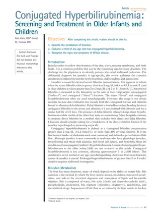
Figure 1. Bilirubin production in the reticuloendothelial system. RBC red blood cell prolonged jaundice to identify
those who have conjugated hyper-
of its constituents within the liver canaliculi and hepa- bilirubinemia and recognize the disorders that may be
tocytes, ultimately creating cholestatic damage to the amenable to early medical intervention (eg, galac-
liver. tosemia, urinary tract infection) or surgery (eg, biliary
Bilirubin is the product of heme breakdown in the atresia, choledochal cyst). In addition, early diagnosis
reticuloendothelial cells of the spleen and liver (Fig. 1). facilitates the institution of necessary nutritional and
The end product of this metabolic pathway is water- medical support to promote optimal growth and devel-
insoluble unconjugated bilirubin, which is bound to opment.
albumin in the circulation. Unconjugated bilirubin is The causes of cholestatic jaundice in the infant vary
taken up and metabolized in the liver to conjugated and can be divided into two primary categories: obstruc-
bilirubin (Fig. 2). Conjugated bilirubin is secreted into tive and hepatocellular. A detailed classification is listed
the biliary system by a specific transporter. Defects in in Table 1. The four most common causes of persistent
bilirubin conjugation cause unconjugated hyperbiliru- cholestatic jaundice in infants are discussed.
binemia (Gilbert syndrome and Crigler-Najjar syn-
dromes I and II). Hepatocellular disease can cause a Extrahepatic Biliary Atresia (EHBA)
mixed unconjugated and conjugated hyperbilirubinemia EHBA is the most common and serious cause of pro-
due to both impaired bilirubin conjugation and canalic- longed cholestatic jaundice in infants. It results from a
ular excretion. Defects in conjugated bilirubin excretion progressive and destructive inflammatory process that
cause isolated conjugated hyperbilirubinemia without affects both the extra- and intrahepatic biliary tree. The
cholestasis (Rotor and Dubin-Johnson syndromes). cause of EHBA has not been identified clearly. Two
Other mutations in membrane transporters of other or- clinical forms have been defined: an embryonic/fetal
ganic anions, such as bile acids, are linked with several form, which constitutes 20% of cases, and a perinatal/
diseases, including cystic fibrosis, adrenoleukodystrophy, acquired type, which comprises the remaining 80% of
and the familial intrahepatic cholestasis syndromes. cases. (1) The embryonic type has an earlier onset, has no
Once bile is excreted from the liver, it is stored in the jaundice-free interval, and is associated with other non-
gallbladder until a meal activates duodenal cholecystoki- hepatic anomalies or syndromic features, such as isolated
nin release and expulsion of gallbladder contents into the cardiovascular and gastrointestinal anomalies (intestinal
intestine. Conjugated bilirubin cannot be reabsorbed by malrotation, preduodenal portal vein, abdominal situs
intestinal epithelial cells and is degraded by intestinal inversus) and splenic anomalies (polysplenia, asplenia).
flora into stercobilin and urobilinogen, which are ex- The acquired type is not associated with other congenital
creted into stool. A small portion of conjugated bilirubin anomalies, usually occurs in an otherwise healthy term
is deconjugated by intestinal beta-glucuronidase. The infant, and has a jaundice-free interval followed by the
unconjugated bilirubin can be reabsorbed into the circu- development of jaundice in the first few postnatal weeks.
lation and returned to the liver, which is known as Both forms share the cardinal features of cholestatic
enterohepatic bilirubin circulation. The amount of bili- jaundice, hepatomegaly, and acholic stools.
rubin reabsorbed normally is very small, but it can be- EHBA was universally fatal before the Kasai hepato-
84 Pediatrics in Review Vol.28 No.3 March 2007
3. gastroenterology conjugated hyperbilirubinemia
rence has decreased as specific dis-
orders that cause a similar clinical
and histologic picture have been
identified (eg, alpha-1-antitrypsin
deficiency, defective bile acid syn-
thesis and transport). The diagno-
sis is made in infants who have pro-
longed cholestatic jaundice and
typical biopsy findings of disrupted
hepatic architecture, multinucle-
ated “giant” hepatocytes, focal he-
patocyte necrosis, expansion of
portal triads with inflammatory in-
filtrate, and extramedullary hema-
topoiesis in addition to the absence
of another disorder. Electron mi-
croscopy often is useful in diagno-
sis.
The prognosis of “idiopathic”
hepatitis is variable and depends on
Figure 2. Bilirubin conjugation. Movement of bilirubin (B) from the circulation into the whether a metabolic or infectious
hepatocyte occurs at the hepatocyte basolateral membrane with the help of a membrane
cause ultimately is diagnosed.
carrier protein. Binding of B to glutathione-S-transferase (GST) facilitates movement into
Jaundice usually resolves by 3 to 4
the rough endoplasmic reticulum (RER), where conjugation with glucuronic acid (GA) is
enabled by bilirubin ceridine diphosphate glucuronosyl transferase 1A1 (UGT1A1). The months of age; persistence of jaun-
conjugated bilirubin is excreted into bile at the hepatocyte canalicular membrane through dice beyond this age warrants addi-
a process mediated by the membrane-bound transporter cMOAT (canalicular multispecific tional evaluation.
organic anion transporter). Alb albumin, cytosol the fluid within a cell that contains the
cell’s oranelles, B-GA conjugated bilirubin (mono- and di-glucuronides). Adapted from Alagille Syndrome (AGS)
Gourley GR. Bilirubin metabolism. In: Walker WA, Goulet OJ, Kleinman RE, et al, eds. AGS also is known as syndromic
Pediatric Gastrointestinal Disease: Pathophysiology, Diagnosis, Management. 4th ed. bile duct paucity or arteriohepatic
Hamilton, Ontario, Canada: BC Decker, Inc; 2004:1344 –1362. dysplasia. It is an autosomally
dominant inherited with low pen-
portoenterostomy was introduced by Dr Morio Kasai in etrance disorder of bile duct paucity that occurs in con-
1959. This procedure establishes bile flow in up to 80% junction with syndromic extrahepatic findings.
of patients if performed prior to 60 days after birth. The The defect in AGS is a mutation of the Jagged 1 (JAG1)
success rate decreases as the infant’s age increases, with gene, which is mapped to chromosome 20p12 and en-
bile flow established in up to 45% of infants 60 to 90 days codes a ligand for the Notch signaling pathway, which is
of age and 10% of infants 90 to 120 days of age. (1) These important in cell fate determination. The diagnosis can
results underscore the importance of diagnosing this be made in the patient who has a marked reduction of
condition early. Approximately one third of patients intrahepatic bile ducts on liver biopsy in association with
require liver transplant in the first postnatal year, one other cardiac, ocular, skeletal, and facial abnormalities.
third require it in their teens, and one third live with The bile duct paucity may not be apparent in early
some liver function after the Kasai procedure into adult- infancy. AGS usually presents in the first 3 postnatal
hood. (1) It is estimated that approximately 50% of months and must be distinguished from biliary atresia
patients who have good results from the initial Kasai and other causes of nonsyndromic paucity. It may be
surgery still become transplant candidates later in life. diagnosed in older children who have persistent choles-
tatic jaundice and in adults after diagnosis in a related
“Idiopathic” Neonatal Hepatitis child.
“Idiopathic” neonatal hepatitis, also known as “giant In the original series described by Alagille in 1975, (2)
cell” hepatitis, used to be considered the most common 15 of 30 patients who had cholestatic jaundice and
cause of neonatal cholestasis. However, its relative occur- hepatic ductular hypoplasia with intact extrahepatic bile
Pediatrics in Review Vol.28 No.3 March 2007 85
4. gastroenterology conjugated hyperbilirubinemia
ducts had other common features.
Differential Diagnosis of Cholestatic
Table 1. These included a characteristic fa-
cies (prominent forehead, deep-set
Jaundice in the Infant eyes with mild hypertelorism,
Obstructive straight nose, and small, pointed
chin), a systolic murmur caused by
● Extrahepatic biliary atresia peripheral pulmonic stenosis, verte-
● Choledochal cyst bral arch defects, growth retarda-
● Spontaneous perforation of the bile duct tion, mild-to-moderate mental re-
● Inspissated bile
● Mass: stone, tumor tardation, and hypogonadism in
boys. Emerick and associates (3)
Hepatocellular studied 92 patients who had AGS
● Idiopathic neonatal hepatitis and found cholestasis in 96%, bile
● Disorders of the intrahepatic bile ducts duct paucity in 85%, cardiac mur-
– Alagille syndrome (arteriohepatic dysplasia/syndromic paucity of the
intrahepatic bile ducts) mur in 97%, vertebral anomalies in
– Nonsyndromic paucity of the intrahepatic bile ducts 51%, characteristic facies in 96%,
– Congenital hepatic fibrosis with bile duct cysts (Caroli disease) eye findings (posterior embryo-
● Metabolic disorders toxon) in 78%, and renal anomalies
– Disorders of amino acid metabolism in 40%. Minor features included
Tyrosinemia
– Disorders of lipid metabolism growth retardation (87%), mental
Gaucher disease retardation (2%), developmental
Niemann-Pick disease delay (16%), and pancreatic insuffi-
Cholesterol ester storage disease (Wolman syndrome) ciency (41%). Alagille and col-
– Disorders of carbohydrate metabolism leagues (4) have recommended that
Galactosemia
Hereditary fructose intolerance the diagnosis be made by confirm-
Glycogen storage disease ing the existence of cholestasis and
– Disorders of bile acid metabolism and transport excretion two of the other four abnormalities.
– Zellweger syndrome and other disorders of peroxisomal metabolism Factors that contribute significantly
– Disorders of bilirubin transport (do not cause cholestatic liver injury) to mortality in AGS include cardiac
Dubin-Johnson syndrome
Rotor syndrome disease (other cardiac defects be-
– Mitochondrial disorders sides peripheral pulmonic stenosis
– Alpha-1-antitrypsin deficiency such as tetralogy of Fallot), intra-
– Cystic fibrosis cranial hemorrhage, and progres-
– Neonatal iron storage disease sive liver disease.
● Endocrine disorders
– Hypothyroidism
– Hypopituitarism and septo-optic dysplasia Alpha-1-Antitrypsin
● Infectious Deficiency
– Sepsis (urinary tract infection, endotoxemia, enterocolitis) Alpha-1-antitrypsin is a member of
– TORCH infections (toxoplasmosis, cytomegalovirus, herpesvirus, rubella, syphilis) the serine protease inhibitor family
– Hepatitis B, non-typeable hepatitis
– Human immunodeficiency virus (the serpins) that protects the con-
● Drugs and Toxins nective tissue from degradation by
– Total parenteral nutrition inhibition of neutrophil elastase,
– Medications cathepsin G, and proteinase 3. Al-
– Fetal alcohol syndrome though lung disease associated with
● Other
– Vascular anomalies alpha-1-antitrypsin deficiency is at-
Budd-Chiari syndrome tributed to markedly reduced con-
Hepatoendothelioma/hemangioma centrations, liver disease results
– Cardiac insufficiency and hypoperfusion from retention of the abnormally
– Chromosomal abnormalities folded protein in the endoplasmic
Trisomy 21
Trisomy 18 reticulum (ER) of the hepatocyte
(the site of synthesis of most alpha-
86 Pediatrics in Review Vol.28 No.3 March 2007
5. gastroenterology conjugated hyperbilirubinemia
1-antitrypsin). Alpha-1-antitrypsin is a polymorphic pro- Conjugated hyperbilirubinemia results from obstruc-
tein that has allelic variants defined by isoelectric focusing tive or hepatocellular causes. Biliary stones and sludge
of the plasma and classified according to the protease can obstruct the common bile duct and cause subsequent
inhibitor (Pi) phenotype. Structural variants that have jaundice. Other causes of obstructive jaundice in this age
normal plasma concentrations or functional activity are group include parasitic infestations (ascaris, liver flukes),
known as PiM alpha-1-antitrypsin. Variants in which primary sclerosing cholangitis, choledochal cyst, and tu-
alpha-1-antitrypsin is not detected are known as null mors. Hepatocellular causes of conjugated hyperbiliru-
allelic variants and when inherited with another null binemia, such as viral or medication-related hepatitis, can
variant, are associated with early development of emphy- result in jaundice. Clinically apparent liver disease from
sema but no liver disease. Variants that have reduced chronic hepatitis B infection has decreased markedly due
alpha-1-antitrypsin activity include the PiZ and PiS. The to the effective implementation of the vaccine for this
PiZ homozygote is the condition associated most com- disorder. Signs of active hepatitis C usually occur in
monly with liver and lung disease. adults and are not discussed in detail in this article.
Alpha-1-antitrypsin deficiency is the most common Wilson disease and autoimmune hepatitis are relatively
genetic cause of acute and chronic liver disease in chil- uncommon causes of hyperbilirubinemia but are two
important entities for the
Alpha-1- antitrypsinofdeficiency ischronic
pediatrician to recognize
early because treatment
the that may prevent progres-
sion is available. Other
most common genetic cause acute and hepatocellular disorders
liver disease in children. are detailed in Table 2.
Wilson Disease
dren and the most common genetically caused disorder Wilson disease is an auto-
necessitating liver transplantation in children. Presenta- somal recessive disorder of copper homeostasis. The
tion of liver disease in ZZ homozygotes is variable. In a affected gene is on chromosome 13 and encodes a highly
review of 44 patients, Volpert and colleagues (5) re- conserved copper-transporting P-type adenosine
ported that the age of diagnosis of alpha-1-antitrypsin triphosphatase (ATP7b) that excretes copper into bile.
deficiency-associated liver disease was younger than 1 Copper is an essential trace element that participates in
month in 10 patients, 1 month to 1 year in 21 patients, cellular respiration, iron oxidation, pigment formation,
and older than 1 year in 13 patients. One of every 10 and antioxidant defense. It is absorbed from copper-rich
patients has prolonged jaundice in infancy, and 1 of 100 foods from the stomach and duodenum, bound to ceru-
develops cirrhosis and requires transplantation. (6) loplasmin in the circulation, and excreted by the liver
Other clinical presentations include neonatal hepatitis into bile.
syndrome, mild elevation of transaminase values in the The prevalence of Wilson disease is 1 in 30,000 and is
toddler, portal hypertension and cirrhosis in the child or equal among all ethnic groups. The condition presents in
adolescent, chronic hepatitis in the adult, and hepatocel- most patients as either hepatic or central nervous system
lular carcinoma in the adult. Diagnosis is made by dem- involvement. Liver disease occurs at an average age of
onstrating an abnormal Pi phenotype (PiZZ) and peri- 10 to 13 years in 45% of patients and rarely is seen before
odic acid-Schiff-positive, diastase-resistant globules in 3 years of age. Some 35% of patients present with neuro-
the ER of hepatocytes. logic signs (tremor, rigidity, dysarthria) a decade older
than patients who have hepatic involvement; 10% present
with psychiatric disturbances (depression, new-onset
Jaundice in the Older Child/Adolescent school problems, impulsive behavior); and 10% present
New-onset cholestatic jaundice in the older child and with other manifestations, including hemolytic anemia,
adolescent always requires additional investigation. It is Fanconi syndrome (glycosuria, aminoaciduria), and car-
essential to fractionate the bilirubin to differentiate cho- diomyopathy. Thus, patients who have Wilson disease
lestatic jaundice from unconjugated hyperbilirubinemia can have a mixed conjugated and unconjugated hyperbi-
due to hemolysis or defective bilirubin conjugation, as lirubinemia, depending on which disease manifestations
occurs in Gilbert syndrome. are present.
Pediatrics in Review Vol.28 No.3 March 2007 87
6. gastroenterology conjugated hyperbilirubinemia
basal ganglia and include parkinsonian symptoms. KF
Differential Diagnosis of
Table 2. rings often are present in patients who have neurologic
symptoms, but may be absent in patients who have liver
Cholestatic Jaundice in the disease.
Older Child and Adolescent Wilson disease is diagnosed in the patient who has
signs and symptoms consistent with the disease in addi-
Obstructive tion to laboratory findings of impaired hepatic copper
● Choledochal cyst metabolism. The serum ceruloplasmin concentration is
● Mass: stone, tumor, parasite decreased because copper is not conjugated to the apo-
Hepatocellular ceruloplasmin synthesized by the hepatocyte. The un-
● Autoimmune conjugated apoceruloplasmin is degraded rapidly. Mea-
– Autoimmune hepatitis surement of urinary copper in a 24-hour collection is
– Primary sclerosing cholangitis increased. A liver biopsy quantitating hepatic copper
– Primary biliary cirrhosis content is a helpful diagnostic tool, and a liver that has
● Disorders of the intrahepatic bile ducts
normal copper content excludes the diagnosis. A slitlamp
– Alagille syndrome (arteriohepatic dysplasia/
syndromic paucity of the intrahepatic bile ducts) examination can be performed to evaluate for KF rings.
– Nonsyndromic paucity of the intrahepatic bile Definitive genetic testing is now available for Wilson
ducts disease.
– Congenital hepatic fibrosis/Caroli disease Therapy is geared toward attaining and maintaining
● Metabolic disorders
normal copper homeostasis. Oral D-penicillamine and
– Wilson Disease
– Disorders of bilirubin transport trientine are copper chelators. Zinc acetate prevents the
– Dubin-Johnson syndrome absorption of copper from the gastrointestinal tract. Pa-
– Rotor syndrome tients are counseled to avoid copper-rich foods such as
– Mitochondrial disorders shellfish, legumes, nuts, chocolate, and liver. Liver trans-
– Alpha-1-antitrypsin deficiency
plantation is the treatment of choice for selected patients
– Cystic fibrosis
– Hemochromatosis who have either advanced liver disease or fulminant liver
● Endocrine disorders failure.
– Hypothyroidism
● Infectious Autoimmune Hepatitis (AIH)
– Sepsis (endotoxemia, enterocolitis)
AIH is an inflammatory hepatitis characterized by the
– Hepatitis A, B, C, E, non-typeable hepatitis
– Human immunodeficiency virus development of pathologic autoantibodies to normal
● Drugs and toxins host proteins and a dense mononuclear infiltrate in the
– Total parenteral nutrition portal tracts in the absence of another cause. AIH can be
– Medications subdivided into two general categories classified by the
● Other
type of autoantibodies produced: anti-nuclear antibody/
– Vascular anomalies
– Budd-Chiari syndrome anti-smooth muscle antibody (ANA/SMA) AIH, and
– Hemangioma anti-liver kidney microsomal antibody 1 (LKM1) AIH.
– Cardiac insufficiency and hypoperfusion In a study of 52 patients who had AIH, investigators
– Chromosomal abnormalities found that the median age at presentation of ANA/SMA
– Trisomy 21
AIH was 10.5 years and that for LKM1 AIH was 7.4
years. (7) Some 75% of patients were female. Three
patterns of presentation were noted:
Hepatic involvement ranges from asymptomatic 1) Acute hepatitis was the pattern in most patients
transaminitis to acute liver failure with jaundice, cirrho- having both types, with nausea, vomiting, anorexia, fa-
sis, hepatic necrosis, and encephalopathy. Liver biopsy tigue, and abdominal pain, followed by jaundice, dark
can show nonspecific findings of steatosis and glycogen urine, and pale stools. Duration of illness varied from
deposition. Micronodular cirrhosis and piecemeal necro- 2 weeks to 2 months in this group.
sis also can be seen. Kaiser-Fleischer (KF) rings, repre- 2) Another group (30%) had the insidious onset of
senting copper deposition in Descemet’s membrane, are disease of longer duration (6 mo to 2 yr), with relapsing
visible on a slitlamp examination of the eye. Neurologic jaundice, progressive fatigue, headache, anorexia, and
symptoms are attributed to copper deposition in the weight loss.
88 Pediatrics in Review Vol.28 No.3 March 2007
7. gastroenterology conjugated hyperbilirubinemia
3) A small percent of patients, who had no prior biliary atresia, choledochal cyst, or gallstone disease.
history of jaundice, had complications of portal hyper- Additionally, the birth and perinatal histories, past med-
tension. ical and surgical histories, family history (including con-
AIH is suspected on the basis of clinical characteristics sanguinity), medication and dietary histories, social ac-
and demonstration of the autoantibodies, as well as an tivity and school performance histories, and travel history
elevated immunoglobulin G value. Definitive diagnosis is should be sought.
made on liver biopsy, where the typical histologic picture
is a dense mononuclear infiltrate invading the hepatic Physical Examination
parenchyma (periportal hepatitis), with periportal necro- The clinician should note if the patient is well- or ill-
sis. In most cases, the disease responds well to immuno- appearing as well as irritable or drowsy. Both signs may
suppressive therapy. Urgent liver transplantation is indi- indicate encephalopathy, infection, or metabolic de-
cated if the patient presents in acute fulminant hepatic rangement. Microcephaly in the infant may indicate con-
failure. genital infection. Recognition of dysmorphism is valu-
able. Eyes should be examined for posterior embryotoxon
Evaluation of the Jaundiced Patient or KF rings. The systolic murmur of peripheral pulmonic
Although jaundice is relatively common in the first stenosis, usually heard in the back as well as the front,
2 weeks after birth and is observed frequently in new- suggests AGS. Hepatomegaly typically is present, but a
borns, jaundice in the older infant and child always is small liver may indicate cirrhosis and end-stage liver
abnormal and requires more investigation. Additional disease. Splenomegaly, ascites, and prominent vascula-
evaluation of children who have conjugated hyperbiliru- ture such as caput medusa suggest portal hypertension
and chronic liver disease.
An infant’s diaper should
The keyjaundice is fractionation of when
evaluating
laboratory test to obtain
the
be examined for pale
stools and dark urine.
Neurologic evaluation
should be undertaken for
ataxia and asterixis.
bilirubin.
Laboratory Tests
The key laboratory test to
binemia and chronic liver disease should involve looking obtain when evaluating jaundice is fractionation of the
for the complications of cholestasis, such as coagulopathy, bilirubin. Indirect or unconjugated hyperbilirubinemia
fat malabsorption, ascites, and encephalopathy, to initiate usually indicates excessive red blood cell destruction at
appropriate therapy. Finally, a child who has conjugated any age. Direct or conjugated hyperbilirubinemia indi-
hyperbilirubinemia or evidence of chronic liver disease cates a hepatobiliary disorder. Hepatic transaminase con-
should be referred to a pediatric gastroenterologist. centrations are elevated in the presence of hepatocellular
injury. The alkaline phosphatase and gamma glutamyl
History transferase values often are increased with obstructive
The age of the patient and history of presentation give conditions. Liver function, including prothrombin time,
important clues to the cause of the jaundice. Some albumin, and cholesterol, should be measured. The remain-
conditions of infantile cholestasis and conjugated hyper- der of the evaluation should be tailored to the specific
bilirubinemia present early in life, such as biliary atresia, patient. Hemolytic anemia can be seen in patients who have
AGS, and inherited metabolic disorders. Others often Wilson disease. Thyroid function tests can be obtained if
manifest beyond infancy, such as AIH and Wilson dis- hypothyroidism is suspected. Other age-specific tests may
ease. Diseases such as cystic fibrosis and alpha-1- be considered, including TORCH (toxoplasmosis, rubella,
antitrypsin deficiency can present as either neonatal cho- cytomegalovirus, herpes simplex) titers, blood and urine
lestasis or later in life as chronic liver disease. cultures, alpha-1-antitrypsin Pi phenotype, iron profile,
Signs such as poor feeding, irritability, and vomiting chloride sweat test, urine-reducing substances (galac-
may be associated with a metabolic condition, such as tosemia), and a metabolic screen in young infants. Testing
galactosemia, or suggest encephalopathy. The presence for Wilson disease (ceruloplasmin) or for AIH is appropriate
of acholic stools suggests an obstructive process such as in older children.
Pediatrics in Review Vol.28 No.3 March 2007 89
8. gastroenterology conjugated hyperbilirubinemia
Imaging min concentrations with supplementation. Management
Real-time ultrasonography is an important diagnostic of the complications of chronic liver disease and portal
tool in the evaluation of the jaundiced patient. The hypertension in conjunction with a pediatric gastroenterol-
absence of a gallbladder on a fasting examination in an ogist is essential. All children, particularly those who have
infant is suggestive but not diagnostic of biliary atresia. chronic liver disease, should be immunized, including both
Ultrasonography may demonstrate gallstones, chole- hepatitis A and B, using the guidelines of the American
dochal cyst, or ascites. A Doppler ultrasonographic study Academy of Pediatrics. It is appropriate to refer any child
of the portal circulation may identify portal hypertension who evidences acute, severe liver disease or a chronic liver
or portal vein thrombosis. Hepatobiliary scintigraphy condition to a pediatric transplant center for evaluation.
with technetium-labeled iminodiacetic acid analogs may
help differentiate obstructive jaundice from nonobstruc-
tive causes. In the case of obstructive jaundice, the he- References
patic uptake of tracer is normal, but there is no intestinal 1. Balistreri WF, Grand R, Hoofhagle JH, et al. Biliary atresia:
excretion. In cases of severe hepatocellular disease, up- current concepts and research directions. Hepatology. 1996;23:
1682–1692
take into the liver is delayed, but tracer eventually is
2. Alagille D, Odievre M, Gautier M, et al. Hepatic ductular
`
excreted into the intestine. Premedication with pheno- hypoplasia associated with characteristic facies, vertebral malforma-
barbital for 3 to 5 days prior to the study enhances biliary tions, retarded physical growth, mental and sexual development,
excretion and imaging of the isotope. and cardiac murmur. J Pediatr. 1975;86:63–71
3. Emerick KM, Rand EB, Goldmuntz E, et al. Features of Alagille
syndrome in 92 patients: frequency and relation to prognosis.
Liver Biopsy Hepatology. 1999;29:822– 829
Ultimately, many patients require a liver biopsy for de- 4. Alagille D, Estrada A, Hadchouel M, et al. Syndromic paucity of
finitive and reliable diagnosis. The liver biopsy can be interlobular bile ducts (Alagille syndrome or arteriohepatic dyspla-
performed percutaneously, with or without ultrasono- sia): review of 80 cases. J Pediatr. 1987;110:195–200
graphic guidance, or surgically. In addition to evaluation 5. Volpert D, Molleston JP, Perlmutter DH. Alpha-1-antitrypsin
deficiency-associated liver disease progresses slowly in some chil-
by an experienced pathologist for specific histologic fea-
dren. J Pediatr Gastroenterol Nutr. 2000;31:258 –263
tures, liver tissue can be used to quantify iron and copper 6. Carrell RW, Lomas DA. Alpha1-antitrypsin deficiency: a model
content and for electron microscopy to detect certain for conformational diseases. N Engl J Med. 2002;346:45–53
metabolic conditions. 7. Mieli-Vergani G, Vergani D. Immunological liver diseases in
children. Semin Liver Dis. 1998;18:271–279
Management of Jaundice
Treatment is directed at the specific underlying disorder,
although persistent cholestasis generally can cause reten- Suggested Reading
Gitlin JD. Wilson disease. Gastroenterology. 2003;125:1868 –1877
tion of bile acids, bilirubin, and cholesterol; decreased
McLin VA, Balistreri WF. Approach to neonatal cholestasis. In:
excretion of bile acids into the intestine with resulting fat Walker WA, Goulet OJ, Kleinman RE, et al, eds. Pediatric
malabsorption; and hepatocellular damage that eventu- Gastrointestinal Disease: Pathophysiology, Diagnosis, Manage-
ally causes portal hypertension and end-stage liver dis- ment. 4th ed. Hamilton, Ontario, Canada: BC Decker Inc;
ease. Some general principles apply to the management 2004:1079 –1091
Moyer V, Freese DK, Whitington PF, et al. Guideline for the
of these consequences independent of the specific cause.
evaluation of cholestatic jaundice in infants: recommendations
These principles include optimizing nutrition by em- of the North American Society for Pediatric Gastroenterology,
ploying the use of medium-chain triglyceride-containing Hepatology and Nutrition. J Pediatr Gastroenterol Nutr. 2004;
formula or supplements and monitoring fat-soluble vita- 39:115–128
90 Pediatrics in Review Vol.28 No.3 March 2007
9. gastroenterology conjugated hyperbilirubinemia
PIR Quiz
Quiz also available online at www.pedsinreview.org.
1. Conjugated hyperbilirubinemia is:
A. A marker of accelerated hemoglobin breakdown.
B. A normal finding in otherwise healthy adolescents.
C. Always linked with cholestasis.
D. Less common than unconjugated hyperbilirubinemia.
E. Synonymous with direct hyperbilirubinemia.
2. A 4-week-old breastfeeding boy is jaundiced and has a total bilirubin concentration of 13 mg/dL
(222.3 mmol/L). The laboratory test that maximizes diagnostic efficiency is:
A. A complete blood count.
B. A reticulocyte count.
C. Bilirubin fractionation.
D. Gamma glutamyl transferase.
E. Hepatic transaminase.
3. A 4-week-old breastfeeding boy has become increasingly jaundiced. The pregnancy was unremarkable.
Delivery was at term, and the infant was appropriate for gestational age. The jaundice was not noted in the
hospital. Findings on the physical examination, other than jaundice, are unremarkable. Today, the total
bilirubin concentration is 13 mg/dL (222.3 mmol/L), with a direct fraction of 6 mg/dL (102.6 mmol/L). Of
the following, the condition that is most likely ruled out by these findings is:
A. Alagille syndrome.
B. Alpha-1-antitrypsin deficiency.
C. Extrahepatic biliary atresia.
D. Neonatal hepatitis.
E. Physiologic jaundice.
4. A previously healthy 15-year-old girl develops jaundice and fatigue. She does not complain of colicky
abdominal pain associated with meals. She has had no known exposure to hepatotoxins. Aside from
jaundice and appearing mildly ill, findings on her physical examination are unremarkable. Initial laboratory
evaluation reveals a bilirubin concentration of 11 mg/dL (188.1 mmol/L) with a direct fraction of 4 mg/dL
(68.4 mmol/L), and elevated hepatic transaminases, but no evidence of Epstein-Barr or hepatitis A, B, or C
virus. As suspected from the history, ultrasonography reveals a normal gallbladder and biliary tree. Serum
ceruloplasmin and autoantibody results are inconclusive. Pi typing reveals PiMM, and the patient proceeds
to liver biopsy. Copper content of the sample is normal. Of the following, the patient is most likely to have:
A. Alpha-1-antitrypsin deficiency.
B. Autoimmune hepatitis.
C. Choledochal cyst.
D. Gilbert syndrome.
E. Wilson disease.
5. Wilson disease is diagnosed definitively through:
A. Genetic testing.
B. Liver biopsy.
C. Serum ceruloplasmin concentrations.
D. Slitlamp examination.
E. Urinary copper excretion.
Pediatrics in Review Vol.28 No.3 March 2007 91