
Recomendados
Recomendados
Mais conteúdo relacionado
Mais procurados
Mais procurados (20)
Semelhante a TTT conference presentation ENGLISH
Semelhante a TTT conference presentation ENGLISH (20)
Mais de Mills Cbst
Mais de Mills Cbst (19)
Último
Último (20)
TTT conference presentation ENGLISH
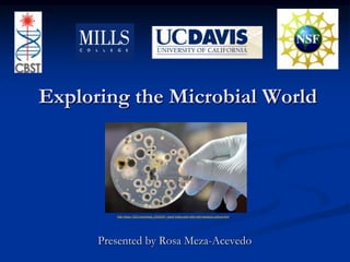
- 1. Exploring the Microbial World http://www.123rf.com/photo_5320347_hand-holds-petri-dish-with-bacteria-culture.html Presented by Rosa Meza-Acevedo
- 2. Center for Biophotonics, Science and Technology (CBST) National Science Foundation UC Davis Mills College
- 4. Illumination
- 5. study of light energy
- 6. Microscopic imagingDefinition: the study of the interaction of light with biological material- where “light” includes all forms of radiant energy whose quantum unit is the photon
- 7. Exploring the Microbial World Sample microbial environment Control microbes Transform E. coli with Green Fluorescent Protein (GFP)
- 8. Culture Sample Media Media contain nutrients Liquid and semisolid forms Luria Broth (LB) Agar Petri plate http://www.bestliveshopping.com/toys-165795011-B004MKHNJK-Pre_made_LB_Agar_Plate_10_plates
- 9. How to make LB agar plates? Materials Erlenmeyer flask Sodium chloride (NaCl) Yeast extract Tryptone Agar powder Deionized (DI) water Sterile Petri plates
- 10. Tools for Sterilization LB Agar http://www.missingfeatures.com/2007/06/25/building-a-better-microwave/ http://www.sz-wholesale.com/shenzhen_China_products/Pressure-Cooker_1.htm Microwave Pressure cooker http://sites.google.com/site/scienceprofonline/Lab-gradeAutoclave2.JPG Autoclave
- 11. Agar handling guidelines Label bottom only with name, date and initials (ex/ LB agar 11/4/11 RM) When pouring, store agar plates right side up When solidified, store in refrigerator up side down in container http://www.benchfly.com/video.php?video=164
- 12. Sampling Microbial Environment Worksheet Introduction Hypothesis Prediction (if/then) Procedure Materials Methods
- 13. How to sample? Sample collection is the most important step! Unidentified organisms Contamination Swab replicates
- 14. II. Controlling Microbes Controlling microbes worksheet Introduction Hypothesis Prediction (if/then) Procedure Materials Method
- 15. Procedure Antibiotics Ampicillin Neomycin Streptomycin Tetracycline Erythromycin Penicillin http://www.debendiagnostics.co.uk/Antibiotic.html
- 16. Antiseptics Iodine Alcohol Disinfectants Sapolio Clorox http://www.bio.miami.edu/hare/culture.html
- 17. Learning Outcomes! At the end of this session, students will have an appreciation of &/or be able to: Introduction to microbiology and bacteriology Differentiate between fungi and bacteria growth characteristics Importance of antibiotic selection and resistance genes Impact of disinfectants and antiseptics in controlling microbes
- 18. III. Transform E. coli with Green Fluorescent Protein (GFP) Transformation Introduce foreign DNA pGLO plasmid GFP
- 19. Green Fluorescent Protein (GFP) Jelly fish Aequoreavictoria Visual marker http://brainwindows.wordpress.com/2008/10/08/2008-nobel-prize-in-chemistry-to-gfp/
- 20. GFP changes genetic makeup
- 21. GFP changes genetic makeup http://askville.amazon.com/genetically-blue-eyed-parents-produce-brown-eyed-child/AnswerViewer.do?requestId=2969837 E. coli glowing colonies E. coli colonies
- 22. Transform E. coli with Green Fluorescent Protein (GFP) Why teach? Important modern biotechnology technique Central framework of molecular biology DNA RNA Protein trait/physical characteristic
- 23. Advantages Compatible with 50 min class periods Serves entire class of 32 students Up to 4 students per group Striking results! pGLO Bacterial Transformation Kit
Notas do Editor
- Talk about me graduate to mills college
- Label molecules so they can see inside the cell … red fluorescent tag that would allow researcher to locate particular molecules using advance microscopes under cells
- Definition: the study of the interaction of light with biological material- where “light” includes all forms of radiant energy whose quantum unit is the photon
- Today’s session includes three experiments. First, we are going to sample microbes from your environment of interest. Second, we are going to control microbes using antiseptics, antibiotics, and disinfectants. Third, I am going to teach you how we can use microbes for our advantage by transforming them with green fluorescent protein (GFP). Now put some gloves on and spray your benches with ethanol to clean the area that you are going to be working on.
- Before we even get into the actual experiment, I have to teach you how to make the media necessary in preparation for all of the three experiments that we are going to cover today. You as teachers are going to be able to do this in your classroom or lab space. The media that you will making is a rich culture sample media that contains nutrients necessary for the growth of invisible organisms that you are going to sample. So, there are different types of media but I’ll teach you how to make the semisolid form. What you see in the right corner is a Luria Broth agar petri plate. Most commonly known as LB plate.
- To make LB Agar plates you can use a beaker, an Erlenmeyer flask, or any glassware resistant to heat of any size or shape as long as it has more then enough space to hold your solution. For instance, if you are making 250 ml LB agar you would have to use a 500 ml flask. Then, you follow the protocol written in your worsheet. Mix all the ingredients, then put foil on top and place it into the pressure cooker. You are going to be pouring the agar into these petri plates that come in packs of sterile sleeves.
- You can use these tools for stereilization of the agar. However, I prefer the pressure cooker if you don’t have an autoclave because it does not require constant monitoring.
- Once placed into the pressure cooker you can use this time to label plates. In front of you, you have two plates labelled with the name of the media called LB agar, the date that it was made, and my initials. Now, using a sharpie you are going to label the bottom of both plates with the area that you are going to be sampling later on, the date and your initials.Another agar handling guidelines is that when pouring LB agar plates, store them right side up. Once they have been solidified, you can store the plates in a refrigerator up side down. In this picture you can see the difference between a plate that has been solifidied and one that has been recently been poured.
- Grab your Experiment I worksheet. As part of your introduction, I have added the terminology used in describing bacterial colonies so you can teach your students the different types of colony shapes, colony margins and colony surface characteristics. Today you are going to be studying an environment of your choice, testing for the presence of bacteria and fungi. Now I want you to think about places that you are interested in sampling. Common places to sample. For instance, bathroom, door handles, a wound or a part of their body.Once you have chosen what to sample, hypothesize about the growth of the specie present in the environment of your choice. Then, make a prediction based on your hypothesis using if/then. Remember that a Hypothesis is a testable statement, an educated guess. A prediction is that if the hypothesis is true you will find lot of microbes on the plate o no microbes at all. For instance, “the janitor has clean the bathroom really well ” is a hypothesis. A prediction, if the janitor has clean the bathroom really well, then I will not find any bacterial colonies growing on my plate.Now let’s get into the procedure. In front of you, you have sterile cotton swabs foiled with aluminium and two LB labelled plates. Hypothesis where students think where microbes can exists … what you think is true “there will be more bacteria on my computer keyboard than on my hands after I wash them” not good too broadTry to get a look into invisible world that was not imagining before microscope take one minute and talk to your neighbor and formulate a hypothesis about what would you expect from the environment that you are sampling from
- Since the sample collection is the most important step, before you actually sample you environment of interest you have to remember thatYou are working with unidentified organisms .. They are too small to seeYou have to prevent contamination from carrying you cotton swab all over until you actually swab the plate. Use the foil that has been covering your sterile cotton swab to cover it if neccesary.I want you to sample two plates identically so they can be replicates. It is important that you do this twice in two different plates bc you are going to need a control sample for your next expermiment. Be careful no to break the surface of the agar and poke holes. Gently grab and spread it around the surface of the plate. Then, you can wait at from 1-3 days depending of the temperature where you store the plates to see results. For instance, we are going to analyze these plates that I have here with me. I’m going to let you take the plates so you can analyze them at home. Also, in the worksheet you are going to be able to find questions in the results and discussion section that you can use as homework for your students.
- Now grab your experiment II worksheet. You can combine Experiment I and II if you want. However, it is important to remember to formulate your hypothesis and prediction for this experiment as well.
- Depending on the size of the LB petri dish, you are going to choose the number of agents of your choice. For instance, in this small plate we are going to be using two agents. You should pick one from a different group.I have brought several antibiotics such as …..
- , antiseptics such as … and disinfectants such as ….Remember that an antibiotic will kill certain bacteria, an antiseptics will kill bacteria in your skin, and disinfectant can be use to clean surface or furniture.So, if you choose an antibiotic agent, dispense the disk onto the surface of the agar. If you choose an antiseptic or disinfectant. Pick up a sterile filter disk using sterile tweezers. Then, dip the filter halfway into one of the chemical solutions. Let the solution diffuse into the disk. Make sure the disk is not too wet before placing it onto the surface of the agar. Remember to keep disks away from the edge of the plate and once they are placed onto the agar, they should not be move.So, grab one of your replicate plates and follow the protocol. The other plate will be used as your control. Once they have been incubated for a few days, you can expect a zone of inhibition, which is represented by the clear area around the disk. If the bacteria is resistant to the agent chosen, then no zone of inhibition will be present. If the bacteria is susceptible to the agent chosen, then a zone of inhibition will be present.In the worksheet you are going to be able to find instruction for calculating the zone of inhibtion as well as questions for homework.
- There are many microbes that are important to your body (mention that)
- Now the third experiment consist on transforming E. coli with Green Fluorescent protein, most commonly known as GFP. When I say transformation that means that we are going to be introducing a foreign DNA into E. coli. This foreign DNA in this case is called plasmid pGLO. This plasmid contains genes such as GFP.
- GFP was isolated from a jelly fish called Aequorea victoria. Since it fluoresces bright green when exposed to blue light, it helps to track a trait inside a cell. GFP is an important tool for many scientistist because is works as a visual marker so we basically can label whatever we want.
- GFP changes the genetic makeup of the DNA. Since proteins are extremely small and cannot be seen normally, when attaching GFP to them it makes a protein fluoresce. Here you can see a 3D version of the protein after it has been translated from the DNA, that’s how the gene normally will look like. However, if we transform the DNA into this GFP gene before the stop codon or just after the promoter in the beginning of the gene, the protein that is coded by the DNA then is coded green.