
Recommended
More Related Content
What's hot
What's hot (20)
Viewers also liked
Viewers also liked (20)
Similar to Section 1, chapter 15: anatomy of the heart
Similar to Section 1, chapter 15: anatomy of the heart (20)
More from Michael Walls
More from Michael Walls (20)
Recently uploaded
Recently uploaded (20)
Section 1, chapter 15: anatomy of the heart
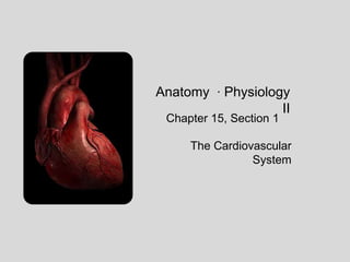
- 1. Anatomy ∙ Physiology II Chapter 15, Section 1 The Cardiovascular System
- 2. FUNCTIONS OF THE HEART It pumps 7000L (1800 gallons) of blood through our body every day. The heart contracts 2.5 billion times in a lifetime.
- 3. The heart is composed of two pumps The systemic circuit carries blood to the body The pulmonary circuit carries blood to the lungs
- 4. the heart is located within the mediastinum It is about the size of a fist (14cm x 9cm) 2/3 of the heart is left of the midline 1/3 2/3
- 5. The heart is posterior to the sternum Base attachment of major vessels 2nd intercostal space Apex Pointed inferior margin 5th intercostal space
- 6. The heart is surrounded by a pericardial membrane. The fibrous pericardium forms a thick outer layer of connective tissue. The parietal pericardium is a serous membrane attached directly to the fibrous layer. A visceral pericardium is a serous membrane that forms the outer layer of the heart wall. The pericardial cavity contains serous fluid.
- 7. The wall of the heart contains 3 layers The epicardium is also called the visceral pericardium The myocardium contains a thick layer of cardiac muscle, with blood vessels and nerves The endocardium is a smooth layer of squamous epithelium that lines the heart chambers and valves
- 8. The heart contains 4 chambers The right atrium receives blood from the body The left atrium receives blood from the lungs The right ventricle pumps blood towards the lungs The left ventricle pumps blood towards the body Interventricular septum
- 9. A pocket on each atrium, called the auricle increases the capacity of the atria.
- 10. blood enters the heart through the great veins The superior vena cava returns blood from the upper body to the heart Four pulmonary veins return blood from the lungs to the heart The coronary sinus returns blood from the myocardium to the heart The inferior vena cava returns blood from the lower body to the heart
- 11. Great arteries carry blood away from the heart The aorta delivers oxygenated blood to the systemic circulation The pulmonary trunk* delivers deoxygenated blood to the lungs * The pulmonary trunk immediately divides into a left and right pulmonary
- 12. Left and right AV valves prevent backflow into the atria. *AV = atrioventricular The tricuspid valve guards the right AV orifice The bicuspid (mitral) valve guards the left AV orifice
- 13. AV valves are anchored to the ventricles by chordae tendineae Chordae tendineae anchored to the cusps papillary muscles Papillary muscles contract to pull the valves tightly shut Mitral Valve Prolapse – cusp of the mitral valve protrudes into atrium. Symptoms include: chest pain, heart palpitations, and fatigue.
- 14. Semilunar valves prevent backflow of blood into the ventricles A pulmonary valve is located at the base of the pulmonary trunk An Aortic valve (not shown) is located at the base of the aorta
- 15. Each cusp of a semilunar valve is shaped like a crescent moon
- 16. path of blood through the right heart 1. Blood enters right atrium through the SVC, IVC, and coronary sinus 1 2. It passes through the tricuspid valve into the right ventricle 4 4 3. Blood is pumped from the right ventricle, through the pulmonary valve, and into the pulmonary trunk. 2 4. Blood passes into the pulmonary arteries towards the lungs 3 1
- 17. path of blood through the left heart 5. Oxygenated blood is returned to the heart through 4 pulmonary veins. 6. Blood enters the left atrium. 9 7. Blood passes through the bicuspid valve into the left ventricle. 8. The left ventricle pumps blood through the aortic valve into the aorta. 9. Blood enters systemic circulation to the tissues throughout the body. 9 9 8 5 5 6 7
Editor's Notes
- Of course 2.5 billion times refers to a person who has lived 100 years old! You're not their yet.
- The pulmonary circuit receives O2 from the lungs, and releases CO2 from the blood.The systemic circuit releases O2 to the tissues , and receives CO2 produced by the cells.
- The mediastinum contains many important structures, including the heart, pericardium, trachea, esophagus, along with blood vessels and nerves.
- The fibrous pericardium is attached to the diaphragm, the sternum, and the vertebrae.The visceral layer is folded over at the great vessels, and gives rise to the parietal layer. Both layers are serous membranes, which means they secrete serous fluid.
- Purkinje fibers are located along the endocardium. They initiate cardiac muscle contractions, which we’ll discuss in the next section.
- Deoxygenated blood from systemic circulation enters the right atrium.The right ventricle pumps the blood into pulmonary circulation, towards the lungs.The left atrium receives oxygenated blood from pulmonary circulation.The left ventricle pumps the oxygenated blood into systemic circulation.
- The superior diaphragm drains systemic circulation from regions above the diaphragm.The inferior diaphragm drains systemic circulation from regions below the diaphragm.
- Notice the pulmonary trunk and pulmonary arteries are blue. The blue indicates deoxygenated blood in these illustrations. The coloring does NOT refer to arteries or veins.
- The AV valves are open when blood fills the ventricles. The AV valves shut as the ventricles contract.
- Semilunar valves are pushed open when the ventricles contract. As they relax, the weight of blood left in the aorta and pulmonary trunk pushes the valves shut.