Exocrine glands histology
•
15 gostaram•5,108 visualizações

Classification of glands. Detailed microscopic structure of exocrine glands. differences between serous and mucus acini. Microscopic structure of Parotid, submandibular and sublingual glands.

Recomendados
Mais conteúdo relacionado
Mais procurados
Mais procurados (20)
Semelhante a Exocrine glands histology
Semelhante a Exocrine glands histology (20)
Mais de Mehul Tandel
Último
Último (20)
Exocrine glands histology
- 1. Glands
- 2. Competencies • AN70.1 - Identify exocrine gland under the microscope & distinguish between serous, mucous and mixed acini • AN52.1 Describe & identify the microanatomical features of ……. Suprarenal gland
- 4. Objectives • Introduction of Glands • General Features • Development • Classifications • Examples • Applied
- 5. Gland - Introduction • Some epithelial cells converted in to specialized cells to perform a secretory function - form glands • Definition: An organ of secretion made up of specialized secretory cells; derived from surface epithelium on which it opens
- 6. General Features of Glands • Can be present as “Discrete Organ” or “ in the layers of viscera” • Epithelial in origin (Derived from surface epithelium) • Functional unit of gland is formed by specialized secretory cells known as – Secretory End Piece • Fluid secreted by glands contain enzymes, mucus, hormones, protein, fat etc…
- 7. General Features of Glands • Rate of secretion is modulated by Nervous & Hormonal influence • Secretory end piece of some exocrine glands is surrounded by star shaped contractile cells that lies between cells & basement membrane –Called Myo-epithelial cells –Share features of both epithelium & muscle cell –Help in expulsion of secretions
- 9. Development of Glands Developed as cords of epithelial cells from the surface of membrane Invaginates in to underlying Connective tissue & Form 2 parts Proximal Part Distal Part
- 10. Development of Glands Form duct Connect secretory end piece with surface of epithelium Proximal Part Distal Part Exocrine Glands Differentiated in to the secretory cells Secretory End Piece Pours the secretion through ducts / directly on surface of the glands or epithelium
- 11. Development of Glands DisappearProximal Part Distal Part Endocrine Glands Form islands of the secretory cells permeated / surrounded by the blood capillaries Pours their secretion in to directly in to blood through the blood capillaries Ductless Gland *
- 12. Based on Site of Secretion Exocrine With Ducts Endocrine Ductless Exocrine + Endocrine Paracrine Classification of Glands Unicellular / Multicellular Cord & Clump type/ Follicular Type Pancreas & Liver APUD & DNES APUD – Amine Precursor Uptake & Decarboxylation DNES - Diffuse NeuroEndocrine System
- 13. Classifications of Exocrine Glands Based on numbers of cell Unicellular Multicellular Goblet Cell
- 14. Multicellular Exocrine Glands 1. Based on branching pattern of ducts Simple No Branching Compound Branched
- 15. Multicellular Exocrine Glands 2. Based on Shape of Secretory End Piece Tubular Alveolar / Acinar Tubulo- alveolar
- 16. Multicellular Exocrine Glands 3. Based on Nature of Secretion Mucous Serous Mixed / Sero-mucous
- 17. Multicellular Exocrine Glands 4. Based on Manner of Secretion Merocrine Apocrine Holocrine Cytocrine
- 18. Multicellular Exocrine Glands 5. Based on Development of gland Ectodermal Endodermal Mesodermal
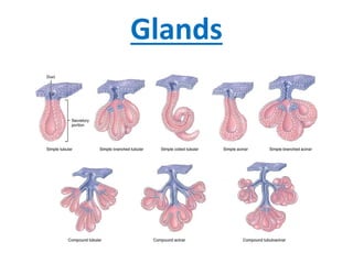
- 19. Exocrine glands Based on number of ducts • Simple(single duct) • Compound(minor & major ducts) Based on shape of secretory piece: • Simple tubular/ alveolar • Simple branched tubular/ alveolar • Simple coiled tubular • Compound tubular/ acinar • Compound tubulo-alveolar / acinar
- 20. • Simple: – Secretion poured to surface by un-branched duct • Compound: – Duct divides in to branches to form elaborated / complex duct system; – Each smaller terminal duct receive secretion from it’s own secretory end piece – These ducts unite to form larger ducts which finally drain on to surface Based on Number of Ducts:
- 21. • Tubular: (Simple / Compound) – Secretory end piece is like tubule – Straight, branched or coiled Based on Shape of Secretory End Piece: Simple Straight Intestinal Crypts Simple Coiled Sweat Gland Simple Branched Uterine gland Fundic & pyloric glands of stomach Compound Cardiac glands of stomach & Brunner’s gland of duodenum
- 22. • Alveolar: (Simple / Compound) – Secretory end piece Flask shaped with large lumen – Un-branched or branched Simple Un-branched Urethral Glands Simple Branched Sebaceous & Tarsal Glands Compound Mammary gland betn puberty & 1st Pregnancy Based on Shape of Secretory End Piece:
- 23. • Acinar: (Mostly Compound) – Secretory end piece Round shaped with small lumen – Mostly branched Compound Acinar Pancreas Parotid Based on Shape of Secretory End Piece:
- 24. • Tubulo – Alveolar / Acinar: – Combination of both Tubular & alveolar / Acinar – Mostly branched Compound Tubulo – Alveolar / Acinar Sublingual gland Submandibular gland Lactating mammary gland Based on Shape of Secretory End Piece:
- 25. Simple Glands
- 26. Compound Glands
- 27. According to Mode of secretion: • Merocrine • Apocrine • Holocrine • Cytocrine Exocrine glands
- 28. • Merocrine: – A.K.A.: Eccrine / Epicrine – Secretion discharged through intact cell membrane – By Exocytosis – No loss of cytoplasm Based on Mode of Secretion: E.G. Protein content of mammary gland Parotid Pancreas Typical & Atypical sweat glands
- 30. • Apocrine: – Apical portion (luminal) of cell disintegrate to discharge its secretion – Nucleus & basal portion remain intact from which cell can regenerate – Partial loss of cytoplasm Based on Mode of Secretion: E.G. Lipid content of mammary gland Ceruminous gland of Ear Moll’s Gland in Eyelid Modified Sweat gland
- 32. • Holocrine: – Entire cell disintegrate to discharge its secretion – Result in death of cell – Complete loss of cytoplasm Based on Mode of Secretion: E.G. Sebaceous gland Tarsal Glands in Eyelid
- 34. • Cytocrine: – Cell are released as secretion Based on Mode of Secretion: E.G. Spermatozoa from Testis Ovum from Ovary Bone Marrow
- 37. According to Nature of secretion: • Serous • Mucous • Mixed Exocrine glands Mucous Serous Mixed
- 38. Serous Thin, watery Proteinaceous Zymogen granules Central rounded Small Lumen Indistinct Darkly stained Enzymatic action Parotid Gland Mucous Thick, viscous Mucopolysaccharides Mucinogen droplets Flat & peripheral Large Lumen Distinct Lighly stained Protection & lubrication Sublingual gland Consistency Content Cytoplasm Nucleus Lumen Cell boundaries H&E Staining Function Examples
- 39. Serous Acinus / Gland • Secretion: Thin, watery, – Rich in enzymes, protein in nature • Cells: Pyramidal & Small with Indistinct boundaries • Cytoplasm: Granular (Zymogen) – Rich in rER – Stain darkly with H/E stain • Nuclei: rounded & basal / central • Size of lumen: Small • Function: Enzymatic • E.G.: Parotid & Lacrimal glands
- 40. • Secretion: Thick Viscous – Which collects in the apical part • Cells: Low Columnar & Large with distinct boundaries • Cytoplasm: Mucinogen droplets – Stain very lightly with H/E stain & – Looks empty • Nuclei: Flattened & basal (mucoid pushes nuclei towards base) • Size of lumen: Large • Function: Protection & Lubrication • E.G.: Sublingual glands Mucous Acinus / Gland
- 41. • Contain both serous & mucous secretory units • Sometimes serous cells form crescentic caps on mucous acini called as Serous Demilunes Mixed Acinus / Gland
- 42. Serous Acini Mucous Acini
- 43. Serous Acini Mucous Acini
- 44. Salivary Glands
- 45. • Exocrine: with Ducts / Externally secretory gland • Secrete its products on to the surface through it’s duct • Produce saliva and pour their secretion in the oral cavity • Contain water, mucus, proteins, salts, salivary amylase(ptyalin) and IgA • Functions: • Moisten food • Lubricate & moisten oral cavity • Initiate digestion of carbohydrates Salivary Glands
- 46. Two types: • Major (paired) • Parotid • Submandibular • Sublingual • Minor • Lingual in tongue • Palatine in palate & tonsil • Labial in lips and • Buccal in cheeks Salivary Glands
- 48. • Consist of 3 components – Secretory end piece – Duct System – Supporting Connective tissues Salivary Glands Parenchyma Stroma Parenchyma
- 49. Structural Organization of Salivary Glands Lobes Lobules Blood Vessels & Nerves Parenchyma Stroma Inter-lobar Septa Capsule Ducts Inter-lobular Septa
- 50. General Architecture of a Salivary Glands • Parenchyma- – Secretory part- Acini / tubules / tubulo-acinar – Conducting part: Ducts- Intralobular, interlobular, main excretory duct • Stroma- – Capsule – Septa (interlobular, interlobar) – Loose inter-lobular connective tissue supporting the parenchyma – Divide the gland into lobes and lobules.
- 51. Salivary Glands :- Parenchyma - Secretory part • Compound tubulo-alveolar glands – Racemose gland • Blind sac composed of secretory cells • Acini / tubules / tubulo- acinar • Based on nature of secretion 3 Types: – Serous – Mucous – Mixed
- 52. Salivary Glands:- Parenchyma -Conducting part Secretory Acinus Intercalated ducts Low cuboidal / Squamous epithelium Secrete HCO3 ¯ & Absorb Cl ¯ Prominent in serous glands
- 53. Parenchyma - Conducting part Secretory Acinus Intercalated duct Simple Columnar epithelium Regulates of water and electrolyte contents of saliva Presence of basal striation Striated duct
- 54. Cell of Striated Duct Infoldings of basal plasma membrane Occupied by mitochondria Basal Striation
- 55. Parenchyma - Conducting part Secretory Acinus Intercalated duct Simple columnar to Pseudo-stratified Columnar Striated duct Inter-lobular duct Stratified Columnar / cuboidal Lobular & Main Excretory duct Near opening Stratified squamous
- 56. Parenchyma - Conducting part Low cuboidal / Squamous epithelium Simple Columnar epithelium Stratified Columnar / cuboidal
- 58. • Development of duct system of the salivary gland depends on the nature of secretion • Predominantly serous secreting gland: well developed duct system –Modification of secretion occurs • Predominantly mucous secreting gland: poorly developed duct system –No modification of secretion Parenchyma - Conducting part
- 59. Salivary Glands - Section Large number of closely packed acini with ducts scattered between them Supported by connective tissue septa containing blood vessels, lymphatics and nerves Homocrine & Heterocrine ?
- 60. Salivary Glands Both acini and ducts surrounded by contractile cells Myo-epithelial cells • Star shaped / Basket shaped around acini & • Fusiform around ducts
- 62. Salivary Glands - Classification • Parotid gland: – Only serous acini • Submandibular & sublingual: – Submandibular: • Predominantly serous secreting • Serous + mucous + mixed acini – Sublingual: • Predominantly mucous secreting • Mucous + mixed acini (Pure serous acini are rare) Purely serous Mixed glands
- 63. Parotid
- 64. Parotid
- 68. Submandibular
- 71. Submandibular
- 74. Clinical • ADENOMA: Benign tumors arising in the gland • ADENOCARCINOMA: Malignant growth in the gland