1. Introduction
Salmonella spp. are enteric pathogens notable for their ability to
cause a range of diseases including gastroenteritis, septicaemia,
osteomyelitis, pneumonia, meningitis, and arthritis1. The food
borne pathogen Salmonella enterica and various members of
the familyEnterobacteriaceae areabletoformbiofilmondifferent
biotic and abiotic surfaces2-3. A bioûlm is a group of
microorganisms that attach to each other and to a biotic or abiotic
surface, resulting in stability and protection from environmental
factors mediated in part by a self-initiated exopolysaccharide
(EPS) matrix4. Biofilms can become a persistent source of
contamination5 with increased ability to colonize and survive in
a harsh condition6. The formation of biofilms involve multiple
processes including initial surface attachment, monolayer
formation, migration to form multilayered microcolonies,
production of extracellular matrix and biofilm maturation with a
three dimensional architecture7. Asmall number of bacterial cells
adhere to the surface, a process facilitated by bacterial motility.
Cells that attach irreversibly to the surface divide, forming
microcolonies, that produce extracellular polymeric substances
(EPS), primarily polysaccharides8. The EPS attaches the cells to
the surface and stabilizes the colonies. With time, attached
Original Article
Bangladesh J Microbiol, Volume 31, Number 1&2, June-December 2014, pp 35-39
bacteria from the biofilm detach and disperse in order to survive
and colonize new niches9.
Understanding the formation of biofilms is important for their
control. Biofilms are less susceptible to antimicrobials than are
planktonic cells10. Bacteria within a biofilm are more resistant to
environmental factors because of limited availability of key
nutrients5,11 andowingtotheextracellularmatrix8.Withinabiofilm
there is reduced diffusion, physiological changes due to reduced
growth rates and production of enzymes which degrade
antimicrobial substances8 leading to increased resistance. Biofilms
are a concern in the food industry as they can lead to illness,
disease outbreaks with subsequent economic losses12. In the
medical field, bacterial biofilms are worrying concerns because
they can occur on the surfaces of medical devices and on tissue
surfaces within compromised organs13. Biofilms grow similarly in
the environment and in industrial systems14. In the present study,
the ability of clinical isolates of Salmonella enterica serovarTyphi
and Paratyphi to form biofilm in vitro was investigated. To our
knowledgethisconstitutesthefirstworld-widereportofthebiofilm
forming ability by clinical Salmonella enterica serovar Paratyphi.
Previous reports on biofilm-forming S. enterica serovar Paratyphi
were based on reference bacteria rather than on clinical isolates.
In vitro Biofilm FormationAbility of Clinical Isolates of Salmonella enterica
SerovarsTyphi and Paratyphi
MaishaMalihaMorium1
, SunjuktaAhsan1*
, M Shahidul Kabir2
, Marufa ZerinAkhter1
andMFaridulIslam3
1Department of Microbiology, University of Dhaka, Dhaka 1000, Bangladesh, 2Department of Microbiology, Stamford University Bangladesh, Dhaka
1217, Bangladesh, 3Department of Microbiology, Square Hospital, Panthapath, Dhaka 1205, Bangladesh
In the present study the ability of clinical isolates of Salmonella enterica serovars Typhi (n = 30) and Paratyphi
A (n = 11) to form biofilm on abiotic surface was investigated. All isolates were found capable of biofilm
formation in a microtitre plate assay. Upon optimization of biofilm formation by the test isolates, Adherence
test medium (ATM) was found to be the best medium for biofilm formation by both S. enterica serovars Typhi
and Paratyphi. Growth was optimized by incubation at 37°C in an orbital shaker set at 150 rpm for 48-72
hours. Biofilms were best detected when washed with PBS (1X), stained with crystal violet (1%) and
subsequently washed with acetone (33%). Optical density (OD) readings were better correlated with growth
at 570 nm when compared to 600 nm. Of the 28 Salmonella Typhi isolates, 17 (61%) were very strong biofilm
producers, 8 (29%) were strong biofilm producers and 3 (11%) were moderate biofilm producers. On the other
hand, out of 13 S. Paratyphi, 9 (69%) were very strong biofilm producers, 3 (23%) were strong biofilm formers
and 1 (8%) was a moderate biofilm producer. None of them were weak biofilm producers. The present study
raises concern from a public health point of view because the ability of the clinical isolates to form biofilm
would indicate their ability of being transmitted from abiotic surface to uninfected host giving rise to
disease.
Keywords: Biofilm, Salmonella enterica Typhi, Paratyphi, Planktonic
*Corresponding author:
Sunjukta Ahsan, Department of Microbiology, University of Dhaka, Dhaka 1000, Bangladesh.
Tel: +880 (02) 9661920-73; E-mail: sunjukta@du.ac.bd
2. MaterialsandMethods
Bacterial isolates
Twenty-eight Salmonella enteric serovar Typhi and 13 S. enterica
serovar Paratyphi clinical isolates obtained from a hospital in
Dhaka City, Bangladesh were used in this study. The isolates
were confirmed by biochemical and serological tests.
Optimization of incubation conditions for maximum biofilm
formation
(i) Media optimization
In spite of the fact that one earlier report has suggested the use
of adherence test medium (ATM) for Salmonella enterica serovar
Typhi15 a number of media were tested for their ability to support
maximum biofilm formation by the test strains. This was deemed
necessary as strain variations may cause differences in
requirement. The media used in this study included tryptic soy
broth (TSB), Mueller-Hinton broth (MHB), Luria-Bertani broth
(LB), Luria-Bertani broth with 5 g/l D-glucose (LB + glucose) and
adherence test media (ATM).
(ii) Incubation condition optimization
In all conditions of optimization, incubation temperature of 37°C
was used. Both static and shaker incubators were used. In shaker,
two different shaking conditions, viz., 120 and 150 rpm were used.
Time of incubation was also varied and biofilm production was
observed after 24, 48 and 72 h incubation.
(iii) Biofilm staining and washing condition optimization
Non-adherentcellswerewashedwithtwodifferentwashingsolution,
viz., double distilled H2O and 1X PBS (phosphate buffer saline).
Biofilmwasstainedwithcrystalvioletattwodifferentconcentrations
of0.03and1%for30min.Todetachbiofilmfromwallofthetube,two
differentsolutionswereused,viz.,80:20=ethanol:acetoneand33%
aceticacid,whichwereappliedfor15min.
(iv) Wave length and absorbance optimization
In case of 80:20 = ethanol:acetone solution absorbance was taken
at 600 nm. In case of 33% acetic acid solution absorbance took at
570 nm.Absorbance was obtained after 24, 48 and 72 h incubation
in ELISAplate reader.
Bacteriological medium
Adherence test medium (ATM) with slight modifications was
adapted following optimization of biofilm formation and medium
was prepared as described earlier15. This medium contained 60
mM NaCl,20mMKCl,111mMglucoseand30mM NaHCO3.The
pH was adjusted to 8.4. To this a supplement containing NH4Cl
(20 mM), Na2HPO4 (40 mM), (NH4)H2PO4 (50 mM), CaCl2 (999
µM), MgCl2 (980 µM), FeCl3 (86 µM) and Na2SO4 (40 mM) was
added separately.
Biofilm assay
The ability of the bacterial strains to form bioûlms in polystyrene
(PS) microtitre plates was evaluated by using the method
described elsewhere16 with some modifications. Colonies from
an overnight plate was grown in adherence test medium (ATM) at
37°Cinanorbitalshaker(120rpm)toobtainaMacFarlandstandard
of 0.5 equivalents of cells. From this, 200 µl of bacterial suspension
was transferred into a well of a sterile 96-well PS microtitre plate
and the plate was then incubated for 48 hours at 37°C, 120 rpm to
allowbiofilmformation.Planktonicbacteriawereremovedandeach
well was washed thrice with phosphate buffered saline (dissolving
8gofNaCl,0.2gofKCl,1.44gofNa2HPO4,0.24gofKH2PO4 in1
l distilled H2O and pH adjusted to 7.4) to remove loosely attached
cells. Subsequently, 200 µl of 1% (wt/vol) crystal violet solution
(Sigma-Aldrich/ Life Science Chemilab SA,Athens, Greece) was
added into each well, and the plate was then incubated for 30 min
at room temperature.After being washed three times with 200 µl of
PBS to remove excess stain, the crystal violet was solubilised in
200 µl of 33% acetate solution. Dye absorbance at 570 nm was
measured using a microtitre plate reader (Sunrise, Tecan,
Männedorf, Switzerland). Each experiment (isolate) was done in
triplicate wells. Salmonella enteric serovar Typhimurium ATCC
14028 was used as a positive control for biofilm formation and
sterileATM were used as negative controls. When the absorbance
values of the crystal violet bound to the sample bacteria was shown
at least twice that of the control it was considered as positive
result for bioûlm formation. Based on the binding of crystal violet
by the biofilm forming bacteria the isolates were categorized as
very strong (VS) biofilm producers, strong (S) biofilm producers
and moderate (M) biofilm producers using a modified convention
described earlier17.
Results
Optimized incubation conditions for biofilm formation
Following optimization, best biofilm production was obtained in
ATM medium at 37°C in 150 rpm shaker after an incubation time
of 48 h.Among the two washing solutions used, 1X PBS was the
better washing solution. For staining of biofilm, 1% crystal violet
worked better than the lower concentration of 0.03% when used
for 30 min. As detachment solution, 33% acetic acid solution
worked better than ethanol:acetone = 80:20. Absorbance was
taken at 570 nm after 48 h of incubation. Figure 1 depicts the
OD600 values when using the optimized conditions for biofilm
formation. The results are mean of four readings and are expressed
as mean ± 1 standard deviation.
Biofilm formation by the S. enterica Typhi and Paratyphi isolates
All isolates of S. enterica Typhi and Paratyphi were tested in
triplicate were found to be capable of forming biofilm to different
extents. The degree of crystal violet retention is an indication of
the number of bacteria in the biofilm which bind to crystal violet.
Hence, the absorbance at 570 nm correlates positively with the
number of biofilm producer. The cut-off OD was taken as two
standard deviations above the mean value of negative control.
Figure 2 represents the mean OD values of the crystal violet
retained by the Salmonella Typhi isolates and the controls and
Figure 3 depicts that retained by S. enterica Paratyphi.
Morium et. al
36
3. 0
0.5
1
1.5
2
2.5
3
ODat570nm
Figure 1. Biofilm formation under optimized condition (ATM
medium incubated at 37°C in an orbital shaker adjusted to 150
rpm and incubated for 48-72 hours. Biofilms were washed with
PBS, 1X, stained with crystal violet, 1% and washed with
acetone, 33%). All experiments were carried out four times.
Results are mean OD570 ± SD.
Figure 2. Absorbance of crystal violet OD570 by clinical isolates
of Salmonella enterica serovar Typhi biofilms in modified ATM.
Results are mean of three readings ± 2 SD.
Figure 3. Absorbance of crystal violet OD570 by clinical isolates of Salmonella enterica serovar Paratyphi biofilms in modified
ATM. Results are mean of three readings ± 2 SD.
Categorization of the isolates on the basis of strength of biofilm
formation
The isolates were classified as follows: non-producing, weak,
moderate, and strong-producing, based on the following optical
density (OD) average values: OD (Isolate) d” OD (Control) =
Non-biofilm-producer; OD (Control) d” OD (Isolate) d” 2 OD
(Control)=Weakbiofilmproducer;2OD(Control)d”OD(Isolate)
d” 4 OD (Control) = Moderate biofilm producer; 4 OD (Control)
d” OD (Isolate) = Strong biofilm producer. We modified these
interpretive criteria by adding yet another category, which we
described as 8 OD (Control) d” OD (Isolate) =Very strong biofilm
producer. In this case, the category ‘strong biofilm producer’
wasdescribedas4OD(Control)d”OD(Isolate)d”8OD(Control)
= Strong biofilm producer. According to this categorization, of
the 28 S. enterica Typhi isolates, 17 (61%) were very strong (VS)
biofilm producers, 8 (29%) were strong (S) biofilm producers and
3 (11%) were moderate (M) biofilm producers (Table 1). On the
other hand, 9 (69%) of the S. enterica Paratyphi were very strong
(VS) biofilm producers, 3 (23%) were strong (S) biofilm formers
and 1 (8%) was a moderate (M) biofilm producer.
Biofilm Formation by Clinical Salmonella
37
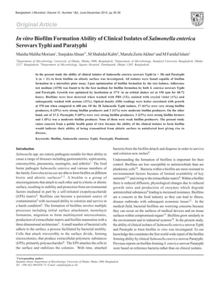
4. Microscopy of a sample biofilm
A sample of the biofilm was observed under the light microscope
(100x) following crystal violet staining. The micrograph indicated
clumps of biofilm bacteria (Figure 4).As negative control, blank
medium was used instead of bacterial culture. The micrograph
which reflected a blank view has not been shown here.
formation for both S. enterica Typhi and Paratyphi was observed
inATM medium at 37°C under vigorous aeration (150 rpm). This
finding is similar to that of Raza et al.15 who modified ATM
medium slightly for optimum biofilm formation by S. enterica
Typhi only.
There are several reports that describe the ability of Salmonella
to form biofilms on abiotic surfaces outside the host, such as
stainless steel19, plastic20, rubber21, glass2, cement22, marble and
granite14.All these surfaces are commonly encountered in farms,
slaughter houses, food industries and kitchens which raise the
risk for public health. It is strongly believed that the ability of
Salmonella to form biofilms on inanimate surfaces contributes
to its survival and persistence in non-host environments and its
transmission to new hosts. However, adhesion by Salmonella is
strain-dependent and probably influenced by surface structures,
such as cell wall and membrane proteins, fimbriae, flagella and
polysaccharides19,23-24 has reported on the ability of reference
S. enterica Typhi and Paratyphi isolates to form biofilm in
microtitre plate.
Conclusion
The significance of the present study lies in the fact that all
isolates were clinical in origin and most of them were very strong
biofilm producers. This raises the chance of formation of biofilms
by clinical Salmonella enterica serovars Typhi and Paratyphi on
abiotic surfaces, a condition which is of public health significance
since attached bacteria on commonly used plastic surfaces can
aid transmission to uninfected hosts and gives rise to disease.
Persistence of such biofilm bacteria on abiotic surfaces may form
the basis of future studies.
References
1. Chiu C, Su L and Chu C. 2004. Salmonella enterica Serotype
Choleraesuis: Epidemiology, pathogenesis, clinical disease, and
treatment. Clin Microbiol Rev. 17(2): 311-322.
2. Prouty A and Gunn J. 2003. Comparative analysis of Salmonella
enterica serovar Typhimurium biofilm formation on gallstones and
on glass. Infect Immun. 71(12): 7154-7158.
3. Ledeboer NA and Jones BD. 2005. Exopolysaccharide sugars
contribute to biofilm formation by Salmonella enterica serovar
Typhimurium on HEp-2 cells and chicken intestinal epithelium. J
Bacteriol. 187(9): 3214-3226.
Table 1. Categorization of the Salmonella enterica Typhi and S. Paratyphi isolates in terms of biofilm forming ability
Isolate Biofilm category OD control/OD sample
Salmonella Typhi isolates (n = 28)
S. Typhimurium VS 8 OD (Control) d” OD (Isolate)
S8,13,14,24,25,27,30,38,40,42,51,52,54,55,56,57,58 VS 8 OD (Control) d” OD (Isolate)
S4, 9, 20, 32, 34, 35, 45 and 49 S 4 OD (Control) d” OD (Isolate) d” 8 OD (Control)
S2,22,23 M 2 OD (Control) d” OD (Isolate) d” 4 OD (control)
Salmonella Paratyphi isolates (n = 13)
S1,10,12,26,28,29,36,37,50 VS 8 OD (Control) d” OD (Isolate)
S11,18,21 S 4 OD (Control) d” OD (Isolate) d” 8 OD (Control)
S19 M 2 OD (Control) d” OD (Isolate) d” 4 OD (Control)
VS = Very strong; S = Strong; M = Moderate.
Discussion
Salmonellae are recognized worldwide as major zoonotic
pathogens for both humans and animals. Most microorganisms
persist in a biofilm ecosystem and not as free-floating organisms.
The ability of Salmonella to attach to food surfaces was the first
published report on food-borne bacterial biofilms18.
In the present study, all investigated S. enterica Typhi and
Paratyphi isolates were found to be capable of forming biofilms.
Different culture media, incubation conditions solutions were
used for biofilm washing and detachment. The best biofilm
Figure 4. Light micrograph of a representative biofilm forming
Salmonella enterica serovar Typhi.
Morium et. al
38
5. 4. Crawford RW, Gibson DL, Kay WW and Gunn JS. 2008. Identification
of a bile-induced exopolysaccharide required for Salmonella biofilm
formation on gallstone surfaces. Infect Immun. 76(11): 5341-5349.
5. Van Houdt R and Michiels CW. 2010. Biofilm formation and the
food industry, a focus on the bacterial outer surface. J Appl Microbiol.
109(4): 1117-1131.
6. Monier JM and Lindow S. 2003. Differential survival of solitary and
aggregated bacterial cells promotes aggregate formation on leaf
surfaces. Proc Natl Acad Sci USA. 100(26): 15977-15982.
7. Steenackers H, Hermans K, Vanderleyden J, Sigrid CJ and de
Keersmaecker. 2012. Salmonella biofilms: An overview on occurrence,
structure, regulation and eradication. Food Res Int. 45: 502-531.
8. Lewandowski Z. 2000. Structure and function of biofilms. In Biofilms:
Recent Advances in Their Study and Control (Evans LV ed), pp 1-17.
Harwood Academic Publishers, Amsterdam.
9. Kaplan JB. 2010. Biofilm dispersal: Mechanisms, clinical implications,
and potential therapeutic uses. J Dent Res. 89(3): 205-218.
10. Merritt JH, Kadouri DE and O’Toole GA. 2005. Growing and analyzing
static biofilms. Cur Prot Microbiol. Chapter 1: Unit 1B.1.
11. Condell O, Iversen C, Cooney S, Power KA, Walsh C, Burgess C and
Fanning S. 2012. Efficacy of biocides used in the modern food industry
to control Salmonella enterica, and links between biocide tolerance
and resistance to clinically relevant antimicrobial compounds. Appl
Environ Microbiol. 78(9): 3087-3097.
12. Crull K, Rohde M, Westphal K, Loessner H, Wolf K, Felipe-López A,
Hensel M and Weiss S. 2011. Biofilm formation by Salmonella
enterica serovar Typhimurium colonizing solid tumours. Cell
Microbiol. 13(8): 1223-1233.
13. Wong HS., et al. 2010. Comparative susceptibility of Salmonella
Typhimurium biofilms of different ages to disinfectants. Biofouling.
26(7): 859-864.
14. Rodrigues D, Teixeira P, Oliveira R and Azeredo J. 2011. Salmonella
enterica Enteritidis biofilm formation and viability on regular and
triclosan-impregnated bench cover materials. J Food Protection.
74(1): 32-37.
15. Raza A, Sarwar Y, Ali A, Jamil A, Haque A and Haque A. 2011. Effect
of biofilm formation on the excretion of Salmonella enterica serovar
Typhi in feces. Int J Infect Dis. 15(11): e747-e752.
16. van Merode AE, van der Mei HC, Busscher HJ, Waar K and Krom BP.
2006. Enterococcus faecalis strains show culture heterogeneity in
cell surface charge. Microbiology. 152(Pt 3): 807-814.
17. Stepanoviæ S, Vukoviæ D, Hola V, Bonaventura GD, Djukiæ S,
Æirkoviæ I and Ruzicka F. 2007. Quantification of biofilm in
microtiter plates: Overview of testing conditions and practical
recommendations for assessment of biofilm production by
staphylococci. APMIS. 115(8): 891-899.
18. Duguid J, Anderson E and Campbell I. 1966. Fimbriae and adhesive
properties in Salmonellae. J Path Bacteriol. 92(1): 107-137.
19. Agarwal R, Singh S, Bhilegaonkar K and Singh V. 2011. Optimization
of microtitre plate assay for the testing of biofilm formation ability
in different Salmonella serotypes. Int Food Res J. 18(4):1493-1498.
20. Iibuchi R, Hara-Kudo Y, Hasegawa A and Kumagai S. 2010. Survival
of Salmonella on a polypropylene surface under dry conditions in
relation to biofilm-formation capability. J Food Protection. 73(8):
1506-1510.
21. Arnold J and Yates I. 2009. Interventions for control of Salmonella:
Clearance of microbial growth from rubber picker fingers. Poultry
Sci. 88(6): 1292-1298.
22. Joseph B, Otta S, Karunasagar I and Karunasagar I. 2001. Biofilm
formation by Salmonella spp. on food contact surfaces and their
sensitivity to sanitizers. Int J Food Microbiol. 64(3): 367-372.
23. Chia T, Goulter R, McMeekin T, Dykes G and Fegan N. 2009.
Attachment of different Salmonella serovars to materials commonly
used in a poultry processing plant. Food Microbiol. 26(8): 853-859.
24. Oliveira K, Oliveira T, Teixeira P, Azeredo J, Henriques M and Oliveira
R. 2006. Comparison of the adhesion ability of different Salmonella
enteritidis serotypes to materials used in kitchens. J Food Protection.
69(10): 2352-2356.
Biofilm Formation by Clinical Salmonella
39