
Recomendados
Mais conteúdo relacionado
Semelhante a Magnetic Resonance Imaging Factsheet.pdf
Semelhante a Magnetic Resonance Imaging Factsheet.pdf (20)
Mais de MabelWright1
Mais de MabelWright1 (17)
Último
Último (20)
Magnetic Resonance Imaging Factsheet.pdf
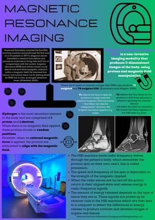
- 1. Raymond Damadian invented the first MRI scanning machine and preformed the first scan on a live patient in 1977 to diagnose cancer. Damadian’s research into sodium and potassium molecules in living cells lead him on to experiment with the nuclear magnetic resonance (NMR) first (where atomic nuclei absorb and emit electromagnetic radiation within a magnetic field). He theorised that tumour and normal tissue can be distinguished by NMR due to their ‘prolonged relaxation times’ (Wakefield, 2000). Magnetic Resonance Imaging Magnetic Resonance imaging (MRI) is a non-invasive imaging modality that produces 3-dimensional images of the body, using protons and magnetic field manipulation Hydrogen is the most abundant element in the body and are comprised of 1 proton and 1 electron. When there is no magnetic field applied, these protons situate in random positions. However, when an external magnetic force is applied, the protons are stimulated to align with the magnetic field. The MRI machine emits radio-frequency waves through the patient’s body, which stimulates the protons spin on their own axis’s, this is called precession. The speed and frequency of the spin is dependent on the strength of the magnetic applied. When the radio waves are turned off the proton return to their aligned state and release energy in radio-frequency signals. The amount of energy released depends on the type of tissue they are in. These signals are picked up by receiver coils in the MRI machine which are then sent to a computer to detect the differences in energy release to produce contrast and detailed images of organs and tissues (National Institute of Biomedical Imaging and Bioengineering, n.d.) There are 2 types of contrast images that MRIs can produce: T1 weighted and T2 weighted MRI: (Kawahara and Nagata, 2021) T1 reflects the time it takes for protons to realign with the external magnetic field (recovery) = the faster the time for realignment/ recovery, the brighter the tissue appears e.g. water T2 reflects the time taken for the protons to shift from spinning in phase to spinning out of phase (decay) = the faster the time for relaxation, the darker the tissue appears on the MRI scan e.g. bone