GHH Muscle Tissues
•
2 likes•678 views

General Histology and Histotechnique 2012 - 2013 Student made handout Credits to the original owner of the pictures used.

Recommended
More Related Content
What's hot
What's hot (20)
Viewers also liked
Similar to GHH Muscle Tissues
Similar to GHH Muscle Tissues (20)
Recently uploaded
Recently uploaded (20)
GHH Muscle Tissues
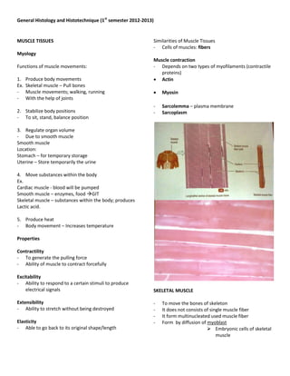
- 1. General Histology and Histotechnique (1st semester 2012-2013) MUSCLE TISSUES Similarities of Muscle Tissues - Cells of muscles: fibers Myology Muscle contraction Functions of muscle movements: - Depends on two types of myofilaments (contractile proteins) 1. Produce body movements Actin Ex. Skeletal muscle – Pull bones - Muscle movements; walking, running Myosin - With the help of joints - Sarcolemma – plasma membrane 2. Stabilize body positions - Sarcoplasm - To sit, stand, balance position 3. Regulate organ volume - Due to smooth muscle Smooth muscle Location: Stomach – for temporary storage Uterine – Store temporarily the urine 4. Move substances within the body Ex. Cardiac muscle - blood will be pumped Smooth muscle – enzymes, food GIT Skeletal muscle – substances within the body; produces Lactic acid. 5. Produce heat - Body movement – Increases temperature Properties Contractility - To generate the pulling force - Ability of muscle to contract forcefully Excitability - Ability to respond to a certain stimuli to produce electrical signals SKELETAL MUSCLE Extensibility - To move the bones of skeleton - Ability to stretch without being destroyed - It does not consists of single muscle fiber - It form multinucleated used muscle fiber Elasticity - Form by diffusion of myoblast - Able to go back to its original shape/length Embryonic cells of skeletal muscle
- 2. General Histology and Histotechnique (1st semester 2012-2013) - Striations are present Dense irregular connective - Nucleus is found at the Peripheral – at the sides tissue - Attached to the bones c. Endomysium I. Connective Tissue components of skeletal muscle - 1 bundle of muscle - Covers the individual muscle fiber 1. Fascia – (Bandage) - Areolar tissue - Fibrous connective tissue that is deep to the skin - Separate muscle fibers from each others - Surrounds muscles and other organs of the body. 2 types: a. Superficial fascia or the subcutaneous layer - separate muscles from skin Made up of: - Areolar connective tissue - Adipose tissue Functions: 1. It provides a pathway for nerves and Blood vessels to enter and exits muscles. 2. Stores most of the triglycerides 3. Serves as good insulating layer; reducing heat loss – due to Connective tissue 4. Protects muscles from physical trauma b. Deep fascia - A dense irregular connective tissue Functions: - Lines the body wall and limbs - Holds muscles together - Allows free movement of muscles - Carries Blood vessels, nerves, Lymphatic vessels - Fills the spaces between muscles 2. Three layers of connective tissue - Extend from the deep fascia to further protect and strengthen skeletal muscle a. Epimysium - Encircles the whole muscle/covers the muscle Basic features of skeletal muscle - Dense irregular connective tissue Nerves and blood vessels b. Perimysium - Each skeletal muscle is supplied by branches of one - Encircles bundles of muscles nerve, one artery, one or more veins Wrapped by Fascicle - Nerves and vessels branch repeatedly 10-100 or more muscle; - Smallest nerve branches serve Individual muscle fibers
- 3. General Histology and Histotechnique (1st semester 2012-2013) Individual muscle fibers Tissue sheaths – extend to from the tendon - attaches muscles to bone. Neuromuscular junction - A place in the body where nerves meet a muscle. - A synapse the motor neuron and the muscle fiber. Muscle attachments - Most skeletal muscles run from one bone to another b. The sarcolemma of the fiber encloses sarcoplasm - One bone will move – other bone remains fixed and myofibrils which are striated Origin – less movable part of the bone Insertion – more movable attachment c. A sac of sarcoplasm is reticulum wraps around each myofibril - Composed of a triad: is a two terminal cisterns and a T (transverse tubule). Histological features Microscopic organization and development a. Embryonic development - Fusion of myoblasts into muscle fibers Myoblast – fused to form the muscle fibers - After fusion, still with satellite cell, immature muscle fiber will lose their potential to divide hence, satellite cell retains it. - It cannot have any cell division Satellite cell – forms the muscle Functions: - Acts as a reserved population of cells - Whenever skeletal muscle is in its normal state it is inactive or quiescent; if it is injured it becomes active. - It continues to divide in order to repair and grow more muscle fibers. - Repair and maintenance of skeletal muscle
- 4. General Histology and Histotechnique (1st semester 2012-2013) - The cytoplasm of muscle fibers - Contains a substantial amount of glycogen and myoglobin Oxygen binding protein which is red colored globins and found only in muscle fibers. 3. Myofibrils - Contractile element of muscles - Contains overlapping thick and thin filaments - With prominent striations Alternating line and dark bands 4. Sarcoplasmic Reticulum - Fluid-filled system of membranous sacs that Structure of a skeletal muscle fiber encircles each myofibrils Function: - Releases Calcium ions to trigger muscle contraction 5. Sarcomere - Basic functional unit of myofibril - Compartment of filaments inside the myofibrils 6. Filaments - Within the myofibrils of two types: a. Thin filaments - 8 nm in diameter - Protein: Myosin b. Thick filaments Sarcoplasmic reticulo Triad - 16 nm in diameter - Protein: Actin 1. Sarcolemma - Muscle fibers plasma membrane which is 7. Mitochondria perforated with thousands of tiny invagination - Small and less numerous skeletal muscle fiber called triad. - For cell respiration Two terminal sisters: - Powerhouse of the cell a. Terminal cisterns - For the synthesis of ATP - Structure madeup of two strands on either strands of T tubule. Zones and bands of sarcomere: b. T tubules - These are tunnels from the surface towards the 1. A-band – anisotropic center of each muscle fiber. - Darker middle portion Function of Triad: - Where thick filaments (myosin) are located To ensure that all parts of the muscle fiber become - Toward the end of the A band is a zone of overlap: excited by an action potential virtually simultaneously. where thick and thin filaments lie side by side 2. Sarcoplasm
- 5. General Histology and Histotechnique (1st semester 2012-2013) 2. I-band – isotropic - Lighter dense area - Where thin filaments (actin) extend - No thick filaments 3. Z-line - Distinct dark line running down the middle of the I- band - Where thin filaments are attached end to end - Zone of apposition (increase in diameter) of actin filaments belonging to two neighboring sarcomeres 4. M-line - So named because it is at the middle of sarcomere - Where thick filaments are attached end-to-end in the center of the A-band - Band of connections between myosin filaments - Supporting proteins that hold thick filaments together at the center of H-zone. 5. H-band - Appears along the middle of A-band, between the free ends of the thin filaments when muscles are stretched. - Zone of myosin filaments only (no overlap with actin filaments) within the A-band CARDIAC MUSCLE II. Cardiac muscle tissue - Principal muscular tissue in the heart wall - Shorter in length and larger in diameter - Exhibit branching which gives an individual fiber and y-shaped appearance - Nucleus centrally located
- 6. General Histology and Histotechnique (1st semester 2012-2013) Histological parts: - Found in wrap around sheets that form part of the 1. Sarcolemma walls of smaller arteries and veins and hollow - Plasma membrane surrounding the cardiac muscle viscera such as the stomach, intestines, uterus, and urinary bladder. 2. Sarcoplasm - The cytoplasm 2. Multiunit smooth muscle tissue - Consists of individual fibers, each with its own 3. Mitochondria motor neuron terminals and with few gap junctions - Larger and more numerous between neighboring fibers - Found in the walls of large arteries, in airways to 4. T-tubules the lungs, in arrector pili muscle that attaches to - Wider and less abundant and there is only one T- hair follicles and muscles of iris that adjust pupil tubule per sarcomere located at the Z-disc. diameter 5. Sarcoplasmic reticulum Histological features: - Few, has limited intracellular reserved of Calcium ions. 1. Endomysium - Surrounds the smooth muscle fiber 6. Myofibril - Contractile unit 2. Have a single centrally located oval nucleus 7. Sarcomeres 3. Sarcoplasm - Functional unit, same zones, bands and lines as the - Contains both thick and thin filaments skeletal muscle. 4. There is no sarcomere SMOOTH MUSCLE 5. Contains intermediate filaments 6. Have a sarcoplasmic reticulum which is scanty 7. Do not have T-tubules 8. Gap junction in visceral smooth muscles is present but not in multiunit smooth muscle. III. Smooth muscle tissue Two types: 1. Visceral smooth muscle tissue - Also known as the single unit smooth muscle tissue