Recomendados

Recomendados
Mais conteúdo relacionado
Mais procurados
Mais procurados (20)
Destaque
Destaque (12)
Semelhante a ACR2016 Wermuth Exosome poster
Semelhante a ACR2016 Wermuth Exosome poster (20)
ACR2016 Wermuth Exosome poster
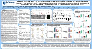
- 1. RNA AND PROTEIN CARGO OF EXOSOMES ISOLATED FROM SERUM OF SYSTEMIC SCLEROSIS PATIENTS INDUCE A PROFIBROTIC PHENOTYPE IN CULTURED NORMAL HUMAN DERMAL FIBROBLASTS: A POTENTIAL MECHANISM FOR THE INITIATION AND PROGRESSION OF A PROFIBROTIC PHENOTYPE IN SSc CONCLUSIONS: The content of profibrotic/antifibrotic miRNA of serum exosomes from SSc patients is different from that of normal serum exosomes and displays an overall profibrotic phenotype. SSc patient- derived exosomes induce a profibrotic phenotype in target cultured normal dermal fibroblasts. The induction of this profibrotic phenotype is mediated by both the RNA and protein content of the exosomes. Since exosomes are involved in intercellular communication these observations suggest a mechanism for transmission of a profibrotic molecular program to normal target cells leading to the extension of the fibrotic process to non-affected tissues. (Supported by NIH Grant AR055660 to SAJ.) Peter. J. Wermuth, Ph.D., Kellan R. Carney, B.S., Sonsoles Piera-Velazquez, Ph.D., and Sergio. A. Jimenez, M.D. Jefferson Institute of Molecular Medicine and Division of Connective Tissue Diseases and Scleroderma Center, Thomas Jefferson University, Philadelphia, Pa.,USA. ABSTRACT Background/Purpose: Exosomes are lipid bilayer-bound microvesicles that contain various macromolecules including numerous microRNA (miRNA) and proteins. Exosomes mediate intercellular communication by fusing and releasing their macromolecular content into target cells. The mechanism of the establishment and progression of a profibrotic phenotype in Systemic Sclerosis (SSc) is not currently well understood. Here, we characterized the miRNA content of exosomes isolated from the serum of SSc patients and analyzed the ability of exosomal RNA and protein components to induce a profibrotic phenotype in normal human dermal fibroblasts in vitro. Methods: Exosomes were isolated from serum from normal individuals and from patients with either limited cutaneous or diffuse cutaneous SSc employing a highly specific polymer precipitation procedure. The isolated exosomes were characterized by Nanosight Particle Tracking Analysis and transmission electron microscopy and the levels of nine pro-fibrotic and nineteen antifibrotic miRNA were assessed by semiquantitative real time PCR. Cultured normal human dermal fibroblasts were incubated with untreated exosomes isolated from the serum of SSc patients or normal donors or with isolated exosomes previously treated with Triton X-100, or with RNAseA, or proteinase K, alone or in combination with Triton X-100 to selectively deplete miRNAs or proteins, respectively. The expression levels of several profibrotic genes in the dermal fibroblasts treated with exosomes isolated from serum of normal individuals or SSc patients were assessed. Results: Nanosight particle tracking analysis and transmission electron microscopy confirmed the isolation of microvesicles in the size range expected for exosomes and an exosome protein array verified that the isolated particles contained exosome-specific proteins and were not contaminated by Golgi-associated proteins. The content of six antifibrotic miRNAs was decreased whereas the content of three profibrotic miRNAs was increased in serum exosomes from both groups of SSc patients compared to normal serum exosomes. Furthermore, the levels of four miRNA were significantly different between the two SSc clinical subsets. Untreated exosomes isolated from the serum of patients with limited or diffuse SSc induced the expression of profibrotic genes in cultured normal human dermal fibroblasts whereas the establishment of this profibrotic phenotype could be partially abrogated by treatment of the exosomes with either RNAse A or proteinase K. Conclusions: The content of profibrotic/antifibrotic miRNA of serum exosomes from SSc patients is different from that of normal serum exosomes and displays an overall profibrotic phenotype. SSc patient- derived exosomes can induce a profibrotic phenotype in target cultured normal dermal fibroblasts. The induction of this profibrotic phenotype is mediated by both the RNA and protein content of the exosomes. Since exosomes are involved in intercellular communication these observations suggest a mechanism for transmission of a profibrotic molecular program to normal target cells leading to the extension of the fibrotic process to non-affected tissues. (Supported by NIH Grant AR055660 to SAJ.) (A) Profibrotic miRNAs with a 2-fold or greater difference in content level in one or both disease subgroups. (B) Antifibrotic miRNAs with a 2-fold or greater difference in content level in one or both disease subgroups. (C) Antifibrotic miRNAs displaying content levels that were significantly different between lcSSc and dcSSc subgroups. Triplicate wells of normal fibroblasts were treated with one of three concentrations of exosomes for each treatment group (N = 2 samples; L and D = 3 samples). Values represent the average change in the levels of gene expression of (A) COL1A1, COL3A1 and FN1; (B) CTGF and TGF-β; and (C) FN-EDA, α-SMA and COMP. Expression levels from the saline-treated fibroblasts were arbitrarily set at the 100% expression level. Values for other samples are expressed relative to this control. Significance was determined by Student’s T-test. Statistical significance: **: p<0.01; ***: p<0.001. Equivalent amounts of exosomes were pre-treated with either saline alone, 0.1% Triton-X100 alone, 20 μg/mL RNase A alone, 100 μg/mL Proteinase K alone, or with 0.1% Triton- X100 + 20 μg/mL Rnase A, or with 0.1% Triton-X100 + 100 μg/mL Proteinase K for 1 hr at 37o C followed by heat inactivation of the enzymes and precipitation with ExoQuick before being added to cultured normal human dermal fibroblasts for 72 h. B Average change in the levels of gene expression of COL1A1 (A), COL3A1 (B), FN1 (C) and FN-EDA (D). Triplicate wells of normal fibroblasts were treated with saline, or with 10 mg exosomes (based on protein concentration) of normal (N = 2 samples) or diffuse SSc (D = 2 samples) exosomes pre-treated with either saline alone (U), Triton-X100 alone (T), RNase A alone (R), Proteinase K alone (P), or with Triton-X100 + Rnase A (T+R) or with Triton-X100 + Proteinase K (T+P). Expression levels from the saline-treated fibroblasts (U) were arbitrarily set at the 100% expression level. Values for N and D samples were averaged and are expressed relative to this control. Significance was determined by Student’s T-test. Statistical significance: **: p<0.01; ***: p<0.001. A B C Results: The isolated exosomes were of the expected size and shape. The content of six profibrotic miRNAs was greater than 2-fold higher whereas the content of ten antifibrotic miRNAs was at least 2-fold lower in SSc exosomes. The levels of eight miRNA were significantly different between the two SSc clinical subsets compared to exosomes from normal serum. Exosomes isolated from SSc patient serum caused a dose related stimulation of profibrotic gene expression in normal dermal fibroblasts, raising them to the levels normally seen in SSc fibroblasts. (A) Size distribution and concentration of purified exosomes. (B) Transmission electron microscopy. Larger vesicles (large arrows) of ~100-120 nm size are interspersed with a population of smaller vesicles (small arrows) of ~40-60 nm (left panel). Enlarged electron micrograph of a representative 100 nm exosome. Scale bars = 100 nm (right panel). (C) Exosome antibody membrane arrays confirming the presence of the exosome-specific proteins CD63, CD81, ALIX, ANXA5 and TSG101 and the absence of the cytosolic cis-Golgi protein GM130. Introduction Exosomes are lipid bilayer-bound microvesicles that contain various macromolecules including numerous microRNA (miRNA) and proteins. Exosomes mediate intercellular communication by fusing and releasing their macromolecular content into target cells. The mechanism of the establishment and progression of a profibrotic phenotype in Systemic Sclerosis (SSc) is not currently well understood. The miRNA contained in blood serum of normal individuals and SSc patients was characterized and their ability to induce a profibrotic phenotype in target fibroblasts was analyzed. Content levels of miRNA present in exosomes isolated from serum of donors with limited and diffuse SSc as assessed by real time PCR. Measurements of particle size and concentration of exosomes isolated from the serum of patients with limited and diffuse cutaneous SSc. Effects of SSc exosomes on the expression of extracellular matrix components, growth factors and myofibroblast-specific genes in cultured normal human dermal fibroblasts. A B C D Effects of pre-treatment of SSc exosomes on their ability to modify the phenotype of target cultured normal human dermal fibroblasts. Strategy for pre-treatment of exosomes for the analysis of effect of exosome RNA and protein components on exosome-mediated alteration of target cell phenotype.