

Recomendados
Mais conteúdo relacionado
Mais procurados
Mais procurados (20)
Destaque
Destaque (15)
Semelhante a Brayner,2005
Semelhante a Brayner,2005 (20)
Último
Último (20)
Brayner,2005
- 1. Micron 36 (2005) 359–367 www.elsevier.com/locate/micron Ultrastructural characterization of the hemocytes of Culex quinquefasciatus (DIPTERA: Culicidae) F.A. Braynera,b,*, H.R.C. Araujoa,b, M.G.S. Cavalcantia,b, L.C. Alvesa,b, C.A. Peixotoa,b,1 ´ a ˜ Departamento de Biologia Celular e Ultraestrutura, Centro de Pesquisas Aggeu Magalhaes (FIOCRUZ), Av. Moraes Rego s/n, Recife 50670-420, Brazil b ´ Laboratorio de Imunopatologia Keizo Asami (LIKA) da Universidade Federal de Pernambuco, Recife, Brazil Received 20 May 2004; revised 30 November 2004; accepted 30 November 2004 Abstract Six hemocytes cell types from Culex quinquefasciatus were identified by light and transmission electron microscopy: They are prohemocytes (9.3%), spherulocytes (1.6%), adipohemocytes (0.8%), oenocytoids (4.6%), plasmatocytes (43.4%) and granulocytes (40.3%). The prohemocytes were the smallest hemocytes encountered in the hemolymph, displaying a large and centrally located nucleus, almost filling the whole cell. The spherulocytes, which were small hemocytes, presented small and numerous spherules with a lamellar pattern and an electron-dense core. Rare adipohemocytes were observed in the C. quinquefasciatus hemolymph, presenting large nucleus with an evident nucleolus, cytoplasm containing rough endoplasmic reticulum (RER), mitochondriae and lipid inclusions. C. quinquefasciatus oenocytoids showed homogeneous cytoplasm with several granules, completely or partially filled with amorphous material. These cells showed abundant smooth endoplasmic reticulum (SER) and dense mitochondriae. By light microscopy analysis we identified two morphological types of plasmatocytes, granular and agranular. However, ultrastructural investigation revealed that the granular cells contained lipid inclusion between RER membranes, instead of membrane-bounded granules. The granulocytes presented a fusiform or circular profile and displayed a unique and very complex process of granules formation, including organization of polysomes inside vesicles that protrude from the Golgi system, synthesis of a proteinaceous material, condensation of the granule matrix and recycling of endoplasmic membranes. Intense endocytic pathways were also observed in the granulocytes. q 2005 Elsevier Ltd. All rights reserved. Keywords: Culex quinquefasciatus (Insecta); Hemocytes; Light microscopy; Electron microscopy; Morphology; Mosquito 1. Introduction immune responses, like phagocytosis, nodulation and encapsulation (Pech and Strand, 2000). However, accord- In insects, the immune system includes both humoral ing to Lavine and Strand (2002) this subdivision of the and cellular components. Humoral defenses involve the insect immune system into cellular and humoral production of antimicrobial peptides (Lowenberger, responses is somewhat arbitrary since humoral factors 2001), reactive free radical intermediates of oxygen or affect hemocyte function and on the other hand, nitrogen (Vass and Nappi, 2001), and the complex hemocytes are an important source of many humoral enzymatic cascades that regulate coagulation or melani- molecules. zation of hemolymph (Muta and Iwanaga, 1996). The hemocytes have the ability to defend insects against In contrast, cellular defense refers to hemocyte-mediated pathogens, parasites and other foreign bodies, which entered in the hemocoel. These defense reactions are mediated by phagocytosis, encapsulation, wound repair and coagulation * Corresponding author. Address: Laboratorio de Imunopatologia Keizo ´ (Lavine and Strand, 2002; Falleiros et al., 2003). The Asami (LIKA) da Universidade Federal de Pernambuco, Recife, Brazil. Tel.: C55 81 3301 2540; fax: C55 81 3453 2449. population of circulating hemocytes is an important tool to E-mail addresses: brayner@cpqam.fiocruz.br (F.A. Brayner), understand the host–parasite interactions, since enhance- cpeixoto@cpqam.fiocruz.br (C.A. Peixoto). ment in the total and differential number of hemocyte may 1 Tel.: C55 81 3301 2557; fax: C55 81 3453 2449. contribute to the protection against a specific parasite 0968-4328/$ - see front matter q 2005 Elsevier Ltd. All rights reserved. (Nappi and Christensen, 1986; Christensen et al., 1989; doi:10.1016/j.micron.2004.11.007 Da Silva et al., 2000).
- 2. 360 F.A. Brayner et al. / Micron 36 (2005) 359–367 Studies by means transmission electron microscopy pH 7.2 and post-fixed with osmium tetroxide in cacodylate (TEM) have produced hemocyte classification by identifi- buffer for 1 h. After dehydration in graded acetone series, cation of seven morphological types: prohemocytes, the cells were embedded in EMBED 812/Araldite (Electron plasmatocytes, granulocytes, spherulocytes, adipohemo- Microscopy Sciences, Fort Washington, PA). cytes, oenocytoids and coagulocytes (Beeman et al., 1983; ˇ Hypsa and Grubhoffer, 1997; Giulianini et al., 2003; 3. Results Falleiros et al., 2003). However, several authors have found between three and four hemocyte types in different ´ Six morphological types of the circulating cells can be genera of mosquitoes (Hernandez et al., 1999; Da Silva recognized in the hemolymph of adult C. quinquefasciatus. et al., 2000; Hillyer and Christensen, 2002; Hillyer et al., They are prohemocytes, spherulocytes, adipohemocytes, 2003). Culex quinquefasciatus is a culicid mosquito that oenocytoids, plasmatocytes and granulocytes. transmits several pathogens, including Wuchereria ban- crofti and arboviruses, such as St Louis encephalitis (SLE) in Western States and Oropouch virus in the north of Brazil 3.1. Prohemocytes (Da Silva et al., 2000). Conversely, there are very few papers about the morphological characterization of hemo- Prohemocytes are the smallest cells encountered in the cytes of C. quinquefasciatus and this is due in large part to hemolymph, displaying a spherical profile with w5–8 mm the small size of these insects, which is difficult to perform in diameter, which represents 9.3% of the total hemocyte manipulative experiments. The aim of the present study was population. The large and centrally located nucleus to characterize by the first time distinct morphological types almost fills the whole cell, so that the cytoplasm of hemocytes of C. quinquefasciatus by transmission occupies only a narrow area around the nucleus electron microscopy. (Figs. 1A and 2A and B). The chromatin is scattered, and in some cells the presence of nucleoli is evident. Only a few organelles can be seen, with a conspicuous 2. Materials and methods development of rough endoplasmic reticulum (RER) and mitochondriae (Fig. 2A and B). 2.1. Insects 3.2. Spherulocytes Laboratory bred C. quinquefasciatus (Recife strain) were used throughout this study. Adults were maintained in cages Spherulocytes shows an oval cell profile with average (30!30!30 cm3) at room temperature (27G3 8C) with diameter of 8–10 mm, displaying a round nucleus and a 85G10% relative humidity and fed with 10% (w/v) sucrose condensed chromatin with a large nucleolus (Fig. 2C). Several solution. small spherules (1–1.5 mm in diameter) (Figs. 1B and 2C and D) containing a lamellar pattern with an electron-dense 2.2. Hemocytes characterization core region (Fig. 2C and D). The cytoplasm also contained other organelles such as RER and mitochondriae (Fig. 2C). For light microscopy (LM), the adult insects were washed The spherulocytes represented 1.6% of the total hemocytes. in PBS and placed on ice (1–2 min) for immobilization. The hemolymph of 10 insects (4-day-old) was obtained by 3.3. Adipohemocytes perfusing the thorax with anticoagulant II solution (Mead et al., 1986) and bled directly on to a glass slide and allowed Adipohemocytes are rare small and elongated cells dry in natural air conditions for 20–30 min. Cells were then measuring 8–15 mm in length. In the adipohemocytes fixed in methanol for 10 min. After natural air-drying of the observed in this study, a round nucleus could be fixative, hemocytes were stained with Giemsa (diluted 1:9 in observed (Fig. 1C). Inside the cytoplasm several large buffered distilled water) for 10–15 min and slides were rapidly lipid vesicles and mitochondriae (Figs. 1C and 2E and F) washed with buffered distilled water (Da Silva et al., 2000; were observed. They were the less frequent hemocytes, Silva et al., 2002). After air drying the slides were dehydrated with 0.8%. and mounted in Entellan. For transmission electron microscopy (TEM), hemo- 3.4. Oenocytoids lymph of at least 300 insects (4-day-old) was collected from a punctured thorax perfused directly with fixation solution Oenocytoids presents a round shape, approximatelly and the obtained hemolymph was pooled and centrifuged at 6–13 mm in diameter, with small and eccentric nucleus 500g for 5 min. The pellet was resuspended and fixed in 4% (Figs. 1D and 3A) or oval with chromatin showing lumps of glutaraldehyde in 0.2 M cacodylate buffer, pH 7.2, over- condensation. The ultrastructure revealed a cytoplasm ˇ night (Hypsa and Grubhoffer, 1997). The samples were rich in vacuoles, some filled with a hetrogeneous electron- washed in 5% sucrose solution in 0.2 M cacodylate buffer, dense material and others completely empties. It presents
- 3. F.A. Brayner et al. / Micron 36 (2005) 359–367 361 Fig. 1. Light microscopy of hemocytes from C quinquefasciatus. (A) A prohemocyte with a large nucleus (thin arrow) containing a prominent nucleolus (arrowhead). (B) A spherulocyte showing an oval profile and numerous spherules within the cytoplasm (thin arrows). (C) An adipohemocyte showing an irregular cell profile and lipid vesicles in the cytoplasm (thin arrows). (D) A oenocytoid with a round eccentric nucleus (thin arrow). (E) A granular plasmatocyte exhibiting a central nucleus with nucleolus (thin arrow). In the cytoplasm, vacuoles (short arrows) and granules (arrowheads) are shown. Observed also a phylopodium process (open arrow). (F) An agranular plasmatocyte spreading a lobular pseudopodia (arrowhead). Note also the eccentric nucleus (thin arrow). (G) A granulocyte displaying a large mass of heterochomatin (long arrow) inside the nucleus. Observe also several granules in the cytoplasm (arrowheads) and a short phylopodium (large arrow). (H) A granulocyte with multiple pseudopodia (short arrow). Observe also the large nucleus and several dense compact granules (arrowheads). BarsZ5 mm.
- 4. 362 F.A. Brayner et al. / Micron 36 (2005) 359–367 Fig. 2. (A–F) Electron micrographs of hemocytes from C. quinquefasciatus. (A and B) Prohemocytes displaying spherical profiles with a large central nucleus (N) containing nucleolus (Nu) and a thin cytoplasm with few organelles. Note lumps of heterochromatin (arrows). Mitochondriae (m) and RER (arrowheads) are also indicated. (C) A spherulocyte with numerous spherules showing a round nucleus with condensed chromatin (N) and nucleolus (Nu), RER complex (thin arrow) and mitochondrion (m). (D) Magnification of the spherules showing a lamellar pattern with an electron-dense core region (arrows). (E–F) Adipohemocytes with oval profile showing large nucleus (N) containing evident nucleolus (Nu). Observe cytoplasm with several lipid vesicles inside (stars) and mitochondriae (m). BarsZ0.5 mm. a homogeneous cytoplasm with abundant SER and dense philopodia and pseudopodia (Fig. 1E and F). The majority mitochondriae. Sparce RER is also present (Fig. 3A). They of the plasmatocytes were mononucleated but some are presented 4.6% of the total circulating hemocytes. binucleated cells were occasionally observed. In electron micrographs agranular plasmatocytes presented a lobated 3.5. Plasmatocytes nucleus with evident nucleoli in a pericentral position (Fig. 3B). The chromatin was finely distributed but some The plasmatocytes were the most cellular types fre- heterochromatin clumps were present. Within the cytoplasm quently observed, representing 43.4%. Two types of several elongated and round mitochondria were observed. plasmatocytes were observed, granular and agranular The reticular cytoplasm showed well-developed RER, plasmatocytes. These cells are very polymorphic, varying Golgi system and some vacuoles. Also patches of smooth from spindle-shaped to round cells, w6–22 mm in diameter. endoplasmic reticulum SER were present at the cell poles The plasma membrane exhibit irregular processes, (Fig. 3B). The granular plasmatocytes showed elongated or
- 5. F.A. Brayner et al. / Micron 36 (2005) 359–367 363 Fig. 3. (A) An oenocytoide showing an eccentric lobated nucleus (N) with clumps of hetrochromatin (short arrows). Several cytoplasmic vacuoles are present, some filled with a heterogeneous electron-dense material (large arrowheads) and others completely empty (thin arrows). Note also electron- dense mitochondriae (arrows), abundant SER (small arrowheads) and few RER (open short arrow). (B) An agranular plasmatocyte showing a lobated nucleus (N) with nucleolus (Nu) in a pericentral position. Some heterochromatin lumps are present (arrows). Within the cytoplasm several elongated and round mitochondria are present (thin arrows). Note the well-developed RER (arrowheads) and isles of SER were presented (stars). (C) A granular plasmatocyte showing elongated nucleus (N) with a scattered mass of heterochromatin. Abundant RER (arrowheads), lipid inclusions located among endoplasmic membranes (thin arrows), and electron-dense inclusions (open arrow) are indicated. Part of adjacent granular plasmatocyte contains lipid inclusions (thin arrow). BarsZ0.5 mm. circular nucleus displaying a scattered mass of hetero- 3.6. Granulocytes chomatin, and in some cells a large nucleolus could be observed (not shown). The cytoplasm was rich in RER and Granulocytes present a circular to fusiform profile Golgi system. The numerous granules seen by LM were w8–13 mm in diameter, which represents 40.3% of the observed by EM with no limiting membrane, similar to total hemocyte population. The plasma membrane is cellular inclusions, showing a homogeneous matrix and irregular displaying pseudopodia and philopodia in its located among endoplasmic membranes. Some of the surface (Fig. 1G and H). In electron micrographs, these inclusions show an electron-dense core, possibly indicating hemocytes show important morphological characteristics. a dual constitution (Fig. 3C). The lobated nucleus has a large mass of heterochomatin and
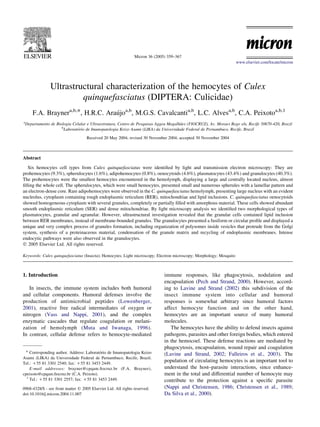
- 6. 364 F.A. Brayner et al. / Micron 36 (2005) 359–367 Fig. 4. A granulocyte showing a lobated nucleus (N) with a large mass of heterochomatin (large short arrows) and an active nucleolus (Nu). Endocytic process with the presence of numerous coated vesicles (short arrows) and coated pits (open short arrow) and mitochondria (m). The cytoplasm contains sparce RER (thin arrows), abundant free ribosomes (small arrows) with formation of numerous polysomes (arrowheads). A well-developed Golgi complex displays vesicle production (G). The following steps of the granule synthesis are indicated. (1) Large vesicles containing engulfed cytoplasm with polysomes (arrowheads), free ribosomes (small arrow) and occasional mitochondria (m). (2) Vesicles containing large empty vacuoles (V) and ribosomes, organized as small polysomes (arrowheads). (3) Initial synthesis of a dense proteinaceous matrix (M) by polysomes (arrowheads). (4) Fusion of multiple polysomes (arrowheads) and formation of the granule matrix (M). (5) Condensation of the granule with production small vesicles in order to eliminate ribosomes and recycle endoplasmic membranes (r). (6) A mature granule inside the cytoplasm. (7) A granule being exocytosed by the cell. BarZ0.5 mm. an active nucleolus and a clear endocytic process with the final stage, the granule expells the ribosomes and produces presence of numerous coated vesicles and coated pits is small vesicles in order to condense the proteinaceous matrix present. Round or elongated mitochondria are also detected. and recycle intracellular membranes. (6) The mature The cytoplasm contains dilated RER, formation of poly- granule is then free inside the cytoplasm. (7) Finally, the somes and abundant ribosomes. Also, a well-developed granule is exocysed by the cell. Golgi complex displays large vesicle production (Fig. 4). Synthesis of a second type of granule is also shown. All the steps of the granule synthesis machinary can be A vesicle containing several sites of membranes of observed, as follows: (1) large vesicles containing engulfed endoplasmic membranes producing a very electron-dense cytoplasm with free ribosomes and occasional mitochon- matrix was observed (Fig. 5). A large electron-dense dria; (2) some vesicles contain large empty vacuoles granule is shown, probably due to the fusion of the products possibly, due to the membrane fusion with other empty of several synthesis sites. vacuoles. Also, ribosomes are organized as numerous large It is important to note that near to this synthesis granule polysomes, some of which show the formation of other machinery several elongated or round mitochondria are small polysomes. (3) Initial synthesis of a proteinaceous present. In addition, in many other granulocytes we found matrix inside the polysomes; (4) fusion of multiple mitochondria inside the vesicle of synthesis of the granule polysomes and formation of the granule matrix. (5) At the (Figs. 4 and 5).
- 7. F.A. Brayner et al. / Micron 36 (2005) 359–367 365 Fig. 5. A granulocyte showing a lobated nucleus (N) with a large mass of heterochromatin (short arrows) and an active nucleolus (Nu). Several elongated mitochondria (m) are also present. The cytoplasm contain sparce RER (thin arrows), abundant free ribosomes (small arrows) and formation of numerous polysomes (arrowheads). The following steps of the granule machinary synthesis are indicated. (1) Not shown. (2) Vesicle containing empty vacuoles (V) and free ribosomes organized as numerous polysomes, some of which are small polysomes (arrowheads). Note the elongated mitochondrion attached to the vesicle membrane (open short arrow). (3) Initial synthesis of a dense proteinaceous matrix by polysomes (M). Note also the multiple polysomes (arrowheads). (4) Fusion of multiple polysomes and formation of the granule matrix (M). Multiple polysomes (arrowheads) are also present. (5) Condensation of granule and recycle of endoplasmic membranes (r). (6) Not shown. (7) A granule exocyted by the cell. Note also a second type of granule synthesis: a vesicle containing several sites of synthesis of very an electrondense matrix (small short arrows) and the formation of a large granule inside (white asterisk). BarZ0.5 mm. 4. Discussion recognized as the spherulocyte. Recently, Hillyer et al. (2003) described only four morphological cell types in C. quinquefasciatus adults possess six different types of Armigeres subalbatus, a natural vector of Japanese ence- hemocytes, which vary considerably in their morphology and phalitis and filarial nematodes: granulocytes, oenocytoids, size, namely: prohemocytes, spherulocytes, adipohemo- adipohemocytes and thrombocytoids. cytes, oenocytoids, plasmatocytes and granulocytes. Silva In the present study, we observed that prohemocytes et al. (2002) studying Anastrepha obliqua instar larva, which displayed unmistakable characteristics such as small size is a member of the Diptera Order, described a similar range of and large nuclear–cytoplasmic ratio, with a morphology morphological types of hemocytes by LM analysis. Analo- identical to that described in previous hemocyte studies gous results were also obtained by Kaaya and Ratcliffe ˇ (Hypsa and Grubhoffer, 1997; Falleiros et al., 2003; (1982) who performed an important morphological com- Giulianini et al., 2003). parison of hemocytes from several medical important Some studies have described spherulocytes as hemocytes- dipterans, including the C. quinquefasciatus. However, in containing large membrane-bound spherules that deform the these LM studies, they did not identify the hemocyte cellular surface. In C. quinquefasciatus, these cells presented
- 8. 366 F.A. Brayner et al. / Micron 36 (2005) 359–367 similar organelles, showing moderated electron density with of a proteinaceous material, and condensation of the an electron dense core, as described for other species granule matrix with recycling of endoplasmic membranes. (Beeman et al., 1983; Falleiros et al., 2003). However, the Synthesis of a second type of granule was also observed, size of spherules of these hemocytes was smaller than those occurring inside vesicles almost free of ribosomes, which described previously and did not protrude from the cellular contained an electron-dense matrix probably composed of surface. Giulianini et al. (2003) observed a similar spherule substances other than proteins. On the other hand, size in Cetonischema aeruginosa larvae (Coleoptera, granular plasmatocytes showed no such machinery for Scarabaeidae). synthesis of granules, instead they display several small Rarely, adipohemocytes were observed in the present lipid inclusions among ER membranes. So, we not agree work, mostly because of the instability of these cells. ˇ with Hypsa and Grubhoffer (1997) since plasmatocytes Besides large lipid vesicles, other cellular organelles could (granular and granular) and granulocytes presented a be readily identified, i.e., scarce RER and mitochondria. completely different morphology, and thus may belong to Conversely, Hillyer et al. (2003) described that these cells a distinct class of hemocytes, however, functional studies were the second most common cells obtained from naive are necessary to confirm this. mosquitoes. In our study, adipohemocytes presented similar size to granulocytes and oenocytoids, whereas Hillyer et al. (2003) found that these cells were w30 mm in diameter, being several times the size of granulocytes and oenocy- toids. Some authors do not regard the adipohemocyte as a Acknowledgements distinct hemocyte type, since they considered their mor- phology to be very similar to that of granulocytes, and also The authors are grateful to Rafael Padilha, Raimundo indicated that these cells are most similar to the fat body ´ Pimentel and Sergio Santos for the precious technical cells (Kaaya and Ratcliffe, 1982). However, we did not support. This work has been supported by Fundacao ¸˜ observe any similarity between these two cellular types and Oswaldo Cruz (FIOCRUZ). therefore further studies are necessary to clarify this controversial matter. Oenocytoids have been described as cells typically References containing a greater number of dense granules located at ˇ the cell periphery (Hyps a and Grubhoffer, 1997; Beeman, S.C., Wilson, M.E., Bulla Jr., L.A., Consigli, R.A., 1983. Giulianini et al., 2003). In EM analyses, we detected Structural characterization of the hemocytes of Plodia interpunctella. that in oenocytoids from C. quinquefasciatus these Journal of Morphology 175, 1–16. granules were completely or partially filled with Christensen, B.M., Huff, B.M., Miranpuri, G.S., Harris, K.L., amorphous material. Christensen, L.A., 1989. Hemocyte population changes during the immune response of Aedes aegypti to inoculated microfilariae of By LM analysis we identified two morphological types Dirofilaria immitis. Journal of Parasitology 75, 119–123. of plasmatocytes, granular and agranular. However, ´ Da Silva, J.B., Albuquerque, C.M.R., Araujo, E.C., Peixoto, C.A., Hurd, H., ultrastructural investigation revealed that the granular 2000. Immune defense mechanisms of Culex quinquefasciatus cells do in fact contain lipid inclusions between RER, (Diptera: Culicidae) against Candida albicans infection. Journal of rather than membrane-bounded granules. In Triatoma Invertebrate Pathology 76, 257–262. ´ Falleiros, A.M.F., Bombonato, M.T.S., Gregorio, E.A., 2003. Ultrastruc- infestans agranular and granular plasmatocytes were tural and quantitative studies of hemocytes in the sugarcane borer, ˇ described by LM and EM analysis (Hypsa and Grubhoffer, Diatraea saccharalis (Lepidoptera: Pyralidae). Brazilian Archives of 1997). These authors demonstrated that granular plasma- Biology and Technology 46, 287–294. tocytes contained granules composed by more or less Giulianini, P.G., Bertolo, F., Battistella, S., Amirante, G.A., 2003. regularly arranged fibrils, through very dense to appar- Ultrastructure of the hemocytes of Cetonischema aeruginosa larvae (Coleoptera, Scarabeidae): involvement of both granulocytes and ently amorphous matrix. According to the Hypsa and ˇ oenocytoids in vivo phagocytosis. Tissue and Cell 35, 243–251. Grubhoffer (1997) terminology, different types of prohe- ´ ´ Hernandez, S., Lanz, H., Rodrıguez, M.H., Torres, J.A., Martınez- ´ mocytes generate two cellular lineages: granular or Palomo, A., Tsutsumi, V., 1999. Morphological and cytochemical agranular plasmocytes. In their opinion, the term granulo- characterization of female Anopheles albimanus (Diptera: Culicidae) cyte is unacceptable, considering that granulocytes are hemocytes. Journal of Medical Entomology 36, 426–434. Hillyer, J.F., Christensen, B.M., 2002. Chracterization of hemocytes from commonly regarded as cells closely related to plasmato- the yellow fever mosquito, A. aegypti. Histochemistry and cell Biology cytes or even differentiated from them. However, in our 313, 117–127. study it was shown that cells termed granulocytes were Hillyer, J.F., Schmidt, S.L., Christensen, B.M., 2003. Hemocyte-mediated quite different from plasmatocytes in several aspects. The phagocytosis and melanization in the mosquito Armigeres subalbatus results of the present work indicate that the process of following immune challenge by bacteria. Cell and Tissue Research 313, 117–127. granule formation in granulocytes is unique and very ˇ Hypsa, V., Grubhoffer, L., 1997. Two hemocyte populations in Triatoma complex, including organization of polysomes insides infestans: ultrastructural and lectin-binding characterization. Folia vesicles, which protrude from the Golgi system, synthesis Parasitologica 44, 62–70.
- 9. F.A. Brayner et al. / Micron 36 (2005) 359–367 367 Kaaya, G.P., Ratcliffe, N.A., 1982. Comparative study of hemocytes and Nappi, A.J., Christensen, B.M., 1986. Hemocyte cell surface changes in associated cells of some medically important Dipterans. Journal of Aedes aegypti in response to microfilariae of Dirofilaria immitis. Morphology 173, 351–365. Journal of Parasitology 72, 875–879. Lavine, M.D., Strand, M.R., 2002. Insect hemocytes and their role in Pech, L.L., Strand, M.R., 2000. Plasmatocytes from the Pseudoplusia immunity. Insect Biochemistry and Molecular Biology 32, 1295–1309. includens induce apoptosis of granular cells. Journal of Insect Lowenberger, C., 2001. Innate immune response of Aedes aegypyi. Insect Physiology 46, 1565–1573. Biochemistry and Molecular Biology 31, 219–229. ˜ Silva, J.E.B., Boleli, I.C., Simoes, Z.L.P., 2002. Hemocytes types and Mead, G.P., Ratcliffe, N.A., Renwrantz, L.R., 1986. The separation of total differential counts in unparasitized and parasitized Anastrepha insect haemocyte types on percoll gradients: methodology and obliqua (Diptera, Tephritidae) larvae. Brazilian Journal of Biology problems. Journal of Insect Physiology 32, 167–177. 62, 689–699. Muta, T., Iwanaga, S., 1996. The role of hemolymph coagulation in innate Vass, E., Nappi, A.J., 2001. Fruit fly immunity. BioEssays 51, 529– immunity. Current Opinion in Immunology 8, 41–47. 535.