SlideShare a Scribd company logo
1 of 7
©Goutam Mallik, Assistant Professor, Pharmacology
Page | 1
TISSUE
 A tissue is a group of cells with similar structure and function. The tissue contributes to the
functioning of the organs in which it is found.
Classification Tissue
Tissues

 Body tissues can be classified into four basic types according to their structure and function.
 Epithelial tissues cover body surfaces and line hollow organs, body cavities and ducts, they also
form glands. This tissue allows the body to interact with both its internal and external
environments.
 Connective tissues protect and support the body and its organs. Various types of connective
tissues bind organs together, store energy reserves as fat and help provide the body with
immunity to disease-causing organisms.
 Muscular tissues are composed of cells specialized for contraction and generation of force. In
the process, muscular tissues generate heat that warms the body. •
 Nervous tissue detects changes in a variety of conditions inside and outside the body and
responds by generating electrical signals called nerve action potentials (nerve impulses) that
activate muscular contractions and glandular secretions.
©Goutam Mallik, Assistant Professor, Pharmacology
Page | 2
Fig: Epithelial Tissue
EPITHELIAL TISSUES
 Epithelial tissue or an epithelium (plural: epithelia), is a sheet of cells that covers a body
surface or lines a body cavity (epithe = laid on, covering).
 Epithelia form boundaries between different environments. For example, the epidermis of the
skin lies between the inside and the outside of the body and epithelium lining the urinary
bladder separates underlying cells of the bladder wall from urine.
 It occurs in the body as:
A. Covering and lining epithelium: Covering and lining epithelium forms the outer layer of the
skin, dips into and lines the open cavities of the cardiovascular system, digestive system,
respiratory systems and covers the walls and organs of the closed ventral body cavities.
B. Glandular epithelium: The function of glandular epithelium is secretion, which is
accomplished by glandular cells that often lie in clusters deep to the covering and lining
epithelium.
EPITHELIAL TISSUE COVERING AND LINING EPITHELIUM
 On the basis of the two characteristics (arrangements of layers and cell shapes), epithelial
tissues are classified into following types :
 Simple epithelium:
(A) Simple squamous epithelium.
(B) Simple cuboidal epithelium.
(C) Simple columnar epithelium (nonciliated and ciliated).
(D) Pseudostratified columnar epithelium (nonciliated and ciliated).
©Goutam Mallik, Assistant Professor, Pharmacology
Page | 3
 Stratified epithelium:
(A) Stratified squamous epithelium (keratinized, when surface cells are dead and become hardened
and nonkeratinized, when surface cells remain alive).
(B) Stratified cuboidal epithelium.
(C) Stratified columnar epithelium.
(D) Transitional epithelium.
Simple epithelium
(A) Simple squamous epithelium:
 Single layer of flat cells that resembles a tiled floor when viewed from apical surface; centrally
located nucleus that is flattened and oval or spherical in shape.
 Location: Most commonly lines the cardiovascular and lymphatic system (heart, blood vessels,
lymphatic vessel linings), where it is known as endothelium and forms the epithelial layer of
serous membranes (peritoneum, pleura, pericardium), where it is called mesothelium. Also
found in air sacs of lungs, glomerular (Bowman’s) capsule of kidneys, inner surface of tympanic
membrane (eardrum).
 Function: Present at sites of filtration (such as blood filtration in kidneys) or diffusion (such as
diffusion of oxygen into blood vessels of lungs) and at site of secretion in serous membranes.
(B) Simple cuboidal epithelium:
 Single layer of cube-shaped cells, round, centrally located nucleus.
 Cuboidal cell shape is obvious when tissue is sectioned and viewed from the side.
 Location: Covers surface of ovary, lines anterior surface of capsule of lens of the eye, forms
pigmented epithelium at posterior surface of retina of the eye. Lines kidney tubules and smaller
ducts of many glands. Makes up secreting portion of some glands, such as thyroid gland and
ducts of some glands such as pancreas.
 Function: Secretion and absorption.
(C) Nonciliated simple columnar epithelium:
 Single layer of nonciliated column like cells with oval nuclei near base of cells, contains
(1) columnar epithelial cells with microvilli at apical surface and
(2) goblet cells.
 Microvilli, finger like cytoplasmic projections, increase surface area of plasma membrane, thus
increasing cell’s rate of absorption.
 Goblet cells are modified columnar epithelial cells that secrete mucus, a slightly sticky fluid, at
their apical surfaces.
©Goutam Mallik, Assistant Professor, Pharmacology
Page | 4
 Location: Lines gastrointestinal tract (from stomach to anus), ducts of many glands and
gallbladder.
 Function: Secretion and absorption, larger columnar cells contain more organelles and thus are
capable of higher level of secretion and absorption than are cuboidal cells.
(D) Ciliated simple columnar epithelium:
 Single layer of ciliated column like cells with oval nuclei near base of cells. Goblet cells are
usually interspersed among ciliated columnar epithelia.
 Location: Lines some bronchioles (small tubes) of respiratory tract, uterine (fallopian) tubes,
uterus, some paranasal sinuses, central canal of spinal cord and ventricles of brain.
 Function: Cilia beat in unity, moving mucus and foreign particles toward throat, where they can
be coughed up and swallowed or spit out. Cilia also help move oocytes expelled from ovaries
through uterine (fallopian) tubes into uterus.
Fig: Complex Derived Epithelial Tissue
©Goutam Mallik, Assistant Professor, Pharmacology
Page | 5
(E) Pseudostratified columnar epithelium:
 Appears to have several layers because cell nuclei are at various levels. All cells are attached to
basement membrane in a single layer, but some cells do not extend to apical surface.
 When viewed from side, these features give false impression of a multi-layered tissue.
 Pseudostratified ciliated columnar epithelium contains cells that extend to surface and secrete
mucus (goblet cells) or bear cilia.
 Pseudostratified nonciliated columnar epithelium contains cells without cilia and lacks goblet
cells.
 Location: Ciliated variety lines airways of most of upper respiratory tract, nonciliated variety
lines larger ducts of many glands, epididymis and part of male urethra.
 Function: Ciliated variety secretes mucus that traps foreign particles, and cilia sweep away
mucus for elimination from body. Nonciliated variety functions in absorption and protection.
Stratified epithelium
(A) Stratified squamous epithelium:
 Two or more layers of cells, cells in apical layer and several layers deep to it are squamous,
cells in deeper layers vary from cuboidal to columnar.
 Tough proteins predominate as cytoplasm is reduced and cells become tough, hard structures
that eventually die.
 At apical layer, after dead cells lose cell junctions they are sloughed off, but they are replaced
continuously as new cells emerge from basal cells.
 Keratinized stratified squamous epithelium develops tough layer of keratin in apical layer of cells
and several layers deep to it.
 Non keratinized stratified squamous epithelium does not contain large amounts of keratin in
apical layer and several layers deep and is constantly moistened by mucus from salivary and
mucous glands, organelles are not replaced.
 Location: Keratinized variety forms superficial layer of skin, nonkeratinized variety lines wet
surfaces (lining of mouth, esophagus, part of epiglottis, part of pharynx and vagina) and covers
tongue.
 Function: Protection against abrasion, water loss, ultraviolet radiation and foreign invasion.
Both types form first line of defense against microbes.
(B) Stratified cuboidal epithelium:
 Two or more layers of cells, cells in apical layer are cube-shaped, fairly rare type.
 Location: Ducts of adult sweat glands and esophageal glands, part of male urethra.
 Function: Protection, limited secretion and absorption.
©Goutam Mallik, Assistant Professor, Pharmacology
Page | 6
(C) Stratified columnar epithelium:
 Basal layers usually consist of shortened, irregularly shaped cells, only apical layer has
columnar cells, uncommon.
 Location: Lines part of urethra, large excretory ducts of some glands, such as esophageal
glands, small areas in anal mucous membrane, part of conjunctiva of eye.
 Function: Protection and secretion.
(D) Transitional epithelium:
 Variable appearance (transitional). In relaxed or unstretched state, looks like stratified cuboidal
epithelium, except apical layer cells tend to be large and rounded.
 As tissue is stretched, cells become flatter, giving the appearance of stratified squamous
epithelium.
 Multiple layers and elasticity make it ideal for lining hollow structures (urinary bladder) subject to
expansion from within.
 Location: Lines urinary bladder and portions of ureters and urethra.
 Function: Allows urinary organs to stretch and maintain protective lining while holding variable
amounts of fluid without rupturing.
GLANDULAR EPITHELIUM
 Two types of glandular epithelium exists:
(A) Exocrine
(B) Endocrine
(A) Epithelial tissue glandular epithelium (Exocrine):
 Secretory products released into ducts that empty onto surface of a covering and lining
epithelium, such as skin surface or lumen of hollow organ.
 Location: Sweat, oil and earwax glands of skin, digestive glands such as salivary glands
(secretes into mouth cavity) and pancreas (secretes into small intestine).
 Function: Produce substances such as sweat to help lower body temperature, oil, earwax,
saliva, or digestive enzymes.
(B) Epithelial tissue glandular epithelium (Endocrine):
 Secretions (hormones) enter interstitial fluid and diffuse directly into bloodstream without flowing
through a duct.
 Location: Examples include pituitary gland at base of brain, pineal gland in brain, thyroid and
parathyroid glands near larynx (voice box), adrenal glands superior to kidneys, pancreas near
stomach, ovaries in pelvic cavity, testes in scrotum, thymus in thoracic cavity.
 Function: Hormones regulate many metabolic and physiological activities to maintain
homeostasis.
©Goutam Mallik, Assistant Professor, Pharmacology
Page | 7
Fig: Summary of Epithelial Tissues
***********************************
Reference
1. Human Anatomy & Physiology-I, by Mrs. Shubhada Mangrulkar & Ms. Nitu L. Wankhede,
TPS Publication
2. Essentials of Human Anatomy & Physiology 5th
Ed by Valerie C. Scanlon & Tina Sanders
3. Gray’s Anatomy, The Anatomical Basis of Clinical Practice 41st
Ed by Elsevier

More Related Content

What's hot (20)

General principles of cell communication
General principles of cell communicationGeneral principles of cell communication
General principles of cell communication
 
Biomedical eng human physiology, introduction to human body
Biomedical eng   human physiology, introduction to human body Biomedical eng   human physiology, introduction to human body
Biomedical eng human physiology, introduction to human body
 
Tissues Project
Tissues ProjectTissues Project
Tissues Project
 
Human Tissues
Human TissuesHuman Tissues
Human Tissues
 
TISSUE AND GLANDS BASIC
TISSUE AND GLANDS BASICTISSUE AND GLANDS BASIC
TISSUE AND GLANDS BASIC
 
Basic cytology
Basic cytologyBasic cytology
Basic cytology
 
Human cell
Human cellHuman cell
Human cell
 
Epithelial Tissue Histology
Epithelial Tissue HistologyEpithelial Tissue Histology
Epithelial Tissue Histology
 
Cell anatomy
Cell anatomyCell anatomy
Cell anatomy
 
Chapter 3
Chapter 3 Chapter 3
Chapter 3
 
A&P2 Cell Tissue
A&P2 Cell TissueA&P2 Cell Tissue
A&P2 Cell Tissue
 
Tissues, types and functions(Anatomy)- Easy explanation
Tissues, types and functions(Anatomy)- Easy explanationTissues, types and functions(Anatomy)- Easy explanation
Tissues, types and functions(Anatomy)- Easy explanation
 
Tissue
TissueTissue
Tissue
 
9098 et et_1.3_system_physiology (1) (1)
9098 et et_1.3_system_physiology (1) (1)9098 et et_1.3_system_physiology (1) (1)
9098 et et_1.3_system_physiology (1) (1)
 
Cell & tissue nursing
Cell & tissue nursingCell & tissue nursing
Cell & tissue nursing
 
Adipose Tissue Histology
Adipose Tissue HistologyAdipose Tissue Histology
Adipose Tissue Histology
 
Anatomy and physiology of human tissues.pptx
Anatomy and physiology of human tissues.pptxAnatomy and physiology of human tissues.pptx
Anatomy and physiology of human tissues.pptx
 
Presentation cell tissue and organ
Presentation cell tissue and organPresentation cell tissue and organ
Presentation cell tissue and organ
 
Body tissues
Body tissuesBody tissues
Body tissues
 
Introduction to Human Anatomy and Physiology
Introduction to Human Anatomy and PhysiologyIntroduction to Human Anatomy and Physiology
Introduction to Human Anatomy and Physiology
 

Similar to Tissues

Introduction of tissue - Epithelial tissue
Introduction of tissue - Epithelial tissueIntroduction of tissue - Epithelial tissue
Introduction of tissue - Epithelial tissueBithikaBaidya
 
study of epithelium.pptx
study of epithelium.pptxstudy of epithelium.pptx
study of epithelium.pptxDinraj Kulkarni
 
Anatomy :O
Anatomy :OAnatomy :O
Anatomy :OSteven S
 
General principles of tissue organization
General principles of tissue organizationGeneral principles of tissue organization
General principles of tissue organizationEneutron
 
2. epithelial-t[1]
2. epithelial-t[1]2. epithelial-t[1]
2. epithelial-t[1]Lama K Banna
 
The Tissue level of organization .pptx
The Tissue level of organization   .pptxThe Tissue level of organization   .pptx
The Tissue level of organization .pptxshraddhaparab530
 
B.Pharm-Ist sem-HAP-Chapter 3-tissue level of organization.pptx
B.Pharm-Ist sem-HAP-Chapter 3-tissue level of organization.pptxB.Pharm-Ist sem-HAP-Chapter 3-tissue level of organization.pptx
B.Pharm-Ist sem-HAP-Chapter 3-tissue level of organization.pptxSheetal Patil
 
1. Human Organization. biology presentationpdf
1. Human Organization. biology presentationpdf1. Human Organization. biology presentationpdf
1. Human Organization. biology presentationpdfTakondwaMitomoni
 
Tissue level of organization.pdf
Tissue level of organization.pdfTissue level of organization.pdf
Tissue level of organization.pdfKeval80
 

Similar to Tissues (20)

Tissues
TissuesTissues
Tissues
 
Introduction of tissue - Epithelial tissue
Introduction of tissue - Epithelial tissueIntroduction of tissue - Epithelial tissue
Introduction of tissue - Epithelial tissue
 
study of epithelium.pptx
study of epithelium.pptxstudy of epithelium.pptx
study of epithelium.pptx
 
Anatomy :O
Anatomy :OAnatomy :O
Anatomy :O
 
General principles of tissue organization
General principles of tissue organizationGeneral principles of tissue organization
General principles of tissue organization
 
Types of tissues
Types of tissues   Types of tissues
Types of tissues
 
Tissues
TissuesTissues
Tissues
 
2. epithelial-t[1]
2. epithelial-t[1]2. epithelial-t[1]
2. epithelial-t[1]
 
The Tissue level of organization .pptx
The Tissue level of organization   .pptxThe Tissue level of organization   .pptx
The Tissue level of organization .pptx
 
B.Pharm-Ist sem-HAP-Chapter 3-tissue level of organization.pptx
B.Pharm-Ist sem-HAP-Chapter 3-tissue level of organization.pptxB.Pharm-Ist sem-HAP-Chapter 3-tissue level of organization.pptx
B.Pharm-Ist sem-HAP-Chapter 3-tissue level of organization.pptx
 
Tissues
TissuesTissues
Tissues
 
TISSUE-GLANDS
 TISSUE-GLANDS TISSUE-GLANDS
TISSUE-GLANDS
 
Tissue: Epithelial tissue
Tissue: Epithelial tissueTissue: Epithelial tissue
Tissue: Epithelial tissue
 
1. Human Organization. biology presentationpdf
1. Human Organization. biology presentationpdf1. Human Organization. biology presentationpdf
1. Human Organization. biology presentationpdf
 
Tissue level of organization.pdf
Tissue level of organization.pdfTissue level of organization.pdf
Tissue level of organization.pdf
 
Tissues
TissuesTissues
Tissues
 
Tissues
Tissues Tissues
Tissues
 
Basic Tissues 1.pptx
Basic Tissues 1.pptxBasic Tissues 1.pptx
Basic Tissues 1.pptx
 
epithelia Tissues.pptx
epithelia Tissues.pptxepithelia Tissues.pptx
epithelia Tissues.pptx
 
3.tissues of body
3.tissues of body3.tissues of body
3.tissues of body
 

Recently uploaded

Unit-V; Pricing (Pharma Marketing Management).pptx
Unit-V; Pricing (Pharma Marketing Management).pptxUnit-V; Pricing (Pharma Marketing Management).pptx
Unit-V; Pricing (Pharma Marketing Management).pptxVishalSingh1417
 
Holdier Curriculum Vitae (April 2024).pdf
Holdier Curriculum Vitae (April 2024).pdfHoldier Curriculum Vitae (April 2024).pdf
Holdier Curriculum Vitae (April 2024).pdfagholdier
 
Mixin Classes in Odoo 17 How to Extend Models Using Mixin Classes
Mixin Classes in Odoo 17  How to Extend Models Using Mixin ClassesMixin Classes in Odoo 17  How to Extend Models Using Mixin Classes
Mixin Classes in Odoo 17 How to Extend Models Using Mixin ClassesCeline George
 
HMCS Max Bernays Pre-Deployment Brief (May 2024).pptx
HMCS Max Bernays Pre-Deployment Brief (May 2024).pptxHMCS Max Bernays Pre-Deployment Brief (May 2024).pptx
HMCS Max Bernays Pre-Deployment Brief (May 2024).pptxEsquimalt MFRC
 
Graduate Outcomes Presentation Slides - English
Graduate Outcomes Presentation Slides - EnglishGraduate Outcomes Presentation Slides - English
Graduate Outcomes Presentation Slides - Englishneillewis46
 
Vishram Singh - Textbook of Anatomy Upper Limb and Thorax.. Volume 1 (1).pdf
Vishram Singh - Textbook of Anatomy  Upper Limb and Thorax.. Volume 1 (1).pdfVishram Singh - Textbook of Anatomy  Upper Limb and Thorax.. Volume 1 (1).pdf
Vishram Singh - Textbook of Anatomy Upper Limb and Thorax.. Volume 1 (1).pdfssuserdda66b
 
Python Notes for mca i year students osmania university.docx
Python Notes for mca i year students osmania university.docxPython Notes for mca i year students osmania university.docx
Python Notes for mca i year students osmania university.docxRamakrishna Reddy Bijjam
 
How to Manage Global Discount in Odoo 17 POS
How to Manage Global Discount in Odoo 17 POSHow to Manage Global Discount in Odoo 17 POS
How to Manage Global Discount in Odoo 17 POSCeline George
 
Micro-Scholarship, What it is, How can it help me.pdf
Micro-Scholarship, What it is, How can it help me.pdfMicro-Scholarship, What it is, How can it help me.pdf
Micro-Scholarship, What it is, How can it help me.pdfPoh-Sun Goh
 
How to Create and Manage Wizard in Odoo 17
How to Create and Manage Wizard in Odoo 17How to Create and Manage Wizard in Odoo 17
How to Create and Manage Wizard in Odoo 17Celine George
 
TỔNG ÔN TẬP THI VÀO LỚP 10 MÔN TIẾNG ANH NĂM HỌC 2023 - 2024 CÓ ĐÁP ÁN (NGỮ Â...
TỔNG ÔN TẬP THI VÀO LỚP 10 MÔN TIẾNG ANH NĂM HỌC 2023 - 2024 CÓ ĐÁP ÁN (NGỮ Â...TỔNG ÔN TẬP THI VÀO LỚP 10 MÔN TIẾNG ANH NĂM HỌC 2023 - 2024 CÓ ĐÁP ÁN (NGỮ Â...
TỔNG ÔN TẬP THI VÀO LỚP 10 MÔN TIẾNG ANH NĂM HỌC 2023 - 2024 CÓ ĐÁP ÁN (NGỮ Â...Nguyen Thanh Tu Collection
 
Basic Civil Engineering first year Notes- Chapter 4 Building.pptx
Basic Civil Engineering first year Notes- Chapter 4 Building.pptxBasic Civil Engineering first year Notes- Chapter 4 Building.pptx
Basic Civil Engineering first year Notes- Chapter 4 Building.pptxDenish Jangid
 
Unit-IV; Professional Sales Representative (PSR).pptx
Unit-IV; Professional Sales Representative (PSR).pptxUnit-IV; Professional Sales Representative (PSR).pptx
Unit-IV; Professional Sales Representative (PSR).pptxVishalSingh1417
 
Jual Obat Aborsi Hongkong ( Asli No.1 ) 085657271886 Obat Penggugur Kandungan...
Jual Obat Aborsi Hongkong ( Asli No.1 ) 085657271886 Obat Penggugur Kandungan...Jual Obat Aborsi Hongkong ( Asli No.1 ) 085657271886 Obat Penggugur Kandungan...
Jual Obat Aborsi Hongkong ( Asli No.1 ) 085657271886 Obat Penggugur Kandungan...ZurliaSoop
 
On National Teacher Day, meet the 2024-25 Kenan Fellows
On National Teacher Day, meet the 2024-25 Kenan FellowsOn National Teacher Day, meet the 2024-25 Kenan Fellows
On National Teacher Day, meet the 2024-25 Kenan FellowsMebane Rash
 
SKILL OF INTRODUCING THE LESSON MICRO SKILLS.pptx
SKILL OF INTRODUCING THE LESSON MICRO SKILLS.pptxSKILL OF INTRODUCING THE LESSON MICRO SKILLS.pptx
SKILL OF INTRODUCING THE LESSON MICRO SKILLS.pptxAmanpreet Kaur
 
The basics of sentences session 3pptx.pptx
The basics of sentences session 3pptx.pptxThe basics of sentences session 3pptx.pptx
The basics of sentences session 3pptx.pptxheathfieldcps1
 
Spellings Wk 3 English CAPS CARES Please Practise
Spellings Wk 3 English CAPS CARES Please PractiseSpellings Wk 3 English CAPS CARES Please Practise
Spellings Wk 3 English CAPS CARES Please PractiseAnaAcapella
 
Dyslexia AI Workshop for Slideshare.pptx
Dyslexia AI Workshop for Slideshare.pptxDyslexia AI Workshop for Slideshare.pptx
Dyslexia AI Workshop for Slideshare.pptxcallscotland1987
 
SOC 101 Demonstration of Learning Presentation
SOC 101 Demonstration of Learning PresentationSOC 101 Demonstration of Learning Presentation
SOC 101 Demonstration of Learning Presentationcamerronhm
 

Recently uploaded (20)

Unit-V; Pricing (Pharma Marketing Management).pptx
Unit-V; Pricing (Pharma Marketing Management).pptxUnit-V; Pricing (Pharma Marketing Management).pptx
Unit-V; Pricing (Pharma Marketing Management).pptx
 
Holdier Curriculum Vitae (April 2024).pdf
Holdier Curriculum Vitae (April 2024).pdfHoldier Curriculum Vitae (April 2024).pdf
Holdier Curriculum Vitae (April 2024).pdf
 
Mixin Classes in Odoo 17 How to Extend Models Using Mixin Classes
Mixin Classes in Odoo 17  How to Extend Models Using Mixin ClassesMixin Classes in Odoo 17  How to Extend Models Using Mixin Classes
Mixin Classes in Odoo 17 How to Extend Models Using Mixin Classes
 
HMCS Max Bernays Pre-Deployment Brief (May 2024).pptx
HMCS Max Bernays Pre-Deployment Brief (May 2024).pptxHMCS Max Bernays Pre-Deployment Brief (May 2024).pptx
HMCS Max Bernays Pre-Deployment Brief (May 2024).pptx
 
Graduate Outcomes Presentation Slides - English
Graduate Outcomes Presentation Slides - EnglishGraduate Outcomes Presentation Slides - English
Graduate Outcomes Presentation Slides - English
 
Vishram Singh - Textbook of Anatomy Upper Limb and Thorax.. Volume 1 (1).pdf
Vishram Singh - Textbook of Anatomy  Upper Limb and Thorax.. Volume 1 (1).pdfVishram Singh - Textbook of Anatomy  Upper Limb and Thorax.. Volume 1 (1).pdf
Vishram Singh - Textbook of Anatomy Upper Limb and Thorax.. Volume 1 (1).pdf
 
Python Notes for mca i year students osmania university.docx
Python Notes for mca i year students osmania university.docxPython Notes for mca i year students osmania university.docx
Python Notes for mca i year students osmania university.docx
 
How to Manage Global Discount in Odoo 17 POS
How to Manage Global Discount in Odoo 17 POSHow to Manage Global Discount in Odoo 17 POS
How to Manage Global Discount in Odoo 17 POS
 
Micro-Scholarship, What it is, How can it help me.pdf
Micro-Scholarship, What it is, How can it help me.pdfMicro-Scholarship, What it is, How can it help me.pdf
Micro-Scholarship, What it is, How can it help me.pdf
 
How to Create and Manage Wizard in Odoo 17
How to Create and Manage Wizard in Odoo 17How to Create and Manage Wizard in Odoo 17
How to Create and Manage Wizard in Odoo 17
 
TỔNG ÔN TẬP THI VÀO LỚP 10 MÔN TIẾNG ANH NĂM HỌC 2023 - 2024 CÓ ĐÁP ÁN (NGỮ Â...
TỔNG ÔN TẬP THI VÀO LỚP 10 MÔN TIẾNG ANH NĂM HỌC 2023 - 2024 CÓ ĐÁP ÁN (NGỮ Â...TỔNG ÔN TẬP THI VÀO LỚP 10 MÔN TIẾNG ANH NĂM HỌC 2023 - 2024 CÓ ĐÁP ÁN (NGỮ Â...
TỔNG ÔN TẬP THI VÀO LỚP 10 MÔN TIẾNG ANH NĂM HỌC 2023 - 2024 CÓ ĐÁP ÁN (NGỮ Â...
 
Basic Civil Engineering first year Notes- Chapter 4 Building.pptx
Basic Civil Engineering first year Notes- Chapter 4 Building.pptxBasic Civil Engineering first year Notes- Chapter 4 Building.pptx
Basic Civil Engineering first year Notes- Chapter 4 Building.pptx
 
Unit-IV; Professional Sales Representative (PSR).pptx
Unit-IV; Professional Sales Representative (PSR).pptxUnit-IV; Professional Sales Representative (PSR).pptx
Unit-IV; Professional Sales Representative (PSR).pptx
 
Jual Obat Aborsi Hongkong ( Asli No.1 ) 085657271886 Obat Penggugur Kandungan...
Jual Obat Aborsi Hongkong ( Asli No.1 ) 085657271886 Obat Penggugur Kandungan...Jual Obat Aborsi Hongkong ( Asli No.1 ) 085657271886 Obat Penggugur Kandungan...
Jual Obat Aborsi Hongkong ( Asli No.1 ) 085657271886 Obat Penggugur Kandungan...
 
On National Teacher Day, meet the 2024-25 Kenan Fellows
On National Teacher Day, meet the 2024-25 Kenan FellowsOn National Teacher Day, meet the 2024-25 Kenan Fellows
On National Teacher Day, meet the 2024-25 Kenan Fellows
 
SKILL OF INTRODUCING THE LESSON MICRO SKILLS.pptx
SKILL OF INTRODUCING THE LESSON MICRO SKILLS.pptxSKILL OF INTRODUCING THE LESSON MICRO SKILLS.pptx
SKILL OF INTRODUCING THE LESSON MICRO SKILLS.pptx
 
The basics of sentences session 3pptx.pptx
The basics of sentences session 3pptx.pptxThe basics of sentences session 3pptx.pptx
The basics of sentences session 3pptx.pptx
 
Spellings Wk 3 English CAPS CARES Please Practise
Spellings Wk 3 English CAPS CARES Please PractiseSpellings Wk 3 English CAPS CARES Please Practise
Spellings Wk 3 English CAPS CARES Please Practise
 
Dyslexia AI Workshop for Slideshare.pptx
Dyslexia AI Workshop for Slideshare.pptxDyslexia AI Workshop for Slideshare.pptx
Dyslexia AI Workshop for Slideshare.pptx
 
SOC 101 Demonstration of Learning Presentation
SOC 101 Demonstration of Learning PresentationSOC 101 Demonstration of Learning Presentation
SOC 101 Demonstration of Learning Presentation
 

Tissues

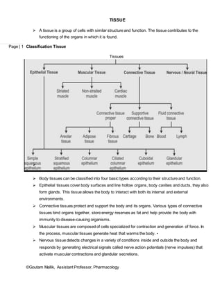
  • 1. ©Goutam Mallik, Assistant Professor, Pharmacology Page | 1 TISSUE  A tissue is a group of cells with similar structure and function. The tissue contributes to the functioning of the organs in which it is found. Classification Tissue Tissues   Body tissues can be classified into four basic types according to their structure and function.  Epithelial tissues cover body surfaces and line hollow organs, body cavities and ducts, they also form glands. This tissue allows the body to interact with both its internal and external environments.  Connective tissues protect and support the body and its organs. Various types of connective tissues bind organs together, store energy reserves as fat and help provide the body with immunity to disease-causing organisms.  Muscular tissues are composed of cells specialized for contraction and generation of force. In the process, muscular tissues generate heat that warms the body. •  Nervous tissue detects changes in a variety of conditions inside and outside the body and responds by generating electrical signals called nerve action potentials (nerve impulses) that activate muscular contractions and glandular secretions.
  • 2. ©Goutam Mallik, Assistant Professor, Pharmacology Page | 2 Fig: Epithelial Tissue EPITHELIAL TISSUES  Epithelial tissue or an epithelium (plural: epithelia), is a sheet of cells that covers a body surface or lines a body cavity (epithe = laid on, covering).  Epithelia form boundaries between different environments. For example, the epidermis of the skin lies between the inside and the outside of the body and epithelium lining the urinary bladder separates underlying cells of the bladder wall from urine.  It occurs in the body as: A. Covering and lining epithelium: Covering and lining epithelium forms the outer layer of the skin, dips into and lines the open cavities of the cardiovascular system, digestive system, respiratory systems and covers the walls and organs of the closed ventral body cavities. B. Glandular epithelium: The function of glandular epithelium is secretion, which is accomplished by glandular cells that often lie in clusters deep to the covering and lining epithelium. EPITHELIAL TISSUE COVERING AND LINING EPITHELIUM  On the basis of the two characteristics (arrangements of layers and cell shapes), epithelial tissues are classified into following types :  Simple epithelium: (A) Simple squamous epithelium. (B) Simple cuboidal epithelium. (C) Simple columnar epithelium (nonciliated and ciliated). (D) Pseudostratified columnar epithelium (nonciliated and ciliated).
  • 3. ©Goutam Mallik, Assistant Professor, Pharmacology Page | 3  Stratified epithelium: (A) Stratified squamous epithelium (keratinized, when surface cells are dead and become hardened and nonkeratinized, when surface cells remain alive). (B) Stratified cuboidal epithelium. (C) Stratified columnar epithelium. (D) Transitional epithelium. Simple epithelium (A) Simple squamous epithelium:  Single layer of flat cells that resembles a tiled floor when viewed from apical surface; centrally located nucleus that is flattened and oval or spherical in shape.  Location: Most commonly lines the cardiovascular and lymphatic system (heart, blood vessels, lymphatic vessel linings), where it is known as endothelium and forms the epithelial layer of serous membranes (peritoneum, pleura, pericardium), where it is called mesothelium. Also found in air sacs of lungs, glomerular (Bowman’s) capsule of kidneys, inner surface of tympanic membrane (eardrum).  Function: Present at sites of filtration (such as blood filtration in kidneys) or diffusion (such as diffusion of oxygen into blood vessels of lungs) and at site of secretion in serous membranes. (B) Simple cuboidal epithelium:  Single layer of cube-shaped cells, round, centrally located nucleus.  Cuboidal cell shape is obvious when tissue is sectioned and viewed from the side.  Location: Covers surface of ovary, lines anterior surface of capsule of lens of the eye, forms pigmented epithelium at posterior surface of retina of the eye. Lines kidney tubules and smaller ducts of many glands. Makes up secreting portion of some glands, such as thyroid gland and ducts of some glands such as pancreas.  Function: Secretion and absorption. (C) Nonciliated simple columnar epithelium:  Single layer of nonciliated column like cells with oval nuclei near base of cells, contains (1) columnar epithelial cells with microvilli at apical surface and (2) goblet cells.  Microvilli, finger like cytoplasmic projections, increase surface area of plasma membrane, thus increasing cell’s rate of absorption.  Goblet cells are modified columnar epithelial cells that secrete mucus, a slightly sticky fluid, at their apical surfaces.
  • 4. ©Goutam Mallik, Assistant Professor, Pharmacology Page | 4  Location: Lines gastrointestinal tract (from stomach to anus), ducts of many glands and gallbladder.  Function: Secretion and absorption, larger columnar cells contain more organelles and thus are capable of higher level of secretion and absorption than are cuboidal cells. (D) Ciliated simple columnar epithelium:  Single layer of ciliated column like cells with oval nuclei near base of cells. Goblet cells are usually interspersed among ciliated columnar epithelia.  Location: Lines some bronchioles (small tubes) of respiratory tract, uterine (fallopian) tubes, uterus, some paranasal sinuses, central canal of spinal cord and ventricles of brain.  Function: Cilia beat in unity, moving mucus and foreign particles toward throat, where they can be coughed up and swallowed or spit out. Cilia also help move oocytes expelled from ovaries through uterine (fallopian) tubes into uterus. Fig: Complex Derived Epithelial Tissue
  • 5. ©Goutam Mallik, Assistant Professor, Pharmacology Page | 5 (E) Pseudostratified columnar epithelium:  Appears to have several layers because cell nuclei are at various levels. All cells are attached to basement membrane in a single layer, but some cells do not extend to apical surface.  When viewed from side, these features give false impression of a multi-layered tissue.  Pseudostratified ciliated columnar epithelium contains cells that extend to surface and secrete mucus (goblet cells) or bear cilia.  Pseudostratified nonciliated columnar epithelium contains cells without cilia and lacks goblet cells.  Location: Ciliated variety lines airways of most of upper respiratory tract, nonciliated variety lines larger ducts of many glands, epididymis and part of male urethra.  Function: Ciliated variety secretes mucus that traps foreign particles, and cilia sweep away mucus for elimination from body. Nonciliated variety functions in absorption and protection. Stratified epithelium (A) Stratified squamous epithelium:  Two or more layers of cells, cells in apical layer and several layers deep to it are squamous, cells in deeper layers vary from cuboidal to columnar.  Tough proteins predominate as cytoplasm is reduced and cells become tough, hard structures that eventually die.  At apical layer, after dead cells lose cell junctions they are sloughed off, but they are replaced continuously as new cells emerge from basal cells.  Keratinized stratified squamous epithelium develops tough layer of keratin in apical layer of cells and several layers deep to it.  Non keratinized stratified squamous epithelium does not contain large amounts of keratin in apical layer and several layers deep and is constantly moistened by mucus from salivary and mucous glands, organelles are not replaced.  Location: Keratinized variety forms superficial layer of skin, nonkeratinized variety lines wet surfaces (lining of mouth, esophagus, part of epiglottis, part of pharynx and vagina) and covers tongue.  Function: Protection against abrasion, water loss, ultraviolet radiation and foreign invasion. Both types form first line of defense against microbes. (B) Stratified cuboidal epithelium:  Two or more layers of cells, cells in apical layer are cube-shaped, fairly rare type.  Location: Ducts of adult sweat glands and esophageal glands, part of male urethra.  Function: Protection, limited secretion and absorption.
  • 6. ©Goutam Mallik, Assistant Professor, Pharmacology Page | 6 (C) Stratified columnar epithelium:  Basal layers usually consist of shortened, irregularly shaped cells, only apical layer has columnar cells, uncommon.  Location: Lines part of urethra, large excretory ducts of some glands, such as esophageal glands, small areas in anal mucous membrane, part of conjunctiva of eye.  Function: Protection and secretion. (D) Transitional epithelium:  Variable appearance (transitional). In relaxed or unstretched state, looks like stratified cuboidal epithelium, except apical layer cells tend to be large and rounded.  As tissue is stretched, cells become flatter, giving the appearance of stratified squamous epithelium.  Multiple layers and elasticity make it ideal for lining hollow structures (urinary bladder) subject to expansion from within.  Location: Lines urinary bladder and portions of ureters and urethra.  Function: Allows urinary organs to stretch and maintain protective lining while holding variable amounts of fluid without rupturing. GLANDULAR EPITHELIUM  Two types of glandular epithelium exists: (A) Exocrine (B) Endocrine (A) Epithelial tissue glandular epithelium (Exocrine):  Secretory products released into ducts that empty onto surface of a covering and lining epithelium, such as skin surface or lumen of hollow organ.  Location: Sweat, oil and earwax glands of skin, digestive glands such as salivary glands (secretes into mouth cavity) and pancreas (secretes into small intestine).  Function: Produce substances such as sweat to help lower body temperature, oil, earwax, saliva, or digestive enzymes. (B) Epithelial tissue glandular epithelium (Endocrine):  Secretions (hormones) enter interstitial fluid and diffuse directly into bloodstream without flowing through a duct.  Location: Examples include pituitary gland at base of brain, pineal gland in brain, thyroid and parathyroid glands near larynx (voice box), adrenal glands superior to kidneys, pancreas near stomach, ovaries in pelvic cavity, testes in scrotum, thymus in thoracic cavity.  Function: Hormones regulate many metabolic and physiological activities to maintain homeostasis.
  • 7. ©Goutam Mallik, Assistant Professor, Pharmacology Page | 7 Fig: Summary of Epithelial Tissues *********************************** Reference 1. Human Anatomy & Physiology-I, by Mrs. Shubhada Mangrulkar & Ms. Nitu L. Wankhede, TPS Publication 2. Essentials of Human Anatomy & Physiology 5th Ed by Valerie C. Scanlon & Tina Sanders 3. Gray’s Anatomy, The Anatomical Basis of Clinical Practice 41st Ed by Elsevier