
Recomendados
Mais conteúdo relacionado
Semelhante a Nervous system Sisay A..pdf
Semelhante a Nervous system Sisay A..pdf (20)
Mais de GedamuDereje
Mais de GedamuDereje (12)
Último
Último (20)
Nervous system Sisay A..pdf
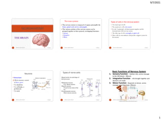
- 1. 9/7/2021 1 THE BRAIN NEUROANATOMY 9/7/2021 Nervous system Sisay A. 1 Nervous system Nervous system The nervous system is composed of organs, principally the brain, spinal cord, nerves, and ganglia. The various activities of the nervous system can be grouped together as three general, overlapping functions: Sensory Integrative Motor The nervous system is composed of organs, principally the brain, spinal cord, nerves, and ganglia. The various activities of the nervous system can be grouped together as three general, overlapping functions: Sensory Integrative Motor 9/7/2021 Nervous system Sisay A. 2 Types of cells in the nervous system Two main types of cells The actual nerve cell is the neuron It is the "conducting" cell that transmits impulses and the structural unit of the nervous system The other type of cell is neuroglia,or glial cell. These cells are nonconductive and provide a support system for the neurons. 9/7/2021 Nervous system Sisay A. 3 Neurons Structure Most neurons consist of three parts The dendrites The cell body, or perikaryon The axon 9/7/2021 Nervous system Sisay A. 4 Types of nerve cells •Based on the size and shape of their processes •Multipolar neurons •E.g- Motor neurons •Bipolar neurons •Found in the retina of the eye •Pseudounipolar neurons •Most sensory neurons 9/7/2021 Nervous system Sisay A. 5 Types of nerve cells 9/7/2021 Nervous system Sisay A. 6
- 2. 9/7/2021 2 Types of glial cells Glial Cell Type Origin Location Main Functions Oligodendrocyte Neural tube Central nervous system Myelin production, electric insulation Schwann cell Neural tube Peripheral nerves Myelin production, electric insulation Astrocyte Neural tube Central nervous system Structural support, repair processes Ependymal cell Neural tube Central nervous system Lining cavities of central nervous system Microglia Bone marrow Central nervous system Macrophagic activity Ganglionic gliocytes Neural tube Peripheral nerves ganglia Support ganglia in the PNS 9/7/2021 Nervous system Sisay A. 7 Covering & protection of CNS Includes The skull –protects the brain The vertebral column - protects the spinal cord The meninges and cerebrospinal fluid protect the central nervous system. Meninges Three connective tissue membranes that lies external to the CNS Dura Mater Arachnoid Mater Pia Mater Covering & protection of CNS Includes The skull –protects the brain The vertebral column - protects the spinal cord The meninges and cerebrospinal fluid protect the central nervous system. Meninges Three connective tissue membranes that lies external to the CNS Dura Mater Arachnoid Mater Pia Mater 9/7/2021 Nervous system Sisay A. 8 Dura Mater Dura Mater Three dural septa extend inward and limit excessive movement of the brain Falx cerebri – fold that dips into the longitudinal fissure Falx cerebelli – runs along the vermis of the cerebellum Tentorium cerebelli – horizontal dural fold extends into the transverse fissure 9/7/2021 Nervous system Sisay A. 9 Arachnoid Mater It is separated from the dura mater by the subdural space Beneath the arachnoid is a wide subarachnoid space filled with CSF and large blood vessels Pia Mater Between the arachnoid and the pia mater is the subarachnoid space. 9/7/2021 Nervous system Sisay A. 10 9/7/2021 Nervous system Sisay A. 11 9/7/2021 Nervous system Sisay A. 12
- 3. 9/7/2021 3 Organization of nervous system Organization of nervous system 9/7/2021 Nervous system Sisay A. 13 14 Subdivisions of Nervous System 9/7/2021 Nervous system Sisay A. THE BRAIN 9/7/2021 Nervous system Sisay A. 15 Regions of the Brain • The four main regions of the brain are: Cerebral hemi- spheres Diencephalon • Thalamus • Hypothalamus Brain stem • Midbrain • Pons • Medulla Cerebellum 9/7/2021 Nervous system Sisay A. 16 Development of Brain Beginning of the 3rd week ectoderm-neural tube- 3 swellings are formed The three primary brain vesicles – forebrain (prosencephalon) – midbrain (mesencephalon) – hindbrain (rhombencephalon) 17 9/7/2021 Nervous system Sisay A. • During the 5th week: Consequently, there are five secondary brain vesicles. 9/7/2021 Nervous system Sisay A. 18
- 4. 9/7/2021 4 Gray & White Matter 19 Microscopically, the CNS contains 2 neural elements: Neuron cell bodies (clusters are known as nuclei) Nerve fibers (axons) in bundles called tracts. Viewed macroscopically, CNS tissues can be distinguished by color: Gray matter consists of somata, dendrites, and unmyelinated axons White matter consists primarily of myelinated axons and dendrites 9/7/2021 Nervous system Sisay A. 9/7/2021 Nervous system Sisay A. 20 The pattern of white & gray matter changes with descent thru the brain stem The cortex disappears, but scattered gray matter nuclei are seen within the white matter At the caudal end of the brain stem the basic pattern is evident 9/7/2021 Nervous system Sisay A. 21 Cerebrum it is divided into two halves called cerebral hemispheres by longitudinal cerebral fissure. They are connected by corpus callosum Separated from cerebellum posteriorly by transverse fissure called tentorium cerebelli. 9/7/2021 Nervous system Sisay A. 22 Cerebrum 9/7/2021 Nervous system Sisay A. 23 Insula Each hemisphere has a covering of gray matter, the cortex and internal masses of white matter, the basal nuclei, and a lateral ventricle. Cerebrum 9/7/2021 Nervous system Sisay A. 24
- 5. 9/7/2021 5 The Cerebral Hemispheres The cerebral hemispheres form the superior part of the brain These two structures account for about 80% of the mass of the brain The two hemispheres cover & obscure the diencephalon & the top of the brain stem 9/7/2021 Nervous system Sisay A. 25 Nearly the entire surface of the cerebral hemispheres is marked by elevated ridges of tissues called gyri separated by shallow grooves called sulci The transverse fissure separates the cerebral hemispheres from the cerebellum below 9/7/2021 Nervous system Sisay A. 26 Lobes of Cerebral Hemispheres Deeper sulci divide each hemisphere into five lobes Frontal lobe Temporal lobe Parietal lobe Occipital lobe Insula (located within the lateral sulcus) Insula 9/7/2021 Nervous system Sisay A. 27 Cerebrum 28 9/7/2021 Nervous system Sisay A. Boundaries of the lobes of the hemisphere The Frontal Lobe lies : in front of the central sulcus above lateral sulcus The Parietal Lobe bounded : Anteriorly: by the central sulcus Posteriorly: parieto occipital sulcus Inferiorly: lateral sulcus 9/7/2021 Nervous system Sisay A. 29 The temporal lobe is bounded: Superiorly: lateral sulcus Posteriorly: parieto-occipital sulcus The occipital lobe: lies behind parieto-occipital sulcus 9/7/2021 Nervous system Sisay A. 30
- 6. 9/7/2021 6 Sulci & Gyri of the Cerebral hemisphere • The surfaces of the hemisphere is formed of grey matter & called cerebral cortex • Each surface is divided by sulci (grooves) into gyri (convolutions) • These infoldings result in marked increase in the surface area of the cerebral cortex without increasing the size of the cerebral hemisphere. 9/7/2021 Nervous system Sisay A. 31 Sulcus of Cerebral Hemispheres Sulci divide lobes of the hemispheres Central sulcus Parieto- occipital sulcus Lateral sulcus Transverse fissure 9/7/2021 Nervous system Sisay A. 32 The sulci & Gyri of the lateral surface Frontal lobe Important sulci: Precentral sulcus: Sup. frontal sulcus Inf. frontal sulcus: Important Gyri: precentral gyrus: superior frontal gyrus: middle frontal gyrus: inferior frontal gyrus 9/7/2021 Nervous system Sisay A. 33 9/7/2021 Nervous system Sisay A. 34 Parietal lobe Important Sulci: The postcentral sulcus: The intra parietal sulcus: Important Gyri The postcentral gyrus: The superior parietal gyrus: The inferior parietal gyrus: The supramarginal gyrus: The angular gyrus : 9/7/2021 Nervous system Sisay A. 35 Temporal lobe Sulci: • The superior temporal sulcus: • The middle temporal sulcus : Gyri: • The sup temporal gyrus: • The middle temporal gyrus: • The inferior temporal gyrus 9/7/2021 Nervous system Sisay A. 36
- 7. 9/7/2021 7 Occipital lobe The main part of the occipital lobe lies on the medial surface Only a small part appears on the lat surface & presents one sulcus called the transverse occipital sulcus 9/7/2021 Nervous system Sisay A. 37 The Insula is hidden in the bottom of the lateral sulcus Function-Memory; integration of other cerebral activities 9/7/2021 Nervous system Sisay A. 38 White matter of cerebral hemispheres Composed of myelinated nerve fibers of different diameters The nerve fibers classified in to three groups. Commissural Association Projection fibers 9/7/2021 Nervous system Sisay A. 39 White matter of cerebral hemispheres Association fibers confined to a given cerebral hemisphere and conduct impulses between neurons within that hemisphere 9/7/2021 Nervous system Sisay A. 40 White matter of cerebral hemispheres Commissural fibers Connect the neurons and gyri of one hemisphere with those of the other They are corpus callosum, anterior commissure, posterior commissure, fornix 9/7/2021 Nervous system Sisay A. 41 White matter of cerebral hemispheres Projecting fibers Form the ascending and descending tracts that transmit impulses From the cerebrum to other parts of the brain and spinal cord From the spinal cord and other parts of the brain to the cerebrum 9/7/2021 Nervous system Sisay A. 42
- 8. 9/7/2021 8 Structure of the cerebral cortex Cerebral cortex forms a complete covering of the cerebral hemispheres Composed of gray matter probably contains in excess of 10 billion neurons The cerebral cortex consists of a mixtures of Nerve cells Nerve fibers Neuroglia Blood vessels 9/7/2021 Nervous system Sisay A. 43 Cerebral Cortex • Research on the structure & function of the brain reveals that there are both specialized & diffuse areas of function • Motor & sensory areas are localized in discrete cortical areas called domains • Many higher mental functions such as memory & language appear to have overlapping domains & are more diffusely located • Broadmann areas are areas of localized function 9/7/2021 Nervous system Sisay A. 44 Cerebral Cortex - Generalizations The cerebral cortex has three types of functional areas Motor areas / control voluntary motor function Sensory areas / provide conscious awareness of sensation Association areas / act mainly to integrate diverse information for purposeful action 9/7/2021 Nervous system Sisay A. 45 functional areas of cerebral hemispheres Cortical areas controlling motor functions lie in the posterior part of the frontal lobes Motor areas include the primary motor cortex premotor cortex Broca’s area front eye field Prefrontal cortex 9/7/2021 Nervous system Sisay A. 46 9/7/2021 Nervous system Sisay A. 47 Frontal Lobe The primary Motor area (area 4) occupies the precentral gyrus Control voluntary contraction of specific muscle Premotor area (area 6): Planning based on past experience Damage results in the loss of the motor skills in that region 9/7/2021 Nervous system Sisay A. 48
- 9. 9/7/2021 9 Broca’s Speach area (area 44,45) in the post part of inf. frontal gyrus formation of words Injury causes loss of ability to produce speech, or expressive aphasia 9/7/2021 Nervous system Sisay A. 49 Frontal eye field (area 8): controls the voluntary movements of the eyes inability to turn the eyes to the opposite side 9/7/2021 Nervous system Sisay A. 50 9/7/2021 Nervous system Sisay A. 51 Prefrontal cortex (areas 9, 10, 11 & 12) for abstract ideas, reasoning & judgment, impulse control, persistence, long term planning Injury may cause mental & personality disorders 9/7/2021 Nervous system Sisay A. 52 9/7/2021 Nervous system Sisay A. 53 Sensory Areas Areas concerned with the conscious awareness of sensation in the parietal, temporal & occipital lobes 9/7/2021 Nervous system Sisay A. 54
- 10. 9/7/2021 10 Parietal Lobe Primary somatosensory cortex (Main sensory area ): area 3,1,2 occupies the postcentral gyrus It receives pain, touch, proprioception, temperature & taste sensations from the opposite ½ of the body Lesion to this area results in loss of tactile discrimination from the opposite ½ of the body • Somatosensory association cortex (areas 5&7 ) integrate & analyze different somatic sensory inputs This loss of integration of sensory impulses is called astereognosis 9/7/2021 Nervous system Sisay A. 55 Temporal Lobe The primary auditory area (41 & 42- reception of sound The secondary auditory or auditory association areas (21 & 22)= interpretation sensory speech area of Wernicke understanding of the written and spoken language 9/7/2021 Nervous system Sisay A. 56 The occipital Lobe The primary visual area (17)-reception of vision The secondary visual areas (18 & 19)= recognize and appreciate what he or she is seeing. 9/7/2021 Nervous system Sisay A. 57 Basal ganglia (basal nuclei) Is applied to a collection of masses of gray matter They are o Corpus striatum, o Amygdaloid nucleus and o Claustrum control unconscious contractions of certain skeletal muscles 9/7/2021 Nervous system Sisay A. 58 9/7/2021 Nervous system Sisay A. 59 The Diencephalon Extends posteriorly to the cerebral aqueduct & anteriorly to Interventricular foramen. includes; the thalamus the hypothalamus, the sub thalamus, the epithalamus. 9/7/2021 Nervous system Sisay A. 60
- 11. 9/7/2021 11 Thalamus Relay center for all sensory impulses, except smell, to the cerebral cortex and performs some sensory interpretation The hypothalamus acts as an autonomic nervous center Cardiovascular regulation Body-temperature regulation Regulation of water and electrolyte balance Regulation of hunger and control of gastrointestinal activity Regulation of sleeping and wakefulness Sexual response Emotions Control of endocrine functions 9/7/2021 Nervous system Sisay A. 61 The ventricles (cavities) of the brain Are series of interconnected, fluid-filled cavities found within the brain Two lateral ventricles, the third ventricle, and the fourth ventricle 9/7/2021 Nervous system Sisay A. 62 Ventricles of the Brain Lateral Third Fourth 9/7/2021 Nervous system Sisay A. 63 Lateral Ventricle Lie within each cerebral hemisphere C-shaped cavity has Body in parietal lobe Anterior horn in frontal lobe Posterior horn in occipital lobe Inferior horn in temporal lobe 9/7/2021 Nervous system Sisay A. 64 Third ventricle Slit like cleft between two thalami Derived from forebrain vesicle 9/7/2021 Nervous system Sisay A. 65 Aqueduct of Sylivius channel connecting 3rd to 4th ventricle Surrounded by a layer of gray matter called central gray no choroid plexus 9/7/2021 Nervous system Sisay A. 66
- 12. 9/7/2021 12 Fourth Ventricle Anterior to cerebellum and posterior to pons and cranial half of medulla oblongata 9/7/2021 Nervous system Sisay A. 67 Communication between ventricles Interventricular foramina (of Monro) Aqueduct of Sylivius Cerebrospinal fluid exits from the fourth ventricle into the subarachnoid space via Foramina of Magndie Foramina of Luschka 9/7/2021 Nervous system Sisay A. 68 CSF Formed in choroid plexus The cerebrospinal fluid then circulates in and around the CNS Reabsorbed through arachnoid villi into the blood in cranial venous sinuses . Formed in choroid plexus The cerebrospinal fluid then circulates in and around the CNS Reabsorbed through arachnoid villi into the blood in cranial venous sinuses . 9/7/2021 Nervous system Sisay A. 69 9/7/2021 Nervous system Sisay A. 70 Brain stem 9/7/2021 Nervous system Sisay A. 71 Brain stem is made up of:- Medulla oblongata, Pons and Midbrain It occupy the posterior cranial fossa Connect spinal cord with forebrain Function:- serves as a conduit for ascending and descending tract connecting spinal cord to forebrain It contain important reflex center( respiratory ,cvs & consciousness) It contain important nuclei of CN 9/7/2021 Nervous system Sisay A. 72
- 13. 9/7/2021 13 Medulla oblongata Gross appearance;- connect pons superiorly & s.c inferior - its upper half expand to the cavity of 4th v. Anterior surface- is a median fissure Pyramid:- contain the motor fibers ; corticobulbarand corticospinal fibers 9/7/2021 Nervous system Sisay A. 73 Medulla oblongata The olive – oval elevation produced by inf. Olivary nuclei. CN XII emerge b/n pyramid & olive Inf .cerebellar peduncles- lie post to olives - connect medulla with cerebellum - CN IX, X & XI emerge b/n ICP and olive. 9/7/2021 Nervous system Sisay A. 74 Medulla oblongata Posterior surface:- sup. half form floor of 4th v. -inf. half is continuous with s.c and posses post. median sulcus Gracile tubercle:- produced by gracile nucleus on each side of median sulcus. Cuneate tubercle:- produced by cuneate nucleus , lateral to Gracile tubercle. 9/7/2021 Nervous system Sisay A. 75 PONS - is a bridge connecting Rt. and Lt. cerebellar hemispheres Lei ant. to cerebellum, connect MO with midbrain Has many transverse fiber and basilary groove CNV emerge in its anterolateral surface. CNVI:- lei in the most inf. Part of pons CNVII :- motor nuclei in the most inf. Part of pons CNVIII :- sensory root in the most inf. Part of pons Post surface form floor of 4thV 9/7/2021 Nervous system Sisay A. 76 Midbrain It connect Diencephalons to the Pons & Cerebellum. Topographically it lies in posterior fossa . Traversed by cerebellar aqueduct Posteriorly has -sup colliculus –visual &-inf colliculus - auditory 9/7/2021 Nervous system Sisay A. 77 9/7/2021 Nervous system Sisay A. 78
- 14. 9/7/2021 14 Midbrain on its lateral side :- - sup brachium pass from sup colliculus to lateral geniculate body - inf brachium connect inf colliculus to medial geniculate body On its anterior part is interpedencular fossa 9/7/2021 Nervous system Sisay A. 79 Midbrain INTERPEDUNCULAR FOSSA Boundaries:- -Optic chiasma anteriorly,anterolaterally optic tract, posterolaterally crus cerebri & posteriorly Pons. Contents - occulomotor nerve - mammilary body -posterior cerebellar aa - tuber cinerium - posterior perforated substance - infundibulum 9/7/2021 Nervous system Sisay A. 80 Cerebellum (Little Brain) Location Posterior cranial fossa Posterior to 4th ventricle, pons & medulla Has two hemispheres & vermis Connected to posterior part of brain stem by three symmetrical bundles of nerve fibers (cerebellar peduncle) 9/7/2021 Nervous system Sisay A. 81 cerebellum Each cerebellar hemisphere has 3 anatomical lobes Anterior Posterior Flocculonodular Has fissures Primary Horizontal Uvulonodula 9/7/2021 Nervous system Sisay A. 82 Structure of Cerebellum Has outer gray and inner white matter Intracellebellar nuclei embedded in white matter 9/7/2021 Nervous system Sisay A. 83 Intracerebellar Nuclei Four masses of gray matter embedded in white matter on each side of the midline Dentate, emboliform, globose and fastigial 9/7/2021 Nervous system Sisay A. 84
- 15. 9/7/2021 15 CEREBELLAR PEDUNCLES Afferent & efferent bundles of nerve fibers Link cerebellum with other parts of CNS Superior CP- Midbrain Middle CP -Pons Largest of the peduncles Inferior CP - M.oblongata, vestibular nuclei, cells of the reticular formation 9/7/2021 Nervous system Sisay A. 85 Functions of cerebellum It serves as: 1. A coordinator of motor activity 2. A regulator of muscle tone 3. Functions to maintain balance and equilibrium 4. Participates in motor planning 5. Plays a role in controlling visceral activity 9/7/2021 Nervous system Sisay A. 86 Functional Areas of Cerebellar Cortex 1. Cortex of Vermis Influence movement of long axis of the body Spinocerebellar tracts sends projections to the fastigial nucleus of the cerebellum, which then sends output to the vestibular nuclei 2. Intermediate Zone CH Receives input from the corticopontocerebellar fibers Receives sensory feedback from the muscles Muscles of distal parts of limbs e.g. hands & feet 3. Lateral Zone CH Receives input from the parietal cortex via pontocerebellar mossy fibers Information regarding the location of the body in the world The large numbers of feedback circuits allow for the integration of this body position information with indications of muscle position, strength, and speed. Planning of sequential movements of entire body& assess movement errors 9/7/2021 Nervous system Sisay A. 87 Blood supply of brain 9/7/2021 Nervous system Sisay A. 88 Circle ofWillis The anterior communicating anterior cerebral internal carotid posterior communicating posterior cerebral basilar arteries 9/7/2021 Nervous system Sisay A. 89 ARTERIES OF SPECIFIC BRAIN AREAS 1. The corpus striatum and the internal capsule are supplied by middle cerebral artery& anterior cerebral artery 2. The thalamus posterior communicating basilar Posterior cerebral 3. The midbrain posterior cerebral superior cerebral basilar arteries 4. Pons basilar anterior, inferior and superior cerebellar arteries 5. Medulla oblongata vertebral, anterior and posterior spinal, posterior inferior cerebellar& basilar 6. Cerebellum Superior cerebellar,anterior inferior cerebellar and, posterior inferior cerebellar arteries 9/7/2021 Nervous system Sisay A. 90
- 16. 9/7/2021 16 Cranial Nerves 9/7/2021 Nervous system Sisay A. 91 There are 12 pairs of cranial nerves - Cranial nerves I and II attach to the forebrain /telencephalon & diencephalon respectively/ - All others attach to the brain stem There are 12 pairs of cranial nerves - Cranial nerves I and II attach to the forebrain /telencephalon & diencephalon respectively/ - All others attach to the brain stem 9/7/2021 Nervous system Sisay A. 92 Functional group of cranial nerves Functional group of cranial nerves Types of cranial nerves I, II,VIII are entirely sensory. III, IV,VI, XI, XII are entirely motor. V,VII, IX, X are mixed. Types of cranial nerves I, II,VIII are entirely sensory. III, IV,VI, XI, XII are entirely motor. V,VII, IX, X are mixed. 9/7/2021 Nervous system Sisay A. 93 Cranial nerve I :Olfactory nerve Cranial nerve I :Olfactory nerve Functions- sense of smell Location of Cell Bodies -Within Sensory Organ or Ganglia (Olfactory mucosa) - Major connections - Mucosa projects to olfactory bulb Functions- sense of smell Location of Cell Bodies -Within Sensory Organ or Ganglia (Olfactory mucosa) - Major connections - Mucosa projects to olfactory bulb 9/7/2021 Nervous system Sisay A. 94 Cranial Nerve II: Optic Nerve Cranial Nerve II: Optic Nerve Contains myelinated axons that arise from the ganglion cells in the retina and passes through the optic papilla to the orbit. The nerve changes its name to optic tract when the fibers have passed through the optic chiasm Contains myelinated axons that arise from the ganglion cells in the retina and passes through the optic papilla to the orbit. The nerve changes its name to optic tract when the fibers have passed through the optic chiasm 9/7/2021 Nervous system Sisay A. 95 Optic tract axons project to the superior colliculus and to the lateral geniculate nucleus within the thalamus, which relays visual information to the cortex. Function - vision Optic tract axons project to the superior colliculus and to the lateral geniculate nucleus within the thalamus, which relays visual information to the cortex. Function - vision Cranial Nerve II: Optic Nerve 9/7/2021 Nervous system Sisay A. 96
- 17. 9/7/2021 17 Cranial Nerve III: Oculomotor Nerve Cranial Nerve III: Oculomotor Nerve Functions - Supply extrinsic mm of the eye (Motor to Medial rectus, superior rectus, inferior rectus, inferior oblique & levator palpebrae superioris muscle ),constrictor pupillae and ciliary mm Location of Cell Bodies – Within midbrain (occulomotor nucleus) & (Edinger-Westphal nucleus). Functions - Supply extrinsic mm of the eye (Motor to Medial rectus, superior rectus, inferior rectus, inferior oblique & levator palpebrae superioris muscle ),constrictor pupillae and ciliary mm Location of Cell Bodies – Within midbrain (occulomotor nucleus) & (Edinger-Westphal nucleus). 9/7/2021 Nervous system Sisay A. 97 Cranial Nerve IV:Trochlear Nerve Cranial Nerve IV:Trochlear Nerve Functions- Motor to Superior oblique muscle Location of Cell Bodies –Within Brain Stem –Trochlear nucleus Functions- Motor to Superior oblique muscle Location of Cell Bodies –Within Brain Stem –Trochlear nucleus 9/7/2021 Nervous system Sisay A. 98 Cranial nerveV :TRIGEMINAL NERVE Cranial nerveV :TRIGEMINAL NERVE Largest , mixed ( have both motor & sensory part) Has 3 divisions: - Ophthalmic, Maxillary, & Mandibular. Motor component of the Trigeminal N Motor nucleus supplies: 1.Muscles of mastication 2.Anterior belly of the digastrics 3.Tensor tympani 4.Tensor veli palatine 5.Mylohyoid Largest , mixed ( have both motor & sensory part) Has 3 divisions: - Ophthalmic, Maxillary, & Mandibular. Motor component of the Trigeminal N Motor nucleus supplies: 1.Muscles of mastication 2.Anterior belly of the digastrics 3.Tensor tympani 4.Tensor veli palatine 5.Mylohyoid 9/7/2021 Nervous system Sisay A. 99 Sensory fibers supplies the face and parts of the scalp Sensory fibers supplies the face and parts of the scalp 9/7/2021 Nervous system Sisay A. 100 Purely motor Emerges from the anterior surface of the brain, in the groove between the lower border of the pons and the medullaoblogata. Finally, it runs forward and supplies the lateral rectus muscle. Purely motor Emerges from the anterior surface of the brain, in the groove between the lower border of the pons and the medullaoblogata. Finally, it runs forward and supplies the lateral rectus muscle. Cranial NerveVI : Abducent Nerve Cranial NerveVI : Abducent Nerve 9/7/2021 Nervous system Sisay A. 101 The facial nerve consists of a motor and a sensory part The two parts emerge at the lower border of the pons in the recess between the olive and the inferior peduncle The facial nerve consists of a motor and a sensory part The two parts emerge at the lower border of the pons in the recess between the olive and the inferior peduncle Facial nerve: CNVII 9/7/2021 Nervous system Sisay A. 102
- 18. 9/7/2021 18 Vestibular Nerve Transmits information about position and movement of the head Its nerve fiber are Central processes of nerve cell located in vestibular ganglion situated in internal acoustic meatus Vestibular Nerve Transmits information about position and movement of the head Its nerve fiber are Central processes of nerve cell located in vestibular ganglion situated in internal acoustic meatus CNVIII:Vestibulocochlear Nerve CNVIII:Vestibulocochlear Nerve 9/7/2021 Nervous system Sisay A. 103 Its nerve fiber are Central processes of nerve cell located in spiral ganglion of the cochlea On reaching the mid brain the fiber terminate on inf. Colliculus or medial geniculate body. Leave posterior cranial fossa via internal acoustic meatus to inner ear. Its nerve fiber are Central processes of nerve cell located in spiral ganglion of the cochlea On reaching the mid brain the fiber terminate on inf. Colliculus or medial geniculate body. Leave posterior cranial fossa via internal acoustic meatus to inner ear. Cochlear Nerve :- conduct impulse concerned with sound Cochlear Nerve :- conduct impulse concerned with sound 9/7/2021 Nervous system Sisay A. 104 Mixed Has 3 nuclei:- two motor & one sensory Main motor supply stylopharyngus msc. Parasympathetic supply – parotid gland Sensory – common & taste sensation Mixed Has 3 nuclei:- two motor & one sensory Main motor supply stylopharyngus msc. Parasympathetic supply – parotid gland Sensory – common & taste sensation CN IX: Glossopharyngeal Nerve CN IX: Glossopharyngeal Nerve 9/7/2021 Nervous system Sisay A. 105 CN X:VAGUS NERVE CN X:VAGUS NERVE Is mixed Has three nucleus Main motor nucleus - Efferent fibers supply constrictor muscles of pharynx and intrinsic mm of larynx. Parasympathetic nucleus - Efferent fibers to abdominal viscera Sensory nucleus- common sensation & taste sensation. Is mixed Has three nucleus Main motor nucleus - Efferent fibers supply constrictor muscles of pharynx and intrinsic mm of larynx. Parasympathetic nucleus - Efferent fibers to abdominal viscera Sensory nucleus- common sensation & taste sensation. 9/7/2021 Nervous system Sisay A. 106 CN XI:Accessory Nerve CN XI:Accessory Nerve Is Purely Motor Has spinal and cranial root. The 2 roots exit via jugular foramen Join vagus and distribute in its pharyngeal and recurrent laryngeal branch to mm of soft palate, pharynx & larynx. Spinal root Supply trapezius and SCM mm Is Purely Motor Has spinal and cranial root. The 2 roots exit via jugular foramen Join vagus and distribute in its pharyngeal and recurrent laryngeal branch to mm of soft palate, pharynx & larynx. Spinal root Supply trapezius and SCM mm 9/7/2021 Nervous system Sisay A. 107 Hypoglossal nerve : CN XII Origin and distribution Is purely motor Arise from medulla oblongata Supply mm of the tongue Origin and distribution Is purely motor Arise from medulla oblongata Supply mm of the tongue 9/7/2021 Nervous system Sisay A. 108
- 19. 9/7/2021 19 Spinal Cord Begin at foramen magnum and ends at L1(adult), L3(child) Surrounded by Meninges CSF Bones Stabilized in position Filum terminale Denticulate ligament Spinal nerve roots 9/7/2021 Nervous system Sisay A. 109 Spinal Cord The spinal cord is divided into 31 segments The distal end of the spinal cord (the conus medullaris) is cone shaped. A fine filament of connective tissue (the pial part of the filum terminale) continues inferiorly from the apex of the conus medullaris 9/7/2021 Nervous system Sisay A. 110 9/7/2021 Nervous system Sisay A. 111 External structure of spinal cord In cross-section,the shape of spinal cord in different regions varies. It has two major swellings or enlargements A cervical enlargement C3-T2 A Lumbosacral enlargement L1-S3 9/7/2021 Nervous system Sisay A. 112 External structure of spinal cord is marked by a number of fissures and sulci The anterior median fissure The posterior median sulcus 9/7/2021 Nervous system Sisay A. 113 Internal structure of spinal cord Has a small central canal surrounded by gray and white matter Gray Matter On cross section it is H shaped It posses Columns The ventral (or anterior) gray column The intermidiolateral gray column The dorsal gray column 9/7/2021 Nervous system Sisay A. 114
- 20. 9/7/2021 20 Structure of Gray Matter Consists of : Mixture of multipolar nerve cells arranged in groups Process of nerve cells Blood vessels Neuroglia 9/7/2021 Nervous system Sisay A. 115 Internal structure of spinal cord White Matter Columns dorsal (also termed posterior), lateral, and ventral (also termed anterior) 9/7/2021 Nervous system Sisay A. 116 Structure of white matter Consist of: Nerve fibers Neuroglia Blood vessels Arrangement of Nerve Fiber in Spinal Cord is termed asTracts Ascending (sensory, posterior) Descending (motor, anterior) Intersegmental 9/7/2021 Nervous system Sisay A. 117 The spinal cord cont… The spinal cord has two main functions Serving as a conduction pathway for impulses going to and from the brain. Serving as a reflex center 9/7/2021 Nervous system Sisay A. 118 Spinal nerves There are 31 pairs spinal nerves All of the spinal nerves are "mixed Formation The anterior root The posterior root Those fromT1 to L2 sympathetic fibers; while S2 to S4 contain parasympathetic fibers. 9/7/2021 Nervous system Sisay A. 119 Peripheral nervous system o Nervous structures outside the brain and spinal cord o Nerves allow the CNS to receive information and take action. o Functional components of the PNS Sensory inputs and motor outputs Motor -Categorized as somatic or visceral Sensory inputs -also classified as general or special 9/7/2021 Nervous system Sisay A. 120
- 21. 9/7/2021 21 9/7/2021 Nervous system Sisay A. 121 Basic structural components of PNS Sensory receptors Motor endings Nerves and ganglia 9/7/2021 Nervous system Sisay A. 122 Spinal Nerves 31 pairs of spinal nerves 8 cervical 12 thoracic 5 lumbar 5 sacral 1 coccygeal 9/7/2021 Nervous system Sisay A. 123 A spinal nerve is a mixed nerve attached to the spinal cord by a posterior root, composed of sensory fibers, and an anterior root, composed of motor fibers A spinal nerve is formed as the fibers from the posterior and anterior roots converge and emerge through an intervertebral foramen. A spinal nerve divides into several branches Posterior ramus, innervates the muscles, joints, and skin of the back along the vertebral column Anterior ramus of a spinal nerve innervates the muscles and skin on the lateral and anterior side of the trunk Except in thoracic nervesT2 throughT12, the anterior rami of the spinal nerves combine and then split again as networks of nerve fibers referred to as nerve plexuses 9/7/2021 Nervous system Sisay A. 124 Innervation of the skin: Dermatomes Dermatome – an area of skin innervated by cutaneous branches of a single spinal nerve. Upper limb – skin is supplied by nerves of the brachial plexus. Lower limb Lumbar nerves – anterior surface Sacral nerves – posterior surface 9/7/2021 Nervous system Sisay A. 125 Branches of spinal Nerves Ventral Ramus -what they innervate depends upon which part of the spinal cord is considered. Thoracic region: form intercostal nerves that innervate the intercostal muscles and the skin over the thorax Remaining spinal nerve ventral rami form five plexuses. Ventral rami of C1-C4= cervical plexus Ventral rami of C5-T1= brachial plexus Ventral rami of L1-L4= lumbar plexus Ventral rami of L4-S4= sacral plexus Ventral rami of S4 and S5= coccygeal plexus 9/7/2021 Nervous system Sisay A. 126
- 22. 9/7/2021 22 Cervical plexus Formed by ventral rami of spinal nerves C1-C4 Motor: Innervates muscles of the neck (SCM, trapezius), laryngeal muscles Sensory: Skin of upper chest, shoulder, neck, and ear 9/7/2021 Nervous system Sisay A. 127 Brachial plexus Formed by ventral rami of spinal nerves C5-T1 Five ventral rami form three trunks that separate into six divisions then form cords that give rise to: Branches/nerves Axillary Radial Musculocutaneous Ulnar Median 9/7/2021 Nervous system Sisay A. 128 Lumbosacral plexus Lumbar plexus: formed by ventral rami of spinal nerves L1-L4 Sacral plexus: formed by ventral rami of L4-S4 Usually considered together because of their close relationship Four major nerves exit and enter lower limb Obturator Femoral Tibial Common fibular (peroneal) 9/7/2021 Nervous system Sisay A. 129 Autonomic nervous system Monitor internal environment of the body Innervates smooth muscle, cardiac muscle, and glands Regulates visceral functions Heart rate, blood pressure, digestion, urination Has afferent, connector and efferent neurons Afferent neurons originate from visceral receptors Efferent pathway comprise – preganglionic and postganglionic neurons Cell bodies of preganglionic neurons are situated in the lateral grey column of sc and motor nuclei of III,VII,IX,& X CN Division – sympathetic -parasympathetic 9/7/2021 Nervous system Sisay A. 130 9/7/2021 Nervous system Sisay A. 131 Sympathetic division Innervate viscera and glands Activated during exercise, excitement, and emergencies Prepare the body for emergency condition< fight or flight> 9/7/2021 Nervous system Sisay A. 132
- 23. 9/7/2021 23 Sympathetic division 9/7/2021 Nervous system Sisay A. 133 The Parasympathetic Division Cranial outflow Comes from the brain Innervates organs of the head, neck, thorax, and abdomen Sacral outflow Supplies remaining abdominal and pelvic organs Concerned with conserving energy “rest and digest” 9/7/2021 Nervous system Sisay A. 134 9/7/2021 Nervous system Sisay A. 135 COMPARISON B/N TWO ANS DIVISION Characteristics Sympathetic Parasympathetic Action Fight or flight Rest & digest Outflow T1-L2/3 CN III, VII, IX,X, S2-S4 Preganglionic fiber Myelinated Myelinated Ganglia Para & Prevertebral Small ganglia Neurotransmitter in ganglia Ach Ach Postganglionic fiber Long non myelinated Short non myelinated Ratio of pre to postganglionic fiber 1:10 1:3 or fewer Neurotransmitter in post ganglionic endings Norepinephrine- most Ach- some, sweat g. Ach Higher control Hypothalamus Hypothalamus 9/7/2021 Nervous system Sisay A. 136 Effect of ANS on organs of the body 9/7/2021 Nervous system Sisay A. 137 9/7/2021 Nervous system Sisay A. 138