2. J. Machi et al. / The American Journal of Surgery 183 (2002) 56 –57 57
factors such as the extent of the inflammation around the with adhesion was noted at the hepatoduodenal ligament
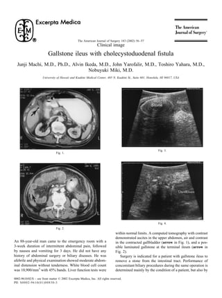
biliary tract, the condition and location of biliary enteric fistula area. Intraoperative ultrasonography showed a contracted
(some fistulas close with fibrous remnant), and the presence or gallbladder containing small stones and air. Cholecystoduo-
absence of biliary obstruction due to bile duct stones. Intraop- denal fistula was identified and accurately localized by ul-
erative cholangiogram can provide valuable information but trasonography, which demonstrated the dynamic movement
may be difficult to perform because cannulation for contrast of fluid and air between the gallbladder and duodenum in
injection is not easy or possible. Intraoperative ultrasonogra- real-time (arrows in Fig. 4). The bile duct was normal in
phy is helpful in this setting, because the ultrasound scanning size (6 to 7 mm) without stones or pneumobilia on ultra-
can be performed without any tissue dissection. sound examination. Because the patient was in a stable
In this patient, laparotomy was performed with the use of condition, cholecystectomy was performed. The fistula was
intraoperative ultrasonography. Near the terminal ileum, detected and excised. The duodenal opening was closed by
there was a palpable stone, confirmed by intraoperative sutures with an omental patch. Although the patient had
ultrasonography (arrow in Fig. 3). The 3.5 ϫ 2 cm stone renal failure and respiratory failure, he recovered from these
was removed through ileotomy. A marked inflammation postoperative complications and was discharged.