SlideShare uma empresa Scribd logo
1 de 11
Baixar para ler offline
neuroradiology solutions
www.abmedica.it
ab medica s.p.a.
Via nerviano, 31 - 20020 Lainate (MI)
tel +39 02 933051 - fax +39 02 93305400
abmedica@abmedica.it
Self-Expandable LEO+:
Intracranial stent designed for the
treatment of wide-neck aneurysm
Braided design allows the fine nitinol
wires to slide smoothly onto each
other to reach perfect vessel
conformability, as well as provide
coil support
The original braided stent
is also compatible with a
coiling microcatheter 1
(VASCO+10, ID: .017”)
LOW PROFILE to gain access to
vessels down to Ø 1,5mm
Avoid microcatheter exchange
original braided stent
so compatible with aArtery
diameter
FINE
BRAIDED
WIRES
HIGHER
RADIAL
FORCE
EASE
OF USE
HIGHER
VISIBILITY
FLARED
ENDS
A
FOR
Mesh size
Stent’s lenght
Original article
Intra-arterial nimodipine for cerebral vasospasm
after subarachnoid haemorrhage: Influence on
clinical course and predictors of clinical outcome
Asma Bashir1
, Morten Andresen1
, Jiri Bartek Jr1,2
, Marie Cortsen3
,
Vagn Eskesen1
and Aase Wagner3
Abstract
Intra-arterial nimodipine (IAN) has shown a promising effect on cerebral vasospasm (CV) after aneurysmal subarachnoid
haemorrhage. At our institution, Rigshospitalet, IAN treatment has been used since 2009, but the short- and long-term
clinical efficacy of IAN has not yet been assessed. The purpose was to evaluate the efficacy and clinical outcome of IAN
treatment of symptomatic CV, and to assess the predictors of clinical outcome. Medical records of 25 patients undergoing a
total of 41 IAN treatment sessions were retrospectively reviewed. Data on angiographic results, blood-flow velocities and the
clinical condition before and after the IAN treatment were recorded. Predictors of the clinical outcome were assessed with a
linear regression model. Positive angiographic response was achieved in 95.1% of 41 IAN treatment sessions. Flow-velocity
measurements showed no relationship with angiographic responses of IAN. The immediate clinical improvement was
observed in three patients (12%). Five patients (20%) had a favourable outcome at discharge and at three-month
follow-up; 10 patients (40%) had a moderate to poor outcome; and the rest (40%) died. Increased number of affected
vessels and number of procedures carried out per patient, and a trend toward an increased delay time from symptomatic CV
to confirming angiographic CV and thus instituting IAN treatment predicted the poor clinical outcome. IAN treatment
appears to be effective in reversing angiographic CV. However, it is not always effective in reversing clinical deterioration,
as several other factors including treatment delay affect the clinical course.
Keywords
Subarachnoid haemorrhage, cerebral vasospasm, intra-arterial nimodipine
Introduction
Cerebral vasospasm (CV) is a feared and potentially life-
threatening complication in patients following aneurys-
mal subarachnoid haemorrhage (SAH). Angiographic
CV is observed in up to 70% of patients, typically
between 4–14 days after the initial bleeding, and leads
to symptomatic brain ischaemia and/or new infarction
on radiographic imaging in about 30% of these
patients.1,2
Treatment of CV and prevention of delayed
cerebral ischaemia (DCI) is therefore a main goal in the
management of patients surviving from SAH. However,
despite maximal contemporary therapy and neurocritical
care, about 30% of these patients die, another 30% sur-
vive with significant disability, and the rest of them
recover with little or no long-term disability.3
Current medical strategies for prevention and
treatment of CV including endovascular therapies
with vasodilators and triple-H (i.e. hypertension, hyper-
volaemia, haemodilution) are suboptimal,2,4–8
as the
exact mechanisms leading to CV are not yet fully
elucidated.2,9
At the moment, nimodipine, a calcium
channel blocker, remains the only drug proved by the
US Food and Drug Administration, which is recom-
mended as the first-line treatment and prevention of
CV and improvement of outcomes after SAH.10,11
Although nimodipine exhibits neuroprotective as well
as vasodilatory benefits,2
some patients deteriorate fur-
ther and develop severe symptomatic CV. In the last
decade, a number of clinical trials have investigated
nimodipine infused directly in the spastic arteries
(intra-arterial nimodipine (IAN)), and found it to be
1
Department of Neurosurgery, Rigshospitalet, Faculty of Health and Medical
Sciences, University of Copenhagen, Blegdamsvej 9, 2100 Copenhagen,
Denmark
2
Department of Clinical Neuroscience, Section for Neurosurgery, Karolinska
Institutet and Department of Neurosurgery, Karolinska University Hospital,
Stockholm, Sweden
3
Department of Radiology, Rigshospitalet, Faculty of Health and Medical
Sciences, University of Copenhagen, Blegdamsvej 9, 2100 Copenhagen,
Denmark
Corresponding author:
Asma Bashir, Department of Neurosurgery, Rigshospitalet, Faculty of Health
and Medical Sciences, University of Copenhagen, Blegdamsvej 9, 2100
Copenhagen, Denmark.
Email: ab@asmabashir.com
The Neuroradiology Journal
2016, Vol. 29(1) 72–81
! The Author(s) 2016
Reprints and permissions:
sagepub.co.uk/journalsPermissions.nav
DOI: 10.1177/1971400915626429
neu.sagepub.com
effective in reversing angiographic CV, although overall
clinical outcome differs through the literature.12–18
At
our institution, Rigshospitalet, IAN treatment has been
used as the method of choice since 2009 whenever endo-
vascular treatment of CV was indicated. However, the
short- and long-term clinical efficacy of IAN has not yet
been assessed.
The purpose of this study was to retrospectively
evaluate the efficacy and clinical outcome of IAN treat-
ment of symptomatic CV, which is refractory to the
haemodynamic treatment, and to assess the predictors
of the clinical outcome.
Materials and methods
Data collection
Between December 2009–May 2012, 325 adults with
evidence of SAH on computerized tomography (CT)
scan were admitted to the neurosurgical intensive care
unit (ICU) at the University Hospital, Rigshospitalet,
in Copenhagen, Denmark. Only patients presenting
with a ruptured intracranial aneurysm on CT angio-
gram (CTA), suffering from symptomatic CV refrac-
tory to the haemodynamic therapy, and receiving
IAN treatment were reviewed. Twenty-eight patients
were identified. Among them, three patients were trea-
ted with a combination of percutaneous transluminal
angioplasty (PTA) and IAN, and were excluded to
avoid confound bias on clinical outcome, as PTA has
also shown to have beneficial effect.19
A total of 25
patients met the inclusion criteria and constituted the
study population. Data on these patients was extracted
from a computerized database and reviewed retrospect-
ively including demographic data, clinical condition at
admission based on World Federation of Neurological
Surgeons (WFNS) grade I–V,20
amount and distribu-
tion of subarachnoid blood detected by admission CT
by using the modified Fisher scale 1–4,21
and aneurysm
location as well as aneurysm treatment. Onset, location
and severity of CV, number of affected vessels, proced-
ure specifics such as dosage of IAN per vessel and per
procedure, and number of repeated procedures were
recorded. To assess the efficacy of IAN treatment,
the clinical response within 24 h (e.g. level of conscious-
ness based on Glasgow Coma Scale (GCS) and neuro-
logical condition), the angiographic response as well as
effects on blood flow-velocity of the middle cerebral
artery (MCA) determined by transcranial Doppler
(TCD) ultrasonography were examined. The clinical
outcome was assessed using the modified Rankin
scale (mRS) score at discharge and at three-month
follow-up.22
Informed consent was obtained from
patients or legal representative for patients with
impaired consciousness at the time of their clinical
work-up. The Board of Health Committee and Data
inspectorate approved the study. All the necessary pre-
cautions were taken to protect the identities of the
patients in the study.
Patient management and monitoring
All patients underwent either surgical or endovascular
obliteration of the ruptured aneurysm within 48 h after
the initial bleeding except seven patients who were hos-
pitalized 3–14 days after onset of SAH. Nimodipine
was administrated, orally or intravenously, in a dose
of 60 mg every 4 h in 21 days according to a standar-
dized SAH-protocol. Maintenance of euvolaemia was
achieved by administration of intravenous isotonic
crystalloid. When flow-velocity of the MCA exceeded
120 cm/s (a TCD cut-off value), triple-H therapy was
initiated for inducing prophylactic hypervolaemia
and elevating the mean arterial pressure by 20%.
Flow-velocity measurements were performed at least
once daily to monitor possible vasospastic activity.
Flow-velocity <120 cm/s was defined as normal,
120–150 cm/s as mild, 150–200 cm/s as moderate and
>200 cm/s as severe. Lindegaard ratio was not measured
routinely in all patients, and is therefore not mentioned
further in the study. The clinical condition was assessed
by continuous neurological examinations including GCS
scores.
Diagnosis of CV and IAN treatment
Cerebral vasospasm was suspected, if patients demon-
strated at least one of the following: (a) a decreased
level of consciousness and/or disorientation with a
decrease of at least two points on the GCS score from
baseline, (b) new focal neurological deficits (cranial
nerve palsy, hemiparesis and/or speech disturbances),
and/or (c) flow-velocity >120 cm/s. Neurological deteri-
oration was the main trigger for suspicion of CV, fol-
lowed by an increase in flow-velocity; the last was
mainly applied to patients with ventilator support.
Possible causes of neurological deterioration such as
presence of ischaemia, re-bleeding or hydrocephalus
on CT scan, electrolyte disturbance, seizures or hypoxia
were eliminated prior to CTA, which was usually per-
formed within 24 h of onset of symptomatic CV.
Degree of angiographic CV was assessed on a digital
subtraction angiography semi-quantitatively and
subjectively and classified as ‘none/mild’ when arterial
narrowing was <25% of the arterial diameter, ‘moder-
ate’ when narrowing was 25–50%, or ‘severe’ when
narrowing >50%.
Patients were found eligible for IAN treatment only
if they showed angiographic CV in territories
compatible with their neurological deficits and/or with
elevated mean flow-velocity. After dilution with 0.9%
saline to obtain a solution of 0.01 mg/ml, nimodipine
was administrated as an infusion over 20–40 minutes
through a microcatheter placed in the affected vessel.
Post-nimodipine angiograms were performed
10–30 min after each session. Two neuroradiologists
evaluated the angiographic results separately, and dis-
agreement was resolved by consensus. Each vessel was
evaluated as ‘responsive’ or ‘non-responsive’, and the
Bashir et al. 73
overall angiographic result was graded as ‘good
response’, ‘no response’ or ‘mixed response’. If persist-
ence or recurrence of CV was suspected, a new CTA
was performed, and IAN treatment was repeated after
the verification of angiographic CV and progressive
neurological deterioration.
Clinical evaluation
Immediate clinical response and flow-velocity measure-
ments within 24h after IAN treatment were recorded.
The change in the clinical condition was graded as
‘improved’, ‘unchanged’ or ‘worsened’. The clinical out-
come at discharge from the hospital and at three-month
follow-up was assessed using the mRS score (no question-
naire).22
A score 2 was considered as a good outcome.
Statistical analysis
Data analysis was performed with the statistical soft-
ware package IBM SPSS version 20.0. Descriptive data
was presented as median values with interquartile range
(IQR) in parentheses, as data was not normally
distributed. A p-value of less than 0.05 was accepted
as statistically significant. Our primary dependent vari-
able was mRS score, which was investigated in a linear
regression model based on following predictor
variables: age, WFNS grade, Fisher scale, aneurysm
location, severity of CV, number of affected vessels,
angiographic response and number of repeated proced-
ures. Significance of time delay in diagnosis of angio-
graphic CV and thus instituting IAN treatment was
assessed as well.
Results
Clinical assessment
Demographic data, WFNS grade, Fisher scale, and
location of aneurysm are presented in Table 1.
Surgical clipping was performed in seven (28%)
patients and endovascular occlusion was performed in
18 (72%) patients. All patients developed neurological
deterioration such as decreased level of consciousness,
hemiparesis, aphasia or other new focal neurological
signs between 4–13 days after onset of SAH (median,
eight days; IQR 6–9 days). Among them, two (8%)
patients already had neurological deficits at the time
of aneurysm treatment and one (4%) due to carotid
artery dissection two days prior to symptomatic CV.
One (4%) patient (no. 17) was admitted to the hospital
14 days after the initial bleeding and already had
developed left-sided arm paresis and leg paralysis due
to CV. In one (4%) patient (no. 15), who was artificially
ventilated, the suspicion of CV was raised by elevated
flow-velocity measurements only, which was the main
indication for angiography. The neurological deterior-
ation did not respond to the haemodynamic treatment.
Onset of occurrence of CV and neurological
deterioration including flow-velocity measurements
are summarized in Table 2.
Angiography and IAN treatment
Diagnostic cerebral angiography was performed within
24 h after onset of symptomatic CV in all patients
except nine (36%), in whom it was performed later
due to their fluctuating neurological condition. The
median time delay from the occurrence of symptomatic
CV to CTA was 14.5 h (IQR, 2.9–38.1 h). In most cases,
angiographic CV diffusely involved all vessels on both
sides of the circulation, while in six cases (24%) only
one side was affected involving 1–3 vessels. Overall, the
degree of CV was severe in 16 (64%) patients and mod-
erate in nine (36%). No one had a mild degree of CV.
In addition, seven (28%) patients showed signs of
incipient hypodensities on their CT scans. Using a
linear regression model with a significance p ¼ 0.048;
adjusted R2
¼ 0.241, severity of CV was significant asso-
ciated with an increased number of affected vessels
(p ¼ 0.004), but not with age (p ¼ 0.50), Fisher scale
(p ¼ 0.85) and aneurysm treatment (p ¼ 0.54). Severity
Table 1. Patient characteristics.
Demographic data
Age, median (years) 52
Female sex, no. (%) 20 (80%)
Risk factors, no. (%)
Hypertension 13 (52%)
Smoking 14 (56%)
Alcohol abuse 3 (12%)
Drug abuse 1 (4%)
WFNS grade I–V, no. (%)
I, II and III 15 (60%)
IV and V 10 (40%)
Fisher grade 1–4, no. (%)
1 and 2 3 (12%)
3 and 4 22 (88%)
Location of aneurysm, no. (%)
ACoA 9 (36%)
ACA 2 (8%)
MCA 4 (16%)
ICA 4 (16%)
PCA 1 (4%)
BA 4 (16%)
PICA 1 (4%)
Surgical or endovascular obliteration, no. (%)
Coils 14 (56%)
Clips 7 (28%)
Stent 3 (12%)
Coils/stent 1 (4%)
ACA: anterior cerebral artery; ACoA: anterior communicating artery; BA:
basilar artery; CV: cerebral vasospasm; ICA: internal cerebral artery; MCA:
middle cerebral artery; PCA: posterior cerebral artery; PICA: posterior infer-
ior cerebral artery; WFNS: World Federation of Neurological Surgeons.
74 The Neuroradiology Journal 29(1)
Table2.Theseverityofcerebralvasospasm(CV)andlocation,neurologicaldeficitincludingflowvelocity(FV),angiographicandclinicalresponsestotheintra-arterialnimodipine(IAN)treatmentand
clinicaloutcomeatdischargeandatthree-monthfollow-up(FU).
Patient/
sessionno.
Onsetof
sCV(day)
Neurological
deficits
Location
ofaCV
Severity
ofaCV
IANdoseper
session(mg)
Angiographic
response
Clinical
response
mRSscoresat
Discharge3-monthFU
1.110Lhemiparesis,5dayslaterRhemiparesis,FV70cm/sLMCA,ACA,ICASevere3.0GoodWorsened
1.2þAphasia,FV178cm/sLMCA,ACA,ICASevere3.0GoodUnchanged
1.3Sedated,FV190cm/sLMCA,ACA,ICASevere3.0MixedresponseUnchanged54
2.19FluctuatingDLC,Larmparesis,FV182cm/sMCAs,ACAs,ICAsSevere4.0GoodWorsened
2.2þSeizures,FV260cm/sACAs,MCASevere4.0GoodWorsened43
3.111FluctuatingDLC,FV187cm/sRMCAModerate3.2NoresponseUnchanged10
4.17Rhemiparesis,facialnervepalsy,visualaxisdeviation,FV
100cm/s
LACA,MCAModerate4.0GoodUnchanged54
5.19FluctuatingDLC,Llegparesis,FV193cm/sPCTSevere4.0GoodWorsened6–
6.15FluctuatingDLC,aphasia,FV180cm/sLMCA,ACAs,ICAsSevere4.0GoodWorsened6–
7.111FluctuatingDLCLMCA,MCAs,ICAs,PCTSevere4.0MixedresponseUnchanged
7.2þFV97cm/sRACA,MCA,ICASevere2.0GoodUnchanged53
8.16FluctuatingDLCaphasia,FV348cm/sACAs,MCAs,ICAs,PCTSevere4.0GoodWorsened
8.2SedatedACAs,MCAs,ICAs,PCTSevere6.0GoodWorsened56
9.15DLC,Lhemiparesisfacialnervepalsy,aphasia,FV
235cm/s
ACAs,MCAs,ICAsSevere6.0GoodWorsened
9.2SedatedACAs,MCAs,ICAsSevere6.0GoodUnchanged53
10.15DLC,Rhemiparesis,aphasiaACAs,MCAs,ICAsSevere4.8GoodUnchanged
10.2DLC,Rhemiparesis,aphasiaACAs,MCAs,ICAsSevere5.0MixedresponseUnchanged51
11.17FluctuatingDLC,Rhemiparesis,aphasiaPCTModerate4.0GoodUnchanged
11.2þVisialaxisdeviationPCTSevere4.0GoodUnchanged43
12.110DLC,FV156cm/sRACA,MCAs,ICAsSevere3.0GoodUnchanged6–
13.111DLC,Rhemiparesis,FV190cm/sACA,MCAs,ICAModerate4.4GoodUnchanged
13.2DLC,RhemiparesisACAs,MCAs,ICAsModerate5.0GoodWorsened6–
14.1a
6DLC,Rhemiparesis,facialnervepalsy,aphasia,FV
230cm/s
ACAs,MCAs,ICAsSevere4.0GoodWorsened6–
15.19Sedated,FV230cm/sACAs,MCAs,ICAsSevere5.0MixedresponseWorsened6–
16.19FluctuatingDLC,FV183cm/sACAsModerate4.5MixedresponseUnchanged32
17.113Lhemiparesis/paralysisACAs,MCAs,ICAsSevere4.0GoodWorsened
17.2þAphasia,FV250cm/sRACA,MCA,ICA,PCTSevere4.0GoodWorsened6–
18.18DLC,aphasia,FV145cm/sACAs,MCAs,ICAsSevere5.0GoodUnchanged
18.2þFV313cm/sLMCA,ICA,ACAsSevere4.0GoodUnchanged
18.3Sedated,FV257cm/sACAs,MCAs,ICAsSevere5.0GoodUnchanged
18.4Sedated,FV290cm/sLACA,MCA,ICASevere4.0MixedresponseUnchanged55
(continued)
Bashir et al. 75
of CV and number of affected vessels including ana-
tomic distribution are presented in Table 2.
Forty-one IAN treatment sessions were performed
in 25 patients (range, 1-4 treatment sessions per
patient), in which 173 vessels were evaluated. The
median time delay from angiographic CV to IAN treat-
ment was 3.0 h (IQR, 2–9.5 h). Median dose of nimodi-
pine was 4 mg per session (range, 2–6 mg per session),
while the largest single dose per vessel was 4 mg
(median, 2 mg; range, 1–4 mg). Complete reversal of
CV was successfully achieved in 31 (75.6%) treatments
(Figure 1); partial reversal was noted in eight (19.5%)
(Figure 2), while two (4.9%) showed no effect. There was
no significant correlation between the degree of CV and
angiographic response to IAN (p ¼ 0.24). Worsening of
CV did not occur during the procedures. No signs
of haemodynamic instability due to IAN infusion or
other procedure-related complications were observed.
However, in one (4%) patient (no. 14), a dosage of
2 mg nimodipine was accidently injected in a few seconds
causing transient drop in blood pressure. Dose per pro-
cedure, number of repeated procedures and angio-
graphic effects are summarized in Table 2.
TCD measurements
Complete results for flow-velocity measurements before
and after IAN treatment within 24h were available for
review in only 13 (32%) of 41 procedures. Mean velocities
decreased by 37cm/s in eight (20%) procedures,
unchanged in one (2%), and increased by 34cm/s in four
(10%). There was no correlation between flow-velocity
measurements and angiographic responses to IAN.
Clinical evaluation within 24 h after treatment, at
discharge and at three-month follow-up
The clinical condition remained unchanged for 12
(48%) patients, although all of them but one showed
angiographic response to IAN treatment. Three (12%)
patients showed clinical improvement based on GCS
scores and/or neurologically, two of them with none
and mixed responses to IAN. Ten (40%) patients
experienced a worsening in their clinical conditions
both consciously and neurologically; among them, six
(24%) had developed new neurological deficits. Two
(8%) had seizures at the time of wake-up call. Three
(12%) patients never regained consciousness and died a
few days later; all three had showed angiographic
response to IAN, two of them with mixed response.
In 12 (48%) patients, new cerebral infarctions were
observed on CT scan hours to a few days after the
IAN treatment.
At discharge, only one (4%) patient showed a good
clinical outcome (mRS 1). Two (8%) patients had a
moderate outcome (mRS 3) and 13 (52%) a poor out-
come (mRS 4–5). Nine (36%) patients died during hos-
pitalization, eight (32%) of them as a consequence of
extensive cerebral infarction secondary to CV (mRS 6).
Table2.Continued
Patient/
sessionno.
Onsetof
sCV(day)
Neurological
deficits
Location
ofaCV
Severity
ofaCV
IANdoseper
session(mg)
Angiographic
response
Clinical
response
mRSscoresat
Discharge3-monthFU
19.18DLC,aphasia,FV226cm/sLMCAModerate4.0NoresponseImproved32
20.15Pronation/extensionpattern,FV110cm/sRACA,MCA,ICAModerate3.0GoodWorsened
20.2þRhemiparalysis,Larmparesis,FV130cm/sRACA,MCA,ICASevere3.0GoodUnchanged53
21.19DLC,FV180cm7sACAs,ICAsSevere4.0GoodImproved42
22.16DLC,aphasiaLACA,ICA,MCAsModerate4.0GoodUnchanged54
23.16DLCLACA,MCAs,ICAsSevere4.0MixedresponseImproved53
24.14DLCACAs,MCAs,ICAs,PCTSevere6.0GoodUnchanged
24.2þRhemiparesis,FV200cm/sACAs,MCAs,ICAs,PCTSevere6.0GoodWorsened6–
25.16DLC,FV197cm/sLICA,ACAs,MCAs,PCTModerate6.0MixedresponseWorsened
25.2þRhemiparesis,extensorplantarreflex,FV237cm/sLACA,MCA,ICA,PCTModerate6.0GoodWorsened6–
ACA:anteriorcerebralartery;aCV:angiographiccerebralvasospasm;DLC:decreasedlevelofconsciousness;FU:follow-up;IAN:intra-arterialnimodipine;ICA:internalcerebralartery;L:leftcirculation;MCA:middlecerebral
artery;PCT:posteriorcirculatoryterritory;R:rightcirculation;sCV:symptomaticcerebralvasospasm.a
ByaccidentIANinjectedintherightICAforafewsecondscausingtransientdropinbloodpressure.Thetreatmentis
repeatedwiththesamedose.
76 The Neuroradiology Journal 29(1)
At the three-month follow-up, another four (16%)
patients showed a favourable outcome (mRS 1–2), six
(24%) had a moderate outcome (mRS 3), and one more
(4%) had died (mRS 6). Data on the clinical evaluation
is presented in Table 2.
Observing a linear regression model (p ¼ 0.007,
adjusted R2
¼ 0.654), significant predictors of a poorer
clinical outcome (mRS 4–6) were an increased number
of affected vessels (p ¼ 0.003) and an increased number
of procedures carried out per patient (p ¼ 0.04), and the
angiographic response of IAN (p ¼ 0.01). Looking at
the time frame from symptomatic CV to confirming
angiographic CV, we saw a trend toward a poorer clin-
ical outcome with increasing time (p ¼ 0.13).
Discussion
Cerebral vasospasm following an aneurysmal SAH
continues to be a leading cause of morbidity and mor-
tality.3
We retrospectively reviewed 25 patients treated
with IAN infusions for medically refractory symptom-
atic CV, and found that IAN treatment was effective in
reducing angiographic CV in 95.1% of 41 treatment
sessions, with complete reversal in 75.6% and partial
reversal in 19.5%. However, the efficacy of treatment
was temporary, and required repeated treatment ses-
sions in 13 (52%) patients, and was not always followed
by clinical improvement. Only three (12%) patients
demonstrated an immediate clinical improvement
after treatment, and only one (4%) had a good clinical
outcome at the time of discharge. Eight (32%) of nine
deceased patients died during hospitalization. At three-
month follow-up, another four (16%) patients showed
a clinical improvement, while one (4%) more had died.
Overall, five (20%) patients had a favourable long-term
outcome (mRS 0–2), 10 (40%) had moderate to poor
outcome (mRS 3–5), and 10 (40%) patients had died
(mRS 6) (Table 2).
Figure 1. Patient no. 24. Lateral angiogram of the internal
cerebral artery (ICA) showing (a) cerebral vasospasm (CV);
(b) after intra-arterial nimodipine (IAN) infusion, an increase
in diameter of the vessels is observed.
Figure 2. Patient no. 12. Anteroposterior angiograms of (a) the left and (b) right internal cerebral arteries (ICAs) showing cerebral
vasospasm (CV). After intra-arterial nimodipine (IAN) infusion, (c) the left side demonstrates an increase in diameter of the vessels, while
(d) the right side shows a mixed response.
Bashir et al. 77
Several clinical trials have evaluated the efficacy of
IAN treatment for symptomatic CV.12–18
These studies
have reported post-IAN angiographic vessel dilatation
in 43.3–82.8% of sessions, clinical improvement in
11.1–88.9% of patients, and 61.1–88.9% with a favour-
able short- or long-term outcome (Table 3). Biondi
et al.12
were the first to assess the efficacy of IAN.
The authors reported post-IAN angiographic response
in 13 (43.3%) of 30 treatment sessions and clinical
improvement in 19 (76%) of 25 patients. Eighteen
(72%) had a favourable long-term outcome (mRS
0–2). Their angiographic results are in line with our
study. We reported complete reversal of angiographic
CV in 75.6% of sessions, and in addition partial rever-
sal of CV was seen in 19.5%. However, our angio-
graphic results did not correlate with the clinical
improvement and the overall clinical outcome was
less satisfactory, compared to Biondi et al.12
Ha¨ nggi
et al.15
investigated the efficacy of IAN in 26 patients
as well. In eight (31%) patients, no angiographic
improvement was seen, and therefore these were
excluded from the study. Using cerebral perfusion CT
(PCT), the authors reported an improved time to peak
and mean transit time a day after the procedure.
Although the efficacy of IAN was transient, and 11
(61%) of 18 patients developed additional infarction,
11 (61%) patients revealed a good clinical condition at
time of discharge, which is remarkable compared to our
study. However, some of these studies are confounded
by the combined use of IAN and papaverine or balloon
angioplasty, which might explain in part for the clinical
improvement reported in the studies.14–16
We are the second group of researchers to evaluate
the influence of time delay in diagnosing and instituting
the IAN treatment after onset of symptomatic CV. In
our study, an increasing time delay from symptomatic
CV to angiographic CV showed a trend toward a
poorer outcome with a median time of 14.5 h. The
median delay time from angiographic CV to the IAN
treatment was 3.0 h. Grotenhuis et al.23
was the first to
report that the IAN treatment was ineffective when
administrated only a few hours after the onset of CV.
However, the authors used only small dosage of nimo-
dipine ranging from 0.068–1.0 mg, which could account
at least in part for the observed lack of effect. Biondi
et al.12
performed the diagnostic CTA and IAN treat-
ment within 24 h of onset of symptomatic CV, with the
mean delay time of 18 Æ 6 h. The dosage of IAN per
vessel consisted of 1–3 mg. Despite time delay in the
diagnostic process, thus instituting the treatment, the
authors demonstrated a clinical improvement in 76%
of patients after IAN. Similarly, Linfante et al.7
inves-
tigated the efficacy of IA nicardipine in 22 patients. A
total of 46 endovascular sessions with intra-arterial
nicardipine were performed within 48 h after onset of
symptoms in 21 patients, and within 72 h in one patient.
At the time of discharge, half of patients were reported
to be independent functionally (mRS 0–2). In our
study, time delays were much less than reported by
Table3.Studieswithcerebralvasospasm(CV)afteraneurysmalsubarachnoidhaemorrhage(SAH)andintra-arterialnimodipine(IAN)treatment.
Author/yearStudydesign
Patient
no.
Session
no.
Doseof
IANpervessel
Angiographic
improvement
Clinical
improvement
Theclinical
outcome(mRS2and
GOS!4)atdischarge
Theclinicaloutcome
(mRS2andGOS!4)
atfollow-up
Biondietal.,20041,2
Retrospective25301–3mg43.3%76%–72%
Choetal.,20111,3
Retrospective421013–6mg82.2%68.3%76.2%84.6%
Dehdashtietal.,201114
Retrospective10151–4mg80%60%–70%
Ha¨nggietal.,200815,a
Prospective18260.8–3.2mg77.3%11.1%61.1%61.1%
HuiandLau,20051,6,b
Retrospective9102–4mg66.6%88.9%88.9%77.8%
Kimetal.,20091,7
Retrospective19531–3mg79.2%68.4%78.9%–
Kimetal.,201218
Prospective29291–3mg82.8%72.4%75.9%–
Ourstudy,2015Retrospective28451–4mg95.6%21.4%10.7%28.6%
GOS:GlasgowOutcomeScale;mRS:modifiedRankinscale.a
Only18of26patientsand22of42proceduresareincludedintheanalysisduetonomeasurableangiographiceffectin20(47.6%)proceduresamongeight(30.7%)
patients.b
Themeanpercentageincreaseinarterialdiameter.Angiographicimprovementincludesresultswithmoderateandmajoreffect.
78 The Neuroradiology Journal 29(1)
Biondi et al.12
and Linfante et al.7
and with a dosage of
IAN per vessel (range, 1–4 mg) similar to Biondi et al.12
However, despite angiographic response in 95.1% of
treatment sessions only a few patients showed clinical
improvement. The discrepancy between angiographic
response and unsatisfactory clinical results could be
explained by the fact that parenchymal damage has
already occurred due to ischaemia resulting from CV
in spite of angiographic response to endovascular treat-
ment with IAN. Furthermore, evidence from a rat
model indicated that reperfusion of parenchyma
injured by an ischaemic insult might lead to further
injury;24–26
this phenomenon could be one of the rea-
sons why the uses of endovascular therapies are asso-
ciated with a poorer outcome despite substantial vessel
dilatation in some instances, as seen in our study.
TCD is often used as a surrogate diagnostic tool to
detect CV. We found no correlation between flow-velo-
cities and angiographic responses to IAN, although
data of TCD measurements was missing in most
cases. Kim et al.17
observed a decrease in flow-velocity
in 29 of 33 sessions. In contrast, Ha¨ nggi et al.15
noticed
a significant increase in flow-velocity one day after IAN
infusion (p ¼ 0.03). Wolf et al.27
was unable to detect
CV with TCD in two patients and reflect the treatment
course in four patients. The interpretation of the TCD
analysis leaves some room for speculation. Estimation
of flow-velocity could vary from minute to minute, and
might be influenced by anatomic details such as vascu-
lar anatomy, vessel diameters and cerebral blood flow.
Influence of pathological conditions such as an
increased intracranial pressure or oedema, or systemic
haemodynamic effects during triple-H therapy such as
changes in cardiac output or blood pressure could
affect the speed and waveform of cerebral blood flow
as well. In addition, the accuracy of this technique is
quite operator-dependent with high intra-variability,
and suboptimal insonation might inhibit the detection
of high flow-velocities.28–30
Vora et al.31
reported that
only very low or very high MCA velocities (e.g.
<120 cm/s or >200 cm/s) negatively or positively pre-
dicted angiographic and symptomatic CV in a reliable
manner, and intermediate velocities had a lower pre-
dictive value and were poorly discriminative.
Vasodilatation by intra-arterial drug infusion also
has been performed with other agents. Papaverine is
the first intra-arterial agent reported to reverse angio-
graphic CV.6
However, it is associated with a consider-
able number of several adverse side effects such as
elevation of intracranial pressure,32
worsening of
CV33
and thrombocytopaenia.34
One study compared
the angiographic effect of papaverine and nimodipine
and reported that there was no significant difference
between these two groups, although the effect of papa-
verine was more pronounced, dilating at least one vessel
in every patient, while nimodipine was ineffective in
16% of patients.35
Intra-arterial nicardipine infusion
has shown to be effective without increasing intracra-
nial pressure.36
However, a transient decrease in blood
pressure and an increase in heart rate may occur.7,36
Intra-arterial verapamil infusion is also reported to
dilate spastic vessels without causing changes in
haemodynamic circulation or intracranial pressure.6
However, it needs to be evaluated in larger series to
confirm its efficacy. PTA is highly effective in relieving
focal CV, and has a continuous effect on narrowed ves-
sels. However, the technique is limited to proximal ves-
sels, and requires expert neurointerventionists due to
the significant risks of artery rupture, dissection and
local thrombosis.19,37
One study compared the clinical
outcome of IAN treatment with PTA, but the authors
did not find significant differences in these two
groups.38
Limitations
The study has several important limitations including
its retrospective design and the small number of
patients due to it being a single centre study, which
may limit generalizability. There was no control
group. It is not clear whether the change in the clinical
condition after IAN infusion was due to the effect of
the treatment or just a natural course. Quantitative
measurement of vessel diameter on pre- and post-
nimodipine angiograms was not available as mm
(Æstandard deviation), but only as Æeffect. TCD data
were missing before and after IAN infusions in several
patients making it more difficult to estimate the abso-
lute correlation between TCD and angiographic results.
Data on monitoring systemic haemodynamic effects
and intracranial pressure during IAN sessions were
not investigated, and therefore we were unable to quan-
tify the effect of IAN on these areas.
Conclusion and perspectives
Although there are several limits in this retrospective
study, we can conclude that IAN treatment appears to
be effective in reversing angiographic CV. However, the
relationship between angiographic and clinical
responses was not convincing, as the treatment is inef-
fective in reversing clinical deterioration caused by
DCI. Several factors such as increased numbers of
affected vessels and procedures carried out per patient
predicted the clinical outcome negatively. In addition, an
increasing time delay in diagnostic process of CV and
thus instituting treatment might contribute to a poorer
clinical outcome. Clearly, early detection and rapid
treatment of CV are needed before ischaemic damage
occurs. Future studies with continued assessment of
cerebral blood flow might help optimize the timing of
IAN treatment and to assess the real efficacy of the IAN
treatment in patients with symptomatic CV after SAH.
Acknowledgements
The study is dedicated to the late Professor Bertil Romner,
Department of Neurosurgery, Rigshospitalet, who originally
conceived and designed the study. A Bashir and the late
Bashir et al. 79
Bertil Romner conceived and designed the study. A Bashir
reviewed patient data and wrote the first and subsequent
drafts of the manuscript. A Wagner and M Cortsen evaluated
pre- and post-angiograms. M Andresen conducted the statis-
tical analysis. J Bartek Jr, M Andresen, A Wagner, M
Cortsen and V Eskesen participated in critical revision and
writing of the article. All authors have seen and approved the
final version.
Funding
The author(s) received no financial support for the research,
authorship, and/or publication of this article.
Conflict of interest
The author(s) declared no potential conflicts of interest with
respect to the research, authorship, and/or publication of this
article.
References
1. Connolly ES, Rabinstein AA, Carhuapoma JR, et al.
Guidelines for the management of aneurysmal subarach-
noid hemorrhage: A guideline for healthcare profes-
sionals from the American Heart Association/American
Stroke Association. Stroke 2012; 43: 1711–1737.
2. Treggiari-Venzi MM, Suter PM and Romand J-A.
Review of medical prevention of vasospasm after aneur-
ysmal subarachnoid hemorrhage: a problem of neuroin-
tensive care. Neurosurgery 2001; 48: 249–262.
3. Washington CW and Zipfel GJ. Detection and monitor-
ing of vasospasm and delayed cerebral ischemia: a review
and assessment of the literature. Neurocrit Care 2011; 15:
312–317.
4. Clouston JE, Numaguchi Y, Zoarski GH, et al.
Intraarterial papaverine infusion for cerebral vasospasm
after subarachnoid hemorrhage. AJNR Am J Neuroradiol
1995; 16: 27–38.
5. Fraticelli AT, Cholley BP, Losser M-R, et al. Milrinone
for the treatment of cerebral vasospasm after aneurysmal
subarachnoid hemorrhage. Stroke 2008; 39: 893–898.
6. Keuskamp J, Murali R and Chao KH. High-dose intraar-
terial verapamil in the treatment of cerebral vasospasm
after aneurysmal subarachnoid hemorrhage. J Neurosurg
2008; 108: 458–463.
7. Linfante I, Delgado-Mederos R, Andreone V, et al.
Angiographic and hemodynamic effect of high concentra-
tion of intraarterial nicardipine in cerebral vasospasm.
Neurosurgery 2008; 63: 1080–1087.
8. Treggiari MM, Walder B, Suter PM, et al. Systematic
review of the prevention of delayed ischemic neurological
deficits with hypertension, hypervolemia, and hemodilu-
tion therapy following subarachnoid hemorrhage.
J Neurosurg 2003; 98: 978–984.
9. Kassell NF, Sasaki T, Colohan AR, et al. Cerebral vaso-
spasm following aneurysmal subarachnoid hemorrhage.
Stroke 1985; 16: 562–572.
10. Barker 2nd, FG and Ogilvy CS. Efficacy of prophylactic
nimodipine for delayed ischemic deficit after subarach-
noid hemorrhage: A metaanalysis. J Neurosurg 1996;
84: 405–414.
11. Koenig MA. Management of delayed cerebral ischemia
after subarachnoid hemorrhage. Continuum (Minneap
Minn) 2012; 18: 579–597.
12. Biondi A, Ricciardi GK, Puybasset L, et al. Intraarterial
nimodipine for the treatment of symptomatic cerebral
vasospasm after aneurysmal subarachnoid hemorrhage:
Preliminary results. AJNR Am J Neuroradiol 2004; 25:
1067–1076.
13. Cho W-S, Kang H-S, Kim JE, et al. Intraarterial nimo-
dipine infusion for cerebral vasospasm in patients with
aneurysmal subarachnoid hemorrhage. Interv
Neuroradiol 2011; 17: 169–178.
14. Dehdashti AR, Binaghi S, Uske A, et al. Intraarterial
nimodipine for the treatment of symptomatic vasospasm
after aneurysmal subarachnoid hemorrhage: A prelimin-
ary study. Neurol India 2011; 59: 810–816.
15. Ha¨ nggi D, Turowski B, Beseoglu K, et al. Intraarterial
nimodipine for severe cerebral vasospasm after aneurys-
mal subarachnoid hemorrhage: Influence on clinical
course and cerebral perfusion. AJNR Am J Neuroradiol
2008; 29: 1053–1060.
16. Hui C and Lau KP. Efficacy of intraarterial nimodipine
in the treatment of cerebral vasospasm complicating sub-
arachnoid haemorrhage. Clin Radiol 2005; 60: 1030–1036.
17. Kim JH, Park IS, Park KB, et al. Intraarterial nimodi-
pine infusion to treat symptomatic cerebral vasospasm
after aneurysmal subarachnoid hemorrhage. J Korean
Neurosurg Soc 2009; 46: 239–244.
18. Kim S-S, Park D-H, Lim D-J, et al. Angiographic fea-
tures and clinical outcomes of intraarterial nimodipine
injection in patients with subarachnoid hemorrhage-
induced vasospasm. J Korean Neurosurg Soc 2012; 52:
172–178.
19. Muizelaar JP, Zwienenberg M, Rudisill NA, et al. The
prophylactic use of transluminal balloon angioplasty in
patients with Fisher grade 3 subarachnoid hemorrhage: A
pilot study. J Neurosurg 1999; 91: 51–58.
20. Drake CG, Hunt WE, Sano K, et al. Report of World
Federation of Neurological Surgeons Committee on a
Universal Subarachnoid Hemorrhage Grading Scale.
J Neurosurg 1988; 68: 985–986.
21. Claassen J, Bernardini GL, Kreiter K, et al. Effect of
cisternal and ventricular blood on risk of delayed cerebral
ischemia after subarachnoid hemorrhage: The Fisher
scale revisited. Stroke 2001; 32: 2012–2020.
22. Banks JL and Marotta CA. Outcomes validity and reli-
ability of the modified Rankin scale: implications for
stroke clinical trials: A literature review and synthesis.
Stroke 2007; 38: 1091–1096.
23. Grotenhuis JA, Bettag W, Fiebach BJO, et al.
Intracarotoid slow bolus injection of nimodipine during
angiography for treatment of cerebral vasospasm after
SAH. A preliminary report. J Neurosurg 1984; 61:
231–2340.
24. Vibulsresth S, Dietrich D, Busto R, et al. Failure of nimo-
dipine to prevent ischemic neuronal damage in rats.
Stroke 1987; 18: 210–216.
25. Gotoh O, Mohamed AA, McCulloch J, et al. Nimodipine
and the haemodynamic and histopathological conse-
quences of middle cerebral artery occlusion in the rat.
J Cereb Blood Flow Metab 1986; 6: 321–331.
26. Snape MF, Baldwin HA, Cross AJ, et al. The effects of
chlormethiazole and nimodipine on cortical infarct area
after focal cerebral ischemia in the rat. Neuroscience 1993;
53: 837–844.
27. Wolf S, Martin H, Landscheidt JF, et al. Continuous
selective intraarterial infusion of nimodipine for therapy
80 The Neuroradiology Journal 29(1)
of refractory cerebral vasospasm. Neurocrit Care 2010;
12: 346–351.
28. Lysakowski C, Walder B, Costanza MC, et al.
Transcranial Doppler versus angiography in patients
with vasospasm due to a ruptured aneurysm. A system-
atic review. Stroke 2001; 32: 2292–2298.
29. Mills JN, Mehta V, Russin J, et al. Advanced imaging
modalities in the detection of cerebral vasospasm. Neurol
Res Int 2013; 2013: 415960. doi: 10.1155/2013/415960.
30. Staalsø JM, Edsen T, Romner B, et al. Transcranial
Doppler velocimetry in aneurysmal subarachnoid haem-
orrhage: intra- and interobserver agreement and relation
to angiographic vasospasm and mortality. Br J Anaesth
2013; 110: 577–585.
31. Vora YY, Suarez-Almazor M, Steinke DE, et al. Role of
transcranial Doppler monitoring in the diagnosis of cere-
bral vasospasm after subarachnoid hemorrhage.
Neurosurgery 1999; 44: 1237–1247. discussion 47–58.
32. McAuliffe W, Townsend M, Eskridge JM, et al.
Intracranial pressure changes induced during papaverine
infusion for treatment of vasospasm. J Neurosurg 1995;
83: 430–434.
33. Clyde BL, Firlik AD, Kaufmann AM, et al. Paradoxical
aggravation of vasospasm with papaverine infusion fol-
lowing aneurysmal subarachnoid hemorrhage. Case
report. J Neurosurg 1996; 84: 690–695.
34. Miller JA, Cross DT, Moran CJ, et al. Severe thrombo-
cytopenia following intraarterial papaverine administra-
tion for treatment of vasospasm. J Neurosurg 1995; 83:
435–437.
35. Kerz T, Boor S, Beyer C, et al. Effect of intraarterial
papaverine or nimodipine on vessel diameter in patients
with cerebral vasospasm after subarachnoid hemorrhage.
Br J Neurosurg 2012; 26: 517–524.
36. Tejada JG, Taylor RA, Ugurel MS, et al. Safety and
feasibility of intra-arterial nicardipine for the treatment
of subarachnoid hemorrhage-associated vasospasm:
Initial clinical experience with high-dose infusions.
AJNR Am J Neuroradiol 2007; 28: 844–848.
37. Linskey ME, Horton JA, Rao GR, et al. Fatal rupture
of the intracranial carotid artery during transluminal
angioplasty for vasospasm induced by subarachnoid
hemorrhage. Case report. J Neurosurg 1991; 74:
985–990.
38. Aburto-Murrieta Y, Marquez-Romero JM, Bonifacio-
Delgadillo D, et al. Endovascular treatment: Balloon
angioplasty versus nimodipine intra-arterial for medically
refractory cerebral vasospasm following aneurysmal sub-
arachnoid hemorrhage. Vasc Endovascular Surg 2012; 46:
460–465.
Bashir et al. 81

Mais conteúdo relacionado

Mais procurados

Transcatheter intraarterial infusion of rt pa for
Transcatheter intraarterial infusion of rt pa forTranscatheter intraarterial infusion of rt pa for
Transcatheter intraarterial infusion of rt pa forHans Garcia
 
Carotid revascularization in cad patients
Carotid revascularization in cad patientsCarotid revascularization in cad patients
Carotid revascularization in cad patientsDIPAK PATADE
 
Intracranial atherosclerotic disease
Intracranial atherosclerotic diseaseIntracranial atherosclerotic disease
Intracranial atherosclerotic diseaseNeurologyKota
 
Austin Journal of Cerebrovascular Disease & Stroke
Austin Journal of Cerebrovascular Disease & StrokeAustin Journal of Cerebrovascular Disease & Stroke
Austin Journal of Cerebrovascular Disease & StrokeAustin Publishing Group
 
Behcet s disease new concepts in vascular involvements
Behcet s disease new concepts in vascular involvementsBehcet s disease new concepts in vascular involvements
Behcet s disease new concepts in vascular involvementsuvcd
 
Terapi Endovaskuler, Standar Baru Manajemen Stroke Iskemik Akut?
Terapi Endovaskuler, Standar Baru Manajemen Stroke Iskemik Akut? Terapi Endovaskuler, Standar Baru Manajemen Stroke Iskemik Akut?
Terapi Endovaskuler, Standar Baru Manajemen Stroke Iskemik Akut? Ersifa Fatimah
 
Large intracoronary thrombus
Large intracoronary thrombusLarge intracoronary thrombus
Large intracoronary thrombusRamachandra Barik
 
Recent trends in management of Myocardial Infarction
Recent trends in management of Myocardial InfarctionRecent trends in management of Myocardial Infarction
Recent trends in management of Myocardial InfarctionParthi Ban
 
Journal club may 2016
Journal club may 2016Journal club may 2016
Journal club may 2016Kunal Mahajan
 
Coronary endarterectomy and patch angioplasty for diffuse coronary artery dis...
Coronary endarterectomy and patch angioplasty for diffuse coronary artery dis...Coronary endarterectomy and patch angioplasty for diffuse coronary artery dis...
Coronary endarterectomy and patch angioplasty for diffuse coronary artery dis...Abdulsalam Taha
 
STICH (Surgical Treatment for Ischemic Heart Failure)
STICH (Surgical Treatment for Ischemic Heart Failure)STICH (Surgical Treatment for Ischemic Heart Failure)
STICH (Surgical Treatment for Ischemic Heart Failure)theheart.org
 
STEMI Late Presentation - Management and practical approach
STEMI Late Presentation - Management and practical approachSTEMI Late Presentation - Management and practical approach
STEMI Late Presentation - Management and practical approachSatyam Rajvanshi
 
Stitch trial
Stitch trialStitch trial
Stitch trialauriom
 

Mais procurados (20)

Transcatheter intraarterial infusion of rt pa for
Transcatheter intraarterial infusion of rt pa forTranscatheter intraarterial infusion of rt pa for
Transcatheter intraarterial infusion of rt pa for
 
Carotid revascularization in cad patients
Carotid revascularization in cad patientsCarotid revascularization in cad patients
Carotid revascularization in cad patients
 
Intracranial atherosclerotic disease
Intracranial atherosclerotic diseaseIntracranial atherosclerotic disease
Intracranial atherosclerotic disease
 
Austin Journal of Cerebrovascular Disease & Stroke
Austin Journal of Cerebrovascular Disease & StrokeAustin Journal of Cerebrovascular Disease & Stroke
Austin Journal of Cerebrovascular Disease & Stroke
 
Behcet s disease new concepts in vascular involvements
Behcet s disease new concepts in vascular involvementsBehcet s disease new concepts in vascular involvements
Behcet s disease new concepts in vascular involvements
 
Terapi Endovaskuler, Standar Baru Manajemen Stroke Iskemik Akut?
Terapi Endovaskuler, Standar Baru Manajemen Stroke Iskemik Akut? Terapi Endovaskuler, Standar Baru Manajemen Stroke Iskemik Akut?
Terapi Endovaskuler, Standar Baru Manajemen Stroke Iskemik Akut?
 
Vsd device closure
Vsd device closureVsd device closure
Vsd device closure
 
Large intracoronary thrombus
Large intracoronary thrombusLarge intracoronary thrombus
Large intracoronary thrombus
 
Left ventricular aneurysm
Left ventricular aneurysmLeft ventricular aneurysm
Left ventricular aneurysm
 
Recent trends in management of Myocardial Infarction
Recent trends in management of Myocardial InfarctionRecent trends in management of Myocardial Infarction
Recent trends in management of Myocardial Infarction
 
Journal club may 2016
Journal club may 2016Journal club may 2016
Journal club may 2016
 
Selective Cerebral Perfusion
Selective Cerebral PerfusionSelective Cerebral Perfusion
Selective Cerebral Perfusion
 
Thromboectomy trial
Thromboectomy trialThromboectomy trial
Thromboectomy trial
 
Analfis
AnalfisAnalfis
Analfis
 
Coronary endarterectomy and patch angioplasty for diffuse coronary artery dis...
Coronary endarterectomy and patch angioplasty for diffuse coronary artery dis...Coronary endarterectomy and patch angioplasty for diffuse coronary artery dis...
Coronary endarterectomy and patch angioplasty for diffuse coronary artery dis...
 
Carotid stenting
Carotid stentingCarotid stenting
Carotid stenting
 
STICH (Surgical Treatment for Ischemic Heart Failure)
STICH (Surgical Treatment for Ischemic Heart Failure)STICH (Surgical Treatment for Ischemic Heart Failure)
STICH (Surgical Treatment for Ischemic Heart Failure)
 
STEMI Late Presentation - Management and practical approach
STEMI Late Presentation - Management and practical approachSTEMI Late Presentation - Management and practical approach
STEMI Late Presentation - Management and practical approach
 
Stitch trial
Stitch trialStitch trial
Stitch trial
 
NO REFLOW
NO REFLOWNO REFLOW
NO REFLOW
 

Semelhante a 1971400915626429

ASandler_JC_07072022_Edoxaban_VKA_TAVR.doc
ASandler_JC_07072022_Edoxaban_VKA_TAVR.docASandler_JC_07072022_Edoxaban_VKA_TAVR.doc
ASandler_JC_07072022_Edoxaban_VKA_TAVR.docAnnaSandler4
 
Abstract world congress
Abstract world congressAbstract world congress
Abstract world congressSergio Pinski
 
Intraoperative Plain Balloon Angioplasty to Augment Creation of Radiocephalic...
Intraoperative Plain Balloon Angioplasty to Augment Creation of Radiocephalic...Intraoperative Plain Balloon Angioplasty to Augment Creation of Radiocephalic...
Intraoperative Plain Balloon Angioplasty to Augment Creation of Radiocephalic...semualkaira
 
Interventional therapies for hypertension
Interventional therapies for hypertensionInterventional therapies for hypertension
Interventional therapies for hypertensionFAARRAG
 
The effects of_rosuvastatin_on_plaque_regression_i
The effects of_rosuvastatin_on_plaque_regression_iThe effects of_rosuvastatin_on_plaque_regression_i
The effects of_rosuvastatin_on_plaque_regression_i19844
 
Left main disease pci vs cabg excel trial 2016
Left main disease   pci vs cabg excel trial 2016Left main disease   pci vs cabg excel trial 2016
Left main disease pci vs cabg excel trial 2016Kunal Mahajan
 
From vertigo to coma basilar artery occlusion بالاتر از سیاهی رنگی نیست
From vertigo to coma basilar artery occlusion بالاتر از سیاهی رنگی نیستFrom vertigo to coma basilar artery occlusion بالاتر از سیاهی رنگی نیست
From vertigo to coma basilar artery occlusion بالاتر از سیاهی رنگی نیستramtinyoung
 
Seminar presentation 7
Seminar presentation 7Seminar presentation 7
Seminar presentation 7SumaiyaShams
 
Michael Knizhnik — Endovascular treatment for patients with critical limb isc...
Michael Knizhnik — Endovascular treatment for patients with critical limb isc...Michael Knizhnik — Endovascular treatment for patients with critical limb isc...
Michael Knizhnik — Endovascular treatment for patients with critical limb isc...Pavel Fedotov
 
Neurosurgical management of ischemic stroke
Neurosurgical management of ischemic strokeNeurosurgical management of ischemic stroke
Neurosurgical management of ischemic strokeDrkedirDekebi
 
22 años d..(1)
22 años d..(1)22 años d..(1)
22 años d..(1)eugenioo
 
Intracranial aneurysm surgery and anesthesia
Intracranial aneurysm surgery and anesthesiaIntracranial aneurysm surgery and anesthesia
Intracranial aneurysm surgery and anesthesiaDrManoj Tripathi
 

Semelhante a 1971400915626429 (20)

ASandler_JC_07072022_Edoxaban_VKA_TAVR.doc
ASandler_JC_07072022_Edoxaban_VKA_TAVR.docASandler_JC_07072022_Edoxaban_VKA_TAVR.doc
ASandler_JC_07072022_Edoxaban_VKA_TAVR.doc
 
Abstract world congress
Abstract world congressAbstract world congress
Abstract world congress
 
Effect of Intracoronary Application of Nicorandil and Tirofiban on No-Reflow ...
Effect of Intracoronary Application of Nicorandil and Tirofiban on No-Reflow ...Effect of Intracoronary Application of Nicorandil and Tirofiban on No-Reflow ...
Effect of Intracoronary Application of Nicorandil and Tirofiban on No-Reflow ...
 
Intraoperative Plain Balloon Angioplasty to Augment Creation of Radiocephalic...
Intraoperative Plain Balloon Angioplasty to Augment Creation of Radiocephalic...Intraoperative Plain Balloon Angioplasty to Augment Creation of Radiocephalic...
Intraoperative Plain Balloon Angioplasty to Augment Creation of Radiocephalic...
 
Intraoperative Plain Balloon Angioplasty to Augment Creation of Radiocephalic...
Intraoperative Plain Balloon Angioplasty to Augment Creation of Radiocephalic...Intraoperative Plain Balloon Angioplasty to Augment Creation of Radiocephalic...
Intraoperative Plain Balloon Angioplasty to Augment Creation of Radiocephalic...
 
Interventional therapies for hypertension
Interventional therapies for hypertensionInterventional therapies for hypertension
Interventional therapies for hypertension
 
The effects of_rosuvastatin_on_plaque_regression_i
The effects of_rosuvastatin_on_plaque_regression_iThe effects of_rosuvastatin_on_plaque_regression_i
The effects of_rosuvastatin_on_plaque_regression_i
 
Endovascular therapy Neuro intervention (MT) in AIS Dr Ganesh.pptx
Endovascular therapy Neuro intervention (MT) in AIS Dr Ganesh.pptxEndovascular therapy Neuro intervention (MT) in AIS Dr Ganesh.pptx
Endovascular therapy Neuro intervention (MT) in AIS Dr Ganesh.pptx
 
Issues in radiological pathology
Issues in radiological pathologyIssues in radiological pathology
Issues in radiological pathology
 
Left main disease pci vs cabg excel trial 2016
Left main disease   pci vs cabg excel trial 2016Left main disease   pci vs cabg excel trial 2016
Left main disease pci vs cabg excel trial 2016
 
From vertigo to coma basilar artery occlusion بالاتر از سیاهی رنگی نیست
From vertigo to coma basilar artery occlusion بالاتر از سیاهی رنگی نیستFrom vertigo to coma basilar artery occlusion بالاتر از سیاهی رنگی نیست
From vertigo to coma basilar artery occlusion بالاتر از سیاهی رنگی نیست
 
oginosawa2002.pdf
oginosawa2002.pdfoginosawa2002.pdf
oginosawa2002.pdf
 
Seminar presentation 7
Seminar presentation 7Seminar presentation 7
Seminar presentation 7
 
Scientific news march 2015 samir rafla
Scientific news march 2015 samir raflaScientific news march 2015 samir rafla
Scientific news march 2015 samir rafla
 
Michael Knizhnik — Endovascular treatment for patients with critical limb isc...
Michael Knizhnik — Endovascular treatment for patients with critical limb isc...Michael Knizhnik — Endovascular treatment for patients with critical limb isc...
Michael Knizhnik — Endovascular treatment for patients with critical limb isc...
 
Neurosurgical management of ischemic stroke
Neurosurgical management of ischemic strokeNeurosurgical management of ischemic stroke
Neurosurgical management of ischemic stroke
 
Journal Reading TCD.pptx
Journal Reading TCD.pptxJournal Reading TCD.pptx
Journal Reading TCD.pptx
 
22 años d..(1)
22 años d..(1)22 años d..(1)
22 años d..(1)
 
Intracranial aneurysm surgery and anesthesia
Intracranial aneurysm surgery and anesthesiaIntracranial aneurysm surgery and anesthesia
Intracranial aneurysm surgery and anesthesia
 
Aneurysm
AneurysmAneurysm
Aneurysm
 

Último

Enjoy ➥8448380779▻ Call Girls In Laxmi Nagar Delhi NCR
Enjoy ➥8448380779▻ Call Girls In Laxmi Nagar Delhi NCREnjoy ➥8448380779▻ Call Girls In Laxmi Nagar Delhi NCR
Enjoy ➥8448380779▻ Call Girls In Laxmi Nagar Delhi NCRalexsharmaa01
 
Escort Service in Al Barsha +971509530047 UAE
Escort Service in Al Barsha +971509530047 UAEEscort Service in Al Barsha +971509530047 UAE
Escort Service in Al Barsha +971509530047 UAEvecevep119
 
Maddie Eastland's Media Studies Music Magazine Research
Maddie Eastland's Media Studies Music Magazine ResearchMaddie Eastland's Media Studies Music Magazine Research
Maddie Eastland's Media Studies Music Magazine ResearchMaddieEastland
 
Aiims Call Girls : ☎ 8527673949, Low rate Call Girls
Aiims Call Girls : ☎ 8527673949, Low rate Call GirlsAiims Call Girls : ☎ 8527673949, Low rate Call Girls
Aiims Call Girls : ☎ 8527673949, Low rate Call Girlsashishs7044
 
Mapeh Music QUARTER FOUR Grade nine haha
Mapeh Music QUARTER FOUR Grade nine hahaMapeh Music QUARTER FOUR Grade nine haha
Mapeh Music QUARTER FOUR Grade nine hahaJoshuaAcido2
 
Roadrunner Motel, Motel/Residence. Tucumcari, NM
Roadrunner Motel, Motel/Residence. Tucumcari, NMRoadrunner Motel, Motel/Residence. Tucumcari, NM
Roadrunner Motel, Motel/Residence. Tucumcari, NMroute66connected
 
Americana Motel, Motel/Residence, Tucumcari, NM
Americana Motel, Motel/Residence, Tucumcari, NMAmericana Motel, Motel/Residence, Tucumcari, NM
Americana Motel, Motel/Residence, Tucumcari, NMroute66connected
 
Russian⚡ Call Girls In Sector 40 Noida✨8375860717⚡Escorts Service
Russian⚡ Call Girls In Sector 40 Noida✨8375860717⚡Escorts ServiceRussian⚡ Call Girls In Sector 40 Noida✨8375860717⚡Escorts Service
Russian⚡ Call Girls In Sector 40 Noida✨8375860717⚡Escorts Servicedoor45step
 
Escort Service in Abu Dhabi +971509530047 UAE
Escort Service in Abu Dhabi +971509530047 UAEEscort Service in Abu Dhabi +971509530047 UAE
Escort Service in Abu Dhabi +971509530047 UAEvecevep119
 
Teepee Curios, Curio shop, Tucumcari, NM
Teepee Curios, Curio shop, Tucumcari, NMTeepee Curios, Curio shop, Tucumcari, NM
Teepee Curios, Curio shop, Tucumcari, NMroute66connected
 
8377087607, Door Step Call Girls In Gaur City (NOIDA) 24/7 Available
8377087607, Door Step Call Girls In Gaur City (NOIDA) 24/7 Available8377087607, Door Step Call Girls In Gaur City (NOIDA) 24/7 Available
8377087607, Door Step Call Girls In Gaur City (NOIDA) 24/7 Availabledollysharma2066
 
Russian⚡ Call Girls In Sector 39 Noida✨8375860717⚡Escorts Service
Russian⚡ Call Girls In Sector 39 Noida✨8375860717⚡Escorts ServiceRussian⚡ Call Girls In Sector 39 Noida✨8375860717⚡Escorts Service
Russian⚡ Call Girls In Sector 39 Noida✨8375860717⚡Escorts Servicedoor45step
 
Call Girls In Sector 63 Noida 8375860717 Escorts Service
Call Girls In Sector 63 Noida 8375860717 Escorts ServiceCall Girls In Sector 63 Noida 8375860717 Escorts Service
Call Girls In Sector 63 Noida 8375860717 Escorts Servicedoor45step
 
Dxb Call Girl +971509430017 Indian Call Girl in Dxb By Dubai Call Girl
Dxb Call Girl +971509430017 Indian Call Girl in Dxb By Dubai Call GirlDxb Call Girl +971509430017 Indian Call Girl in Dxb By Dubai Call Girl
Dxb Call Girl +971509430017 Indian Call Girl in Dxb By Dubai Call GirlYinisingh
 
Kristy Soto's Industrial design Portfolio
Kristy Soto's Industrial design PortfolioKristy Soto's Industrial design Portfolio
Kristy Soto's Industrial design PortfolioKristySoto
 
Escort Service in Al Rigga +971509530047 UAE
Escort Service in Al Rigga +971509530047 UAEEscort Service in Al Rigga +971509530047 UAE
Escort Service in Al Rigga +971509530047 UAEvecevep119
 
Indian High Profile Call Girls In Sector 18 Noida 8375860717 Escorts Service
Indian High Profile Call Girls In Sector 18 Noida 8375860717 Escorts ServiceIndian High Profile Call Girls In Sector 18 Noida 8375860717 Escorts Service
Indian High Profile Call Girls In Sector 18 Noida 8375860717 Escorts Servicedoor45step
 
The Masque of the Red Death Storyboard 2023
The Masque of the Red Death Storyboard 2023The Masque of the Red Death Storyboard 2023
The Masque of the Red Death Storyboard 2023magalybtapia
 
Subway Stand OFF storyboard by Raquel Acosta
Subway Stand OFF storyboard by Raquel AcostaSubway Stand OFF storyboard by Raquel Acosta
Subway Stand OFF storyboard by Raquel Acostaracosta58
 
Escort Service in Al Qusais +971509530047 UAE
Escort Service in Al Qusais +971509530047 UAEEscort Service in Al Qusais +971509530047 UAE
Escort Service in Al Qusais +971509530047 UAEvecevep119
 

Último (20)

Enjoy ➥8448380779▻ Call Girls In Laxmi Nagar Delhi NCR
Enjoy ➥8448380779▻ Call Girls In Laxmi Nagar Delhi NCREnjoy ➥8448380779▻ Call Girls In Laxmi Nagar Delhi NCR
Enjoy ➥8448380779▻ Call Girls In Laxmi Nagar Delhi NCR
 
Escort Service in Al Barsha +971509530047 UAE
Escort Service in Al Barsha +971509530047 UAEEscort Service in Al Barsha +971509530047 UAE
Escort Service in Al Barsha +971509530047 UAE
 
Maddie Eastland's Media Studies Music Magazine Research
Maddie Eastland's Media Studies Music Magazine ResearchMaddie Eastland's Media Studies Music Magazine Research
Maddie Eastland's Media Studies Music Magazine Research
 
Aiims Call Girls : ☎ 8527673949, Low rate Call Girls
Aiims Call Girls : ☎ 8527673949, Low rate Call GirlsAiims Call Girls : ☎ 8527673949, Low rate Call Girls
Aiims Call Girls : ☎ 8527673949, Low rate Call Girls
 
Mapeh Music QUARTER FOUR Grade nine haha
Mapeh Music QUARTER FOUR Grade nine hahaMapeh Music QUARTER FOUR Grade nine haha
Mapeh Music QUARTER FOUR Grade nine haha
 
Roadrunner Motel, Motel/Residence. Tucumcari, NM
Roadrunner Motel, Motel/Residence. Tucumcari, NMRoadrunner Motel, Motel/Residence. Tucumcari, NM
Roadrunner Motel, Motel/Residence. Tucumcari, NM
 
Americana Motel, Motel/Residence, Tucumcari, NM
Americana Motel, Motel/Residence, Tucumcari, NMAmericana Motel, Motel/Residence, Tucumcari, NM
Americana Motel, Motel/Residence, Tucumcari, NM
 
Russian⚡ Call Girls In Sector 40 Noida✨8375860717⚡Escorts Service
Russian⚡ Call Girls In Sector 40 Noida✨8375860717⚡Escorts ServiceRussian⚡ Call Girls In Sector 40 Noida✨8375860717⚡Escorts Service
Russian⚡ Call Girls In Sector 40 Noida✨8375860717⚡Escorts Service
 
Escort Service in Abu Dhabi +971509530047 UAE
Escort Service in Abu Dhabi +971509530047 UAEEscort Service in Abu Dhabi +971509530047 UAE
Escort Service in Abu Dhabi +971509530047 UAE
 
Teepee Curios, Curio shop, Tucumcari, NM
Teepee Curios, Curio shop, Tucumcari, NMTeepee Curios, Curio shop, Tucumcari, NM
Teepee Curios, Curio shop, Tucumcari, NM
 
8377087607, Door Step Call Girls In Gaur City (NOIDA) 24/7 Available
8377087607, Door Step Call Girls In Gaur City (NOIDA) 24/7 Available8377087607, Door Step Call Girls In Gaur City (NOIDA) 24/7 Available
8377087607, Door Step Call Girls In Gaur City (NOIDA) 24/7 Available
 
Russian⚡ Call Girls In Sector 39 Noida✨8375860717⚡Escorts Service
Russian⚡ Call Girls In Sector 39 Noida✨8375860717⚡Escorts ServiceRussian⚡ Call Girls In Sector 39 Noida✨8375860717⚡Escorts Service
Russian⚡ Call Girls In Sector 39 Noida✨8375860717⚡Escorts Service
 
Call Girls In Sector 63 Noida 8375860717 Escorts Service
Call Girls In Sector 63 Noida 8375860717 Escorts ServiceCall Girls In Sector 63 Noida 8375860717 Escorts Service
Call Girls In Sector 63 Noida 8375860717 Escorts Service
 
Dxb Call Girl +971509430017 Indian Call Girl in Dxb By Dubai Call Girl
Dxb Call Girl +971509430017 Indian Call Girl in Dxb By Dubai Call GirlDxb Call Girl +971509430017 Indian Call Girl in Dxb By Dubai Call Girl
Dxb Call Girl +971509430017 Indian Call Girl in Dxb By Dubai Call Girl
 
Kristy Soto's Industrial design Portfolio
Kristy Soto's Industrial design PortfolioKristy Soto's Industrial design Portfolio
Kristy Soto's Industrial design Portfolio
 
Escort Service in Al Rigga +971509530047 UAE
Escort Service in Al Rigga +971509530047 UAEEscort Service in Al Rigga +971509530047 UAE
Escort Service in Al Rigga +971509530047 UAE
 
Indian High Profile Call Girls In Sector 18 Noida 8375860717 Escorts Service
Indian High Profile Call Girls In Sector 18 Noida 8375860717 Escorts ServiceIndian High Profile Call Girls In Sector 18 Noida 8375860717 Escorts Service
Indian High Profile Call Girls In Sector 18 Noida 8375860717 Escorts Service
 
The Masque of the Red Death Storyboard 2023
The Masque of the Red Death Storyboard 2023The Masque of the Red Death Storyboard 2023
The Masque of the Red Death Storyboard 2023
 
Subway Stand OFF storyboard by Raquel Acosta
Subway Stand OFF storyboard by Raquel AcostaSubway Stand OFF storyboard by Raquel Acosta
Subway Stand OFF storyboard by Raquel Acosta
 
Escort Service in Al Qusais +971509530047 UAE
Escort Service in Al Qusais +971509530047 UAEEscort Service in Al Qusais +971509530047 UAE
Escort Service in Al Qusais +971509530047 UAE
 

1971400915626429

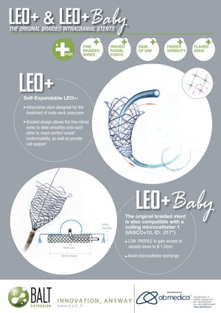
  • 1. neuroradiology solutions www.abmedica.it ab medica s.p.a. Via nerviano, 31 - 20020 Lainate (MI) tel +39 02 933051 - fax +39 02 93305400 abmedica@abmedica.it Self-Expandable LEO+: Intracranial stent designed for the treatment of wide-neck aneurysm Braided design allows the fine nitinol wires to slide smoothly onto each other to reach perfect vessel conformability, as well as provide coil support The original braided stent is also compatible with a coiling microcatheter 1 (VASCO+10, ID: .017”) LOW PROFILE to gain access to vessels down to Ø 1,5mm Avoid microcatheter exchange original braided stent so compatible with aArtery diameter FINE BRAIDED WIRES HIGHER RADIAL FORCE EASE OF USE HIGHER VISIBILITY FLARED ENDS A FOR Mesh size Stent’s lenght
  • 2. Original article Intra-arterial nimodipine for cerebral vasospasm after subarachnoid haemorrhage: Influence on clinical course and predictors of clinical outcome Asma Bashir1 , Morten Andresen1 , Jiri Bartek Jr1,2 , Marie Cortsen3 , Vagn Eskesen1 and Aase Wagner3 Abstract Intra-arterial nimodipine (IAN) has shown a promising effect on cerebral vasospasm (CV) after aneurysmal subarachnoid haemorrhage. At our institution, Rigshospitalet, IAN treatment has been used since 2009, but the short- and long-term clinical efficacy of IAN has not yet been assessed. The purpose was to evaluate the efficacy and clinical outcome of IAN treatment of symptomatic CV, and to assess the predictors of clinical outcome. Medical records of 25 patients undergoing a total of 41 IAN treatment sessions were retrospectively reviewed. Data on angiographic results, blood-flow velocities and the clinical condition before and after the IAN treatment were recorded. Predictors of the clinical outcome were assessed with a linear regression model. Positive angiographic response was achieved in 95.1% of 41 IAN treatment sessions. Flow-velocity measurements showed no relationship with angiographic responses of IAN. The immediate clinical improvement was observed in three patients (12%). Five patients (20%) had a favourable outcome at discharge and at three-month follow-up; 10 patients (40%) had a moderate to poor outcome; and the rest (40%) died. Increased number of affected vessels and number of procedures carried out per patient, and a trend toward an increased delay time from symptomatic CV to confirming angiographic CV and thus instituting IAN treatment predicted the poor clinical outcome. IAN treatment appears to be effective in reversing angiographic CV. However, it is not always effective in reversing clinical deterioration, as several other factors including treatment delay affect the clinical course. Keywords Subarachnoid haemorrhage, cerebral vasospasm, intra-arterial nimodipine Introduction Cerebral vasospasm (CV) is a feared and potentially life- threatening complication in patients following aneurys- mal subarachnoid haemorrhage (SAH). Angiographic CV is observed in up to 70% of patients, typically between 4–14 days after the initial bleeding, and leads to symptomatic brain ischaemia and/or new infarction on radiographic imaging in about 30% of these patients.1,2 Treatment of CV and prevention of delayed cerebral ischaemia (DCI) is therefore a main goal in the management of patients surviving from SAH. However, despite maximal contemporary therapy and neurocritical care, about 30% of these patients die, another 30% sur- vive with significant disability, and the rest of them recover with little or no long-term disability.3 Current medical strategies for prevention and treatment of CV including endovascular therapies with vasodilators and triple-H (i.e. hypertension, hyper- volaemia, haemodilution) are suboptimal,2,4–8 as the exact mechanisms leading to CV are not yet fully elucidated.2,9 At the moment, nimodipine, a calcium channel blocker, remains the only drug proved by the US Food and Drug Administration, which is recom- mended as the first-line treatment and prevention of CV and improvement of outcomes after SAH.10,11 Although nimodipine exhibits neuroprotective as well as vasodilatory benefits,2 some patients deteriorate fur- ther and develop severe symptomatic CV. In the last decade, a number of clinical trials have investigated nimodipine infused directly in the spastic arteries (intra-arterial nimodipine (IAN)), and found it to be 1 Department of Neurosurgery, Rigshospitalet, Faculty of Health and Medical Sciences, University of Copenhagen, Blegdamsvej 9, 2100 Copenhagen, Denmark 2 Department of Clinical Neuroscience, Section for Neurosurgery, Karolinska Institutet and Department of Neurosurgery, Karolinska University Hospital, Stockholm, Sweden 3 Department of Radiology, Rigshospitalet, Faculty of Health and Medical Sciences, University of Copenhagen, Blegdamsvej 9, 2100 Copenhagen, Denmark Corresponding author: Asma Bashir, Department of Neurosurgery, Rigshospitalet, Faculty of Health and Medical Sciences, University of Copenhagen, Blegdamsvej 9, 2100 Copenhagen, Denmark. Email: ab@asmabashir.com The Neuroradiology Journal 2016, Vol. 29(1) 72–81 ! The Author(s) 2016 Reprints and permissions: sagepub.co.uk/journalsPermissions.nav DOI: 10.1177/1971400915626429 neu.sagepub.com
  • 3. effective in reversing angiographic CV, although overall clinical outcome differs through the literature.12–18 At our institution, Rigshospitalet, IAN treatment has been used as the method of choice since 2009 whenever endo- vascular treatment of CV was indicated. However, the short- and long-term clinical efficacy of IAN has not yet been assessed. The purpose of this study was to retrospectively evaluate the efficacy and clinical outcome of IAN treat- ment of symptomatic CV, which is refractory to the haemodynamic treatment, and to assess the predictors of the clinical outcome. Materials and methods Data collection Between December 2009–May 2012, 325 adults with evidence of SAH on computerized tomography (CT) scan were admitted to the neurosurgical intensive care unit (ICU) at the University Hospital, Rigshospitalet, in Copenhagen, Denmark. Only patients presenting with a ruptured intracranial aneurysm on CT angio- gram (CTA), suffering from symptomatic CV refrac- tory to the haemodynamic therapy, and receiving IAN treatment were reviewed. Twenty-eight patients were identified. Among them, three patients were trea- ted with a combination of percutaneous transluminal angioplasty (PTA) and IAN, and were excluded to avoid confound bias on clinical outcome, as PTA has also shown to have beneficial effect.19 A total of 25 patients met the inclusion criteria and constituted the study population. Data on these patients was extracted from a computerized database and reviewed retrospect- ively including demographic data, clinical condition at admission based on World Federation of Neurological Surgeons (WFNS) grade I–V,20 amount and distribu- tion of subarachnoid blood detected by admission CT by using the modified Fisher scale 1–4,21 and aneurysm location as well as aneurysm treatment. Onset, location and severity of CV, number of affected vessels, proced- ure specifics such as dosage of IAN per vessel and per procedure, and number of repeated procedures were recorded. To assess the efficacy of IAN treatment, the clinical response within 24 h (e.g. level of conscious- ness based on Glasgow Coma Scale (GCS) and neuro- logical condition), the angiographic response as well as effects on blood flow-velocity of the middle cerebral artery (MCA) determined by transcranial Doppler (TCD) ultrasonography were examined. The clinical outcome was assessed using the modified Rankin scale (mRS) score at discharge and at three-month follow-up.22 Informed consent was obtained from patients or legal representative for patients with impaired consciousness at the time of their clinical work-up. The Board of Health Committee and Data inspectorate approved the study. All the necessary pre- cautions were taken to protect the identities of the patients in the study. Patient management and monitoring All patients underwent either surgical or endovascular obliteration of the ruptured aneurysm within 48 h after the initial bleeding except seven patients who were hos- pitalized 3–14 days after onset of SAH. Nimodipine was administrated, orally or intravenously, in a dose of 60 mg every 4 h in 21 days according to a standar- dized SAH-protocol. Maintenance of euvolaemia was achieved by administration of intravenous isotonic crystalloid. When flow-velocity of the MCA exceeded 120 cm/s (a TCD cut-off value), triple-H therapy was initiated for inducing prophylactic hypervolaemia and elevating the mean arterial pressure by 20%. Flow-velocity measurements were performed at least once daily to monitor possible vasospastic activity. Flow-velocity <120 cm/s was defined as normal, 120–150 cm/s as mild, 150–200 cm/s as moderate and >200 cm/s as severe. Lindegaard ratio was not measured routinely in all patients, and is therefore not mentioned further in the study. The clinical condition was assessed by continuous neurological examinations including GCS scores. Diagnosis of CV and IAN treatment Cerebral vasospasm was suspected, if patients demon- strated at least one of the following: (a) a decreased level of consciousness and/or disorientation with a decrease of at least two points on the GCS score from baseline, (b) new focal neurological deficits (cranial nerve palsy, hemiparesis and/or speech disturbances), and/or (c) flow-velocity >120 cm/s. Neurological deteri- oration was the main trigger for suspicion of CV, fol- lowed by an increase in flow-velocity; the last was mainly applied to patients with ventilator support. Possible causes of neurological deterioration such as presence of ischaemia, re-bleeding or hydrocephalus on CT scan, electrolyte disturbance, seizures or hypoxia were eliminated prior to CTA, which was usually per- formed within 24 h of onset of symptomatic CV. Degree of angiographic CV was assessed on a digital subtraction angiography semi-quantitatively and subjectively and classified as ‘none/mild’ when arterial narrowing was <25% of the arterial diameter, ‘moder- ate’ when narrowing was 25–50%, or ‘severe’ when narrowing >50%. Patients were found eligible for IAN treatment only if they showed angiographic CV in territories compatible with their neurological deficits and/or with elevated mean flow-velocity. After dilution with 0.9% saline to obtain a solution of 0.01 mg/ml, nimodipine was administrated as an infusion over 20–40 minutes through a microcatheter placed in the affected vessel. Post-nimodipine angiograms were performed 10–30 min after each session. Two neuroradiologists evaluated the angiographic results separately, and dis- agreement was resolved by consensus. Each vessel was evaluated as ‘responsive’ or ‘non-responsive’, and the Bashir et al. 73
  • 4. overall angiographic result was graded as ‘good response’, ‘no response’ or ‘mixed response’. If persist- ence or recurrence of CV was suspected, a new CTA was performed, and IAN treatment was repeated after the verification of angiographic CV and progressive neurological deterioration. Clinical evaluation Immediate clinical response and flow-velocity measure- ments within 24h after IAN treatment were recorded. The change in the clinical condition was graded as ‘improved’, ‘unchanged’ or ‘worsened’. The clinical out- come at discharge from the hospital and at three-month follow-up was assessed using the mRS score (no question- naire).22 A score 2 was considered as a good outcome. Statistical analysis Data analysis was performed with the statistical soft- ware package IBM SPSS version 20.0. Descriptive data was presented as median values with interquartile range (IQR) in parentheses, as data was not normally distributed. A p-value of less than 0.05 was accepted as statistically significant. Our primary dependent vari- able was mRS score, which was investigated in a linear regression model based on following predictor variables: age, WFNS grade, Fisher scale, aneurysm location, severity of CV, number of affected vessels, angiographic response and number of repeated proced- ures. Significance of time delay in diagnosis of angio- graphic CV and thus instituting IAN treatment was assessed as well. Results Clinical assessment Demographic data, WFNS grade, Fisher scale, and location of aneurysm are presented in Table 1. Surgical clipping was performed in seven (28%) patients and endovascular occlusion was performed in 18 (72%) patients. All patients developed neurological deterioration such as decreased level of consciousness, hemiparesis, aphasia or other new focal neurological signs between 4–13 days after onset of SAH (median, eight days; IQR 6–9 days). Among them, two (8%) patients already had neurological deficits at the time of aneurysm treatment and one (4%) due to carotid artery dissection two days prior to symptomatic CV. One (4%) patient (no. 17) was admitted to the hospital 14 days after the initial bleeding and already had developed left-sided arm paresis and leg paralysis due to CV. In one (4%) patient (no. 15), who was artificially ventilated, the suspicion of CV was raised by elevated flow-velocity measurements only, which was the main indication for angiography. The neurological deterior- ation did not respond to the haemodynamic treatment. Onset of occurrence of CV and neurological deterioration including flow-velocity measurements are summarized in Table 2. Angiography and IAN treatment Diagnostic cerebral angiography was performed within 24 h after onset of symptomatic CV in all patients except nine (36%), in whom it was performed later due to their fluctuating neurological condition. The median time delay from the occurrence of symptomatic CV to CTA was 14.5 h (IQR, 2.9–38.1 h). In most cases, angiographic CV diffusely involved all vessels on both sides of the circulation, while in six cases (24%) only one side was affected involving 1–3 vessels. Overall, the degree of CV was severe in 16 (64%) patients and mod- erate in nine (36%). No one had a mild degree of CV. In addition, seven (28%) patients showed signs of incipient hypodensities on their CT scans. Using a linear regression model with a significance p ¼ 0.048; adjusted R2 ¼ 0.241, severity of CV was significant asso- ciated with an increased number of affected vessels (p ¼ 0.004), but not with age (p ¼ 0.50), Fisher scale (p ¼ 0.85) and aneurysm treatment (p ¼ 0.54). Severity Table 1. Patient characteristics. Demographic data Age, median (years) 52 Female sex, no. (%) 20 (80%) Risk factors, no. (%) Hypertension 13 (52%) Smoking 14 (56%) Alcohol abuse 3 (12%) Drug abuse 1 (4%) WFNS grade I–V, no. (%) I, II and III 15 (60%) IV and V 10 (40%) Fisher grade 1–4, no. (%) 1 and 2 3 (12%) 3 and 4 22 (88%) Location of aneurysm, no. (%) ACoA 9 (36%) ACA 2 (8%) MCA 4 (16%) ICA 4 (16%) PCA 1 (4%) BA 4 (16%) PICA 1 (4%) Surgical or endovascular obliteration, no. (%) Coils 14 (56%) Clips 7 (28%) Stent 3 (12%) Coils/stent 1 (4%) ACA: anterior cerebral artery; ACoA: anterior communicating artery; BA: basilar artery; CV: cerebral vasospasm; ICA: internal cerebral artery; MCA: middle cerebral artery; PCA: posterior cerebral artery; PICA: posterior infer- ior cerebral artery; WFNS: World Federation of Neurological Surgeons. 74 The Neuroradiology Journal 29(1)
  • 5. Table2.Theseverityofcerebralvasospasm(CV)andlocation,neurologicaldeficitincludingflowvelocity(FV),angiographicandclinicalresponsestotheintra-arterialnimodipine(IAN)treatmentand clinicaloutcomeatdischargeandatthree-monthfollow-up(FU). Patient/ sessionno. Onsetof sCV(day) Neurological deficits Location ofaCV Severity ofaCV IANdoseper session(mg) Angiographic response Clinical response mRSscoresat Discharge3-monthFU 1.110Lhemiparesis,5dayslaterRhemiparesis,FV70cm/sLMCA,ACA,ICASevere3.0GoodWorsened 1.2þAphasia,FV178cm/sLMCA,ACA,ICASevere3.0GoodUnchanged 1.3Sedated,FV190cm/sLMCA,ACA,ICASevere3.0MixedresponseUnchanged54 2.19FluctuatingDLC,Larmparesis,FV182cm/sMCAs,ACAs,ICAsSevere4.0GoodWorsened 2.2þSeizures,FV260cm/sACAs,MCASevere4.0GoodWorsened43 3.111FluctuatingDLC,FV187cm/sRMCAModerate3.2NoresponseUnchanged10 4.17Rhemiparesis,facialnervepalsy,visualaxisdeviation,FV 100cm/s LACA,MCAModerate4.0GoodUnchanged54 5.19FluctuatingDLC,Llegparesis,FV193cm/sPCTSevere4.0GoodWorsened6– 6.15FluctuatingDLC,aphasia,FV180cm/sLMCA,ACAs,ICAsSevere4.0GoodWorsened6– 7.111FluctuatingDLCLMCA,MCAs,ICAs,PCTSevere4.0MixedresponseUnchanged 7.2þFV97cm/sRACA,MCA,ICASevere2.0GoodUnchanged53 8.16FluctuatingDLCaphasia,FV348cm/sACAs,MCAs,ICAs,PCTSevere4.0GoodWorsened 8.2SedatedACAs,MCAs,ICAs,PCTSevere6.0GoodWorsened56 9.15DLC,Lhemiparesisfacialnervepalsy,aphasia,FV 235cm/s ACAs,MCAs,ICAsSevere6.0GoodWorsened 9.2SedatedACAs,MCAs,ICAsSevere6.0GoodUnchanged53 10.15DLC,Rhemiparesis,aphasiaACAs,MCAs,ICAsSevere4.8GoodUnchanged 10.2DLC,Rhemiparesis,aphasiaACAs,MCAs,ICAsSevere5.0MixedresponseUnchanged51 11.17FluctuatingDLC,Rhemiparesis,aphasiaPCTModerate4.0GoodUnchanged 11.2þVisialaxisdeviationPCTSevere4.0GoodUnchanged43 12.110DLC,FV156cm/sRACA,MCAs,ICAsSevere3.0GoodUnchanged6– 13.111DLC,Rhemiparesis,FV190cm/sACA,MCAs,ICAModerate4.4GoodUnchanged 13.2DLC,RhemiparesisACAs,MCAs,ICAsModerate5.0GoodWorsened6– 14.1a 6DLC,Rhemiparesis,facialnervepalsy,aphasia,FV 230cm/s ACAs,MCAs,ICAsSevere4.0GoodWorsened6– 15.19Sedated,FV230cm/sACAs,MCAs,ICAsSevere5.0MixedresponseWorsened6– 16.19FluctuatingDLC,FV183cm/sACAsModerate4.5MixedresponseUnchanged32 17.113Lhemiparesis/paralysisACAs,MCAs,ICAsSevere4.0GoodWorsened 17.2þAphasia,FV250cm/sRACA,MCA,ICA,PCTSevere4.0GoodWorsened6– 18.18DLC,aphasia,FV145cm/sACAs,MCAs,ICAsSevere5.0GoodUnchanged 18.2þFV313cm/sLMCA,ICA,ACAsSevere4.0GoodUnchanged 18.3Sedated,FV257cm/sACAs,MCAs,ICAsSevere5.0GoodUnchanged 18.4Sedated,FV290cm/sLACA,MCA,ICASevere4.0MixedresponseUnchanged55 (continued) Bashir et al. 75
  • 6. of CV and number of affected vessels including ana- tomic distribution are presented in Table 2. Forty-one IAN treatment sessions were performed in 25 patients (range, 1-4 treatment sessions per patient), in which 173 vessels were evaluated. The median time delay from angiographic CV to IAN treat- ment was 3.0 h (IQR, 2–9.5 h). Median dose of nimodi- pine was 4 mg per session (range, 2–6 mg per session), while the largest single dose per vessel was 4 mg (median, 2 mg; range, 1–4 mg). Complete reversal of CV was successfully achieved in 31 (75.6%) treatments (Figure 1); partial reversal was noted in eight (19.5%) (Figure 2), while two (4.9%) showed no effect. There was no significant correlation between the degree of CV and angiographic response to IAN (p ¼ 0.24). Worsening of CV did not occur during the procedures. No signs of haemodynamic instability due to IAN infusion or other procedure-related complications were observed. However, in one (4%) patient (no. 14), a dosage of 2 mg nimodipine was accidently injected in a few seconds causing transient drop in blood pressure. Dose per pro- cedure, number of repeated procedures and angio- graphic effects are summarized in Table 2. TCD measurements Complete results for flow-velocity measurements before and after IAN treatment within 24h were available for review in only 13 (32%) of 41 procedures. Mean velocities decreased by 37cm/s in eight (20%) procedures, unchanged in one (2%), and increased by 34cm/s in four (10%). There was no correlation between flow-velocity measurements and angiographic responses to IAN. Clinical evaluation within 24 h after treatment, at discharge and at three-month follow-up The clinical condition remained unchanged for 12 (48%) patients, although all of them but one showed angiographic response to IAN treatment. Three (12%) patients showed clinical improvement based on GCS scores and/or neurologically, two of them with none and mixed responses to IAN. Ten (40%) patients experienced a worsening in their clinical conditions both consciously and neurologically; among them, six (24%) had developed new neurological deficits. Two (8%) had seizures at the time of wake-up call. Three (12%) patients never regained consciousness and died a few days later; all three had showed angiographic response to IAN, two of them with mixed response. In 12 (48%) patients, new cerebral infarctions were observed on CT scan hours to a few days after the IAN treatment. At discharge, only one (4%) patient showed a good clinical outcome (mRS 1). Two (8%) patients had a moderate outcome (mRS 3) and 13 (52%) a poor out- come (mRS 4–5). Nine (36%) patients died during hos- pitalization, eight (32%) of them as a consequence of extensive cerebral infarction secondary to CV (mRS 6). Table2.Continued Patient/ sessionno. Onsetof sCV(day) Neurological deficits Location ofaCV Severity ofaCV IANdoseper session(mg) Angiographic response Clinical response mRSscoresat Discharge3-monthFU 19.18DLC,aphasia,FV226cm/sLMCAModerate4.0NoresponseImproved32 20.15Pronation/extensionpattern,FV110cm/sRACA,MCA,ICAModerate3.0GoodWorsened 20.2þRhemiparalysis,Larmparesis,FV130cm/sRACA,MCA,ICASevere3.0GoodUnchanged53 21.19DLC,FV180cm7sACAs,ICAsSevere4.0GoodImproved42 22.16DLC,aphasiaLACA,ICA,MCAsModerate4.0GoodUnchanged54 23.16DLCLACA,MCAs,ICAsSevere4.0MixedresponseImproved53 24.14DLCACAs,MCAs,ICAs,PCTSevere6.0GoodUnchanged 24.2þRhemiparesis,FV200cm/sACAs,MCAs,ICAs,PCTSevere6.0GoodWorsened6– 25.16DLC,FV197cm/sLICA,ACAs,MCAs,PCTModerate6.0MixedresponseWorsened 25.2þRhemiparesis,extensorplantarreflex,FV237cm/sLACA,MCA,ICA,PCTModerate6.0GoodWorsened6– ACA:anteriorcerebralartery;aCV:angiographiccerebralvasospasm;DLC:decreasedlevelofconsciousness;FU:follow-up;IAN:intra-arterialnimodipine;ICA:internalcerebralartery;L:leftcirculation;MCA:middlecerebral artery;PCT:posteriorcirculatoryterritory;R:rightcirculation;sCV:symptomaticcerebralvasospasm.a ByaccidentIANinjectedintherightICAforafewsecondscausingtransientdropinbloodpressure.Thetreatmentis repeatedwiththesamedose. 76 The Neuroradiology Journal 29(1)
  • 7. At the three-month follow-up, another four (16%) patients showed a favourable outcome (mRS 1–2), six (24%) had a moderate outcome (mRS 3), and one more (4%) had died (mRS 6). Data on the clinical evaluation is presented in Table 2. Observing a linear regression model (p ¼ 0.007, adjusted R2 ¼ 0.654), significant predictors of a poorer clinical outcome (mRS 4–6) were an increased number of affected vessels (p ¼ 0.003) and an increased number of procedures carried out per patient (p ¼ 0.04), and the angiographic response of IAN (p ¼ 0.01). Looking at the time frame from symptomatic CV to confirming angiographic CV, we saw a trend toward a poorer clin- ical outcome with increasing time (p ¼ 0.13). Discussion Cerebral vasospasm following an aneurysmal SAH continues to be a leading cause of morbidity and mor- tality.3 We retrospectively reviewed 25 patients treated with IAN infusions for medically refractory symptom- atic CV, and found that IAN treatment was effective in reducing angiographic CV in 95.1% of 41 treatment sessions, with complete reversal in 75.6% and partial reversal in 19.5%. However, the efficacy of treatment was temporary, and required repeated treatment ses- sions in 13 (52%) patients, and was not always followed by clinical improvement. Only three (12%) patients demonstrated an immediate clinical improvement after treatment, and only one (4%) had a good clinical outcome at the time of discharge. Eight (32%) of nine deceased patients died during hospitalization. At three- month follow-up, another four (16%) patients showed a clinical improvement, while one (4%) more had died. Overall, five (20%) patients had a favourable long-term outcome (mRS 0–2), 10 (40%) had moderate to poor outcome (mRS 3–5), and 10 (40%) patients had died (mRS 6) (Table 2). Figure 1. Patient no. 24. Lateral angiogram of the internal cerebral artery (ICA) showing (a) cerebral vasospasm (CV); (b) after intra-arterial nimodipine (IAN) infusion, an increase in diameter of the vessels is observed. Figure 2. Patient no. 12. Anteroposterior angiograms of (a) the left and (b) right internal cerebral arteries (ICAs) showing cerebral vasospasm (CV). After intra-arterial nimodipine (IAN) infusion, (c) the left side demonstrates an increase in diameter of the vessels, while (d) the right side shows a mixed response. Bashir et al. 77
  • 8. Several clinical trials have evaluated the efficacy of IAN treatment for symptomatic CV.12–18 These studies have reported post-IAN angiographic vessel dilatation in 43.3–82.8% of sessions, clinical improvement in 11.1–88.9% of patients, and 61.1–88.9% with a favour- able short- or long-term outcome (Table 3). Biondi et al.12 were the first to assess the efficacy of IAN. The authors reported post-IAN angiographic response in 13 (43.3%) of 30 treatment sessions and clinical improvement in 19 (76%) of 25 patients. Eighteen (72%) had a favourable long-term outcome (mRS 0–2). Their angiographic results are in line with our study. We reported complete reversal of angiographic CV in 75.6% of sessions, and in addition partial rever- sal of CV was seen in 19.5%. However, our angio- graphic results did not correlate with the clinical improvement and the overall clinical outcome was less satisfactory, compared to Biondi et al.12 Ha¨ nggi et al.15 investigated the efficacy of IAN in 26 patients as well. In eight (31%) patients, no angiographic improvement was seen, and therefore these were excluded from the study. Using cerebral perfusion CT (PCT), the authors reported an improved time to peak and mean transit time a day after the procedure. Although the efficacy of IAN was transient, and 11 (61%) of 18 patients developed additional infarction, 11 (61%) patients revealed a good clinical condition at time of discharge, which is remarkable compared to our study. However, some of these studies are confounded by the combined use of IAN and papaverine or balloon angioplasty, which might explain in part for the clinical improvement reported in the studies.14–16 We are the second group of researchers to evaluate the influence of time delay in diagnosing and instituting the IAN treatment after onset of symptomatic CV. In our study, an increasing time delay from symptomatic CV to angiographic CV showed a trend toward a poorer outcome with a median time of 14.5 h. The median delay time from angiographic CV to the IAN treatment was 3.0 h. Grotenhuis et al.23 was the first to report that the IAN treatment was ineffective when administrated only a few hours after the onset of CV. However, the authors used only small dosage of nimo- dipine ranging from 0.068–1.0 mg, which could account at least in part for the observed lack of effect. Biondi et al.12 performed the diagnostic CTA and IAN treat- ment within 24 h of onset of symptomatic CV, with the mean delay time of 18 Æ 6 h. The dosage of IAN per vessel consisted of 1–3 mg. Despite time delay in the diagnostic process, thus instituting the treatment, the authors demonstrated a clinical improvement in 76% of patients after IAN. Similarly, Linfante et al.7 inves- tigated the efficacy of IA nicardipine in 22 patients. A total of 46 endovascular sessions with intra-arterial nicardipine were performed within 48 h after onset of symptoms in 21 patients, and within 72 h in one patient. At the time of discharge, half of patients were reported to be independent functionally (mRS 0–2). In our study, time delays were much less than reported by Table3.Studieswithcerebralvasospasm(CV)afteraneurysmalsubarachnoidhaemorrhage(SAH)andintra-arterialnimodipine(IAN)treatment. Author/yearStudydesign Patient no. Session no. Doseof IANpervessel Angiographic improvement Clinical improvement Theclinical outcome(mRS2and GOS!4)atdischarge Theclinicaloutcome (mRS2andGOS!4) atfollow-up Biondietal.,20041,2 Retrospective25301–3mg43.3%76%–72% Choetal.,20111,3 Retrospective421013–6mg82.2%68.3%76.2%84.6% Dehdashtietal.,201114 Retrospective10151–4mg80%60%–70% Ha¨nggietal.,200815,a Prospective18260.8–3.2mg77.3%11.1%61.1%61.1% HuiandLau,20051,6,b Retrospective9102–4mg66.6%88.9%88.9%77.8% Kimetal.,20091,7 Retrospective19531–3mg79.2%68.4%78.9%– Kimetal.,201218 Prospective29291–3mg82.8%72.4%75.9%– Ourstudy,2015Retrospective28451–4mg95.6%21.4%10.7%28.6% GOS:GlasgowOutcomeScale;mRS:modifiedRankinscale.a Only18of26patientsand22of42proceduresareincludedintheanalysisduetonomeasurableangiographiceffectin20(47.6%)proceduresamongeight(30.7%) patients.b Themeanpercentageincreaseinarterialdiameter.Angiographicimprovementincludesresultswithmoderateandmajoreffect. 78 The Neuroradiology Journal 29(1)
  • 9. Biondi et al.12 and Linfante et al.7 and with a dosage of IAN per vessel (range, 1–4 mg) similar to Biondi et al.12 However, despite angiographic response in 95.1% of treatment sessions only a few patients showed clinical improvement. The discrepancy between angiographic response and unsatisfactory clinical results could be explained by the fact that parenchymal damage has already occurred due to ischaemia resulting from CV in spite of angiographic response to endovascular treat- ment with IAN. Furthermore, evidence from a rat model indicated that reperfusion of parenchyma injured by an ischaemic insult might lead to further injury;24–26 this phenomenon could be one of the rea- sons why the uses of endovascular therapies are asso- ciated with a poorer outcome despite substantial vessel dilatation in some instances, as seen in our study. TCD is often used as a surrogate diagnostic tool to detect CV. We found no correlation between flow-velo- cities and angiographic responses to IAN, although data of TCD measurements was missing in most cases. Kim et al.17 observed a decrease in flow-velocity in 29 of 33 sessions. In contrast, Ha¨ nggi et al.15 noticed a significant increase in flow-velocity one day after IAN infusion (p ¼ 0.03). Wolf et al.27 was unable to detect CV with TCD in two patients and reflect the treatment course in four patients. The interpretation of the TCD analysis leaves some room for speculation. Estimation of flow-velocity could vary from minute to minute, and might be influenced by anatomic details such as vascu- lar anatomy, vessel diameters and cerebral blood flow. Influence of pathological conditions such as an increased intracranial pressure or oedema, or systemic haemodynamic effects during triple-H therapy such as changes in cardiac output or blood pressure could affect the speed and waveform of cerebral blood flow as well. In addition, the accuracy of this technique is quite operator-dependent with high intra-variability, and suboptimal insonation might inhibit the detection of high flow-velocities.28–30 Vora et al.31 reported that only very low or very high MCA velocities (e.g. <120 cm/s or >200 cm/s) negatively or positively pre- dicted angiographic and symptomatic CV in a reliable manner, and intermediate velocities had a lower pre- dictive value and were poorly discriminative. Vasodilatation by intra-arterial drug infusion also has been performed with other agents. Papaverine is the first intra-arterial agent reported to reverse angio- graphic CV.6 However, it is associated with a consider- able number of several adverse side effects such as elevation of intracranial pressure,32 worsening of CV33 and thrombocytopaenia.34 One study compared the angiographic effect of papaverine and nimodipine and reported that there was no significant difference between these two groups, although the effect of papa- verine was more pronounced, dilating at least one vessel in every patient, while nimodipine was ineffective in 16% of patients.35 Intra-arterial nicardipine infusion has shown to be effective without increasing intracra- nial pressure.36 However, a transient decrease in blood pressure and an increase in heart rate may occur.7,36 Intra-arterial verapamil infusion is also reported to dilate spastic vessels without causing changes in haemodynamic circulation or intracranial pressure.6 However, it needs to be evaluated in larger series to confirm its efficacy. PTA is highly effective in relieving focal CV, and has a continuous effect on narrowed ves- sels. However, the technique is limited to proximal ves- sels, and requires expert neurointerventionists due to the significant risks of artery rupture, dissection and local thrombosis.19,37 One study compared the clinical outcome of IAN treatment with PTA, but the authors did not find significant differences in these two groups.38 Limitations The study has several important limitations including its retrospective design and the small number of patients due to it being a single centre study, which may limit generalizability. There was no control group. It is not clear whether the change in the clinical condition after IAN infusion was due to the effect of the treatment or just a natural course. Quantitative measurement of vessel diameter on pre- and post- nimodipine angiograms was not available as mm (Æstandard deviation), but only as Æeffect. TCD data were missing before and after IAN infusions in several patients making it more difficult to estimate the abso- lute correlation between TCD and angiographic results. Data on monitoring systemic haemodynamic effects and intracranial pressure during IAN sessions were not investigated, and therefore we were unable to quan- tify the effect of IAN on these areas. Conclusion and perspectives Although there are several limits in this retrospective study, we can conclude that IAN treatment appears to be effective in reversing angiographic CV. However, the relationship between angiographic and clinical responses was not convincing, as the treatment is inef- fective in reversing clinical deterioration caused by DCI. Several factors such as increased numbers of affected vessels and procedures carried out per patient predicted the clinical outcome negatively. In addition, an increasing time delay in diagnostic process of CV and thus instituting treatment might contribute to a poorer clinical outcome. Clearly, early detection and rapid treatment of CV are needed before ischaemic damage occurs. Future studies with continued assessment of cerebral blood flow might help optimize the timing of IAN treatment and to assess the real efficacy of the IAN treatment in patients with symptomatic CV after SAH. Acknowledgements The study is dedicated to the late Professor Bertil Romner, Department of Neurosurgery, Rigshospitalet, who originally conceived and designed the study. A Bashir and the late Bashir et al. 79
  • 10. Bertil Romner conceived and designed the study. A Bashir reviewed patient data and wrote the first and subsequent drafts of the manuscript. A Wagner and M Cortsen evaluated pre- and post-angiograms. M Andresen conducted the statis- tical analysis. J Bartek Jr, M Andresen, A Wagner, M Cortsen and V Eskesen participated in critical revision and writing of the article. All authors have seen and approved the final version. Funding The author(s) received no financial support for the research, authorship, and/or publication of this article. Conflict of interest The author(s) declared no potential conflicts of interest with respect to the research, authorship, and/or publication of this article. References 1. Connolly ES, Rabinstein AA, Carhuapoma JR, et al. Guidelines for the management of aneurysmal subarach- noid hemorrhage: A guideline for healthcare profes- sionals from the American Heart Association/American Stroke Association. Stroke 2012; 43: 1711–1737. 2. Treggiari-Venzi MM, Suter PM and Romand J-A. Review of medical prevention of vasospasm after aneur- ysmal subarachnoid hemorrhage: a problem of neuroin- tensive care. Neurosurgery 2001; 48: 249–262. 3. Washington CW and Zipfel GJ. Detection and monitor- ing of vasospasm and delayed cerebral ischemia: a review and assessment of the literature. Neurocrit Care 2011; 15: 312–317. 4. Clouston JE, Numaguchi Y, Zoarski GH, et al. Intraarterial papaverine infusion for cerebral vasospasm after subarachnoid hemorrhage. AJNR Am J Neuroradiol 1995; 16: 27–38. 5. Fraticelli AT, Cholley BP, Losser M-R, et al. Milrinone for the treatment of cerebral vasospasm after aneurysmal subarachnoid hemorrhage. Stroke 2008; 39: 893–898. 6. Keuskamp J, Murali R and Chao KH. High-dose intraar- terial verapamil in the treatment of cerebral vasospasm after aneurysmal subarachnoid hemorrhage. J Neurosurg 2008; 108: 458–463. 7. Linfante I, Delgado-Mederos R, Andreone V, et al. Angiographic and hemodynamic effect of high concentra- tion of intraarterial nicardipine in cerebral vasospasm. Neurosurgery 2008; 63: 1080–1087. 8. Treggiari MM, Walder B, Suter PM, et al. Systematic review of the prevention of delayed ischemic neurological deficits with hypertension, hypervolemia, and hemodilu- tion therapy following subarachnoid hemorrhage. J Neurosurg 2003; 98: 978–984. 9. Kassell NF, Sasaki T, Colohan AR, et al. Cerebral vaso- spasm following aneurysmal subarachnoid hemorrhage. Stroke 1985; 16: 562–572. 10. Barker 2nd, FG and Ogilvy CS. Efficacy of prophylactic nimodipine for delayed ischemic deficit after subarach- noid hemorrhage: A metaanalysis. J Neurosurg 1996; 84: 405–414. 11. Koenig MA. Management of delayed cerebral ischemia after subarachnoid hemorrhage. Continuum (Minneap Minn) 2012; 18: 579–597. 12. Biondi A, Ricciardi GK, Puybasset L, et al. Intraarterial nimodipine for the treatment of symptomatic cerebral vasospasm after aneurysmal subarachnoid hemorrhage: Preliminary results. AJNR Am J Neuroradiol 2004; 25: 1067–1076. 13. Cho W-S, Kang H-S, Kim JE, et al. Intraarterial nimo- dipine infusion for cerebral vasospasm in patients with aneurysmal subarachnoid hemorrhage. Interv Neuroradiol 2011; 17: 169–178. 14. Dehdashti AR, Binaghi S, Uske A, et al. Intraarterial nimodipine for the treatment of symptomatic vasospasm after aneurysmal subarachnoid hemorrhage: A prelimin- ary study. Neurol India 2011; 59: 810–816. 15. Ha¨ nggi D, Turowski B, Beseoglu K, et al. Intraarterial nimodipine for severe cerebral vasospasm after aneurys- mal subarachnoid hemorrhage: Influence on clinical course and cerebral perfusion. AJNR Am J Neuroradiol 2008; 29: 1053–1060. 16. Hui C and Lau KP. Efficacy of intraarterial nimodipine in the treatment of cerebral vasospasm complicating sub- arachnoid haemorrhage. Clin Radiol 2005; 60: 1030–1036. 17. Kim JH, Park IS, Park KB, et al. Intraarterial nimodi- pine infusion to treat symptomatic cerebral vasospasm after aneurysmal subarachnoid hemorrhage. J Korean Neurosurg Soc 2009; 46: 239–244. 18. Kim S-S, Park D-H, Lim D-J, et al. Angiographic fea- tures and clinical outcomes of intraarterial nimodipine injection in patients with subarachnoid hemorrhage- induced vasospasm. J Korean Neurosurg Soc 2012; 52: 172–178. 19. Muizelaar JP, Zwienenberg M, Rudisill NA, et al. The prophylactic use of transluminal balloon angioplasty in patients with Fisher grade 3 subarachnoid hemorrhage: A pilot study. J Neurosurg 1999; 91: 51–58. 20. Drake CG, Hunt WE, Sano K, et al. Report of World Federation of Neurological Surgeons Committee on a Universal Subarachnoid Hemorrhage Grading Scale. J Neurosurg 1988; 68: 985–986. 21. Claassen J, Bernardini GL, Kreiter K, et al. Effect of cisternal and ventricular blood on risk of delayed cerebral ischemia after subarachnoid hemorrhage: The Fisher scale revisited. Stroke 2001; 32: 2012–2020. 22. Banks JL and Marotta CA. Outcomes validity and reli- ability of the modified Rankin scale: implications for stroke clinical trials: A literature review and synthesis. Stroke 2007; 38: 1091–1096. 23. Grotenhuis JA, Bettag W, Fiebach BJO, et al. Intracarotoid slow bolus injection of nimodipine during angiography for treatment of cerebral vasospasm after SAH. A preliminary report. J Neurosurg 1984; 61: 231–2340. 24. Vibulsresth S, Dietrich D, Busto R, et al. Failure of nimo- dipine to prevent ischemic neuronal damage in rats. Stroke 1987; 18: 210–216. 25. Gotoh O, Mohamed AA, McCulloch J, et al. Nimodipine and the haemodynamic and histopathological conse- quences of middle cerebral artery occlusion in the rat. J Cereb Blood Flow Metab 1986; 6: 321–331. 26. Snape MF, Baldwin HA, Cross AJ, et al. The effects of chlormethiazole and nimodipine on cortical infarct area after focal cerebral ischemia in the rat. Neuroscience 1993; 53: 837–844. 27. Wolf S, Martin H, Landscheidt JF, et al. Continuous selective intraarterial infusion of nimodipine for therapy 80 The Neuroradiology Journal 29(1)
  • 11. of refractory cerebral vasospasm. Neurocrit Care 2010; 12: 346–351. 28. Lysakowski C, Walder B, Costanza MC, et al. Transcranial Doppler versus angiography in patients with vasospasm due to a ruptured aneurysm. A system- atic review. Stroke 2001; 32: 2292–2298. 29. Mills JN, Mehta V, Russin J, et al. Advanced imaging modalities in the detection of cerebral vasospasm. Neurol Res Int 2013; 2013: 415960. doi: 10.1155/2013/415960. 30. Staalsø JM, Edsen T, Romner B, et al. Transcranial Doppler velocimetry in aneurysmal subarachnoid haem- orrhage: intra- and interobserver agreement and relation to angiographic vasospasm and mortality. Br J Anaesth 2013; 110: 577–585. 31. Vora YY, Suarez-Almazor M, Steinke DE, et al. Role of transcranial Doppler monitoring in the diagnosis of cere- bral vasospasm after subarachnoid hemorrhage. Neurosurgery 1999; 44: 1237–1247. discussion 47–58. 32. McAuliffe W, Townsend M, Eskridge JM, et al. Intracranial pressure changes induced during papaverine infusion for treatment of vasospasm. J Neurosurg 1995; 83: 430–434. 33. Clyde BL, Firlik AD, Kaufmann AM, et al. Paradoxical aggravation of vasospasm with papaverine infusion fol- lowing aneurysmal subarachnoid hemorrhage. Case report. J Neurosurg 1996; 84: 690–695. 34. Miller JA, Cross DT, Moran CJ, et al. Severe thrombo- cytopenia following intraarterial papaverine administra- tion for treatment of vasospasm. J Neurosurg 1995; 83: 435–437. 35. Kerz T, Boor S, Beyer C, et al. Effect of intraarterial papaverine or nimodipine on vessel diameter in patients with cerebral vasospasm after subarachnoid hemorrhage. Br J Neurosurg 2012; 26: 517–524. 36. Tejada JG, Taylor RA, Ugurel MS, et al. Safety and feasibility of intra-arterial nicardipine for the treatment of subarachnoid hemorrhage-associated vasospasm: Initial clinical experience with high-dose infusions. AJNR Am J Neuroradiol 2007; 28: 844–848. 37. Linskey ME, Horton JA, Rao GR, et al. Fatal rupture of the intracranial carotid artery during transluminal angioplasty for vasospasm induced by subarachnoid hemorrhage. Case report. J Neurosurg 1991; 74: 985–990. 38. Aburto-Murrieta Y, Marquez-Romero JM, Bonifacio- Delgadillo D, et al. Endovascular treatment: Balloon angioplasty versus nimodipine intra-arterial for medically refractory cerebral vasospasm following aneurysmal sub- arachnoid hemorrhage. Vasc Endovascular Surg 2012; 46: 460–465. Bashir et al. 81