The facial nerve is the longest nerve in the bony canal. It contains both sensory and motor fibers and innervates the muscles of facial expression. The nerve develops from the second branchial arch and has nuclei in the lower pons connected to four nuclei. It exits the skull through the stylomastoid foramen and divides into branches in the parotid gland. The zygomatic and buccal branches are at risk during surgery on the zygomatic arch and cheek. Facial nerve paralysis can occur from lesions at different levels and have varying clinical presentations such as in Bell's palsy. Care must be taken during parotid and temporal bone surgeries due to the nerve's anatomy.
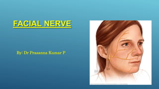
3. INTRODUCTION
• Longest nerve in the bony canal
• Mixed nerve contains both sensory and
motor fibers.
• Sensory root is called nerves intermedius.
• Nerve of 2nd brachial arch
• Nerve of Facial Expression
3
4. .
EMBRYOLOGY
The development of the facial nerve begins in the third
week.
The facial nerve becomes the nerve of the second
branchial arch, and
for this reason it will supply all the elements that derive
from it
the stapes, the styloid process of the temporal bone, the
stylohyoid
ligament, also to the muscles of the stapes, stylohyoid,
posterior belly of
the digastric.
5. NUCLEI
The fibres of nerve are connected to four nuclei
situated in lower pons.
1. Motor Nucleus or
Branchiomotor
2. Superior Salivatory Nucleus
3. Lacrimatory nucleus
4. Nucleus of the tractus solitarus
(gustatory)
6. FUNCTIONAL COMPONENTS
1. Special visceral or branchial motor (SVE) -
muscle of facial expression and elevates
hyoid bone.
2. . General visceral (GVE) – secretomotor to
submandibular and sublingual salivary
gland ,lacrimal gland and gland of nose ,
palate.
3. General visceral afferent (GVA)
4. Special visceral (SVA) – carries taste
sensations from palate and anterior 2/3 rd of
tongue except from vallate papillae
5. General somatic (GSA) - innervate a part of
the skin of the ear
7. COURSE & RELATION
The two roots of facial nerve are attached to the lateral part of lower
border of pons just medial to 8th cranial nerve.
The two roots runs laterally & forwards ,with 8th cranial nerve to reach
internal acoustic meatus.
In the meatus the motor root lies in a groove on 8th nerve with the sensory root
intervening.
At the bottom meatus ,the two roots fuse to form a single trunk which lies in
petrous temporal bone. 7
8. Enters its own canal, named the “facial canal.
Within the canal course can be divided into 3 parts by 2
bends.
The first bend at the junction of 1st &2nd part is sharp. It
lies over the anterosuperior part of the promontory and is
also called the genu.
The 2nd bend is gradual and lies between the promontory
&the aditus to the mastoid antrum.
8
9. • The facial nerve leaves the skull by passing
through the stylomastoid foramen.
• In its extracranial route it crosses the lateral side of
base of styloid process.
It enters the posteromedial surface of parotid gland ,
runs forwards through the gland crossing the
retromandibular vein & external carotid artery.
Behind the neck of mandible , it divides into its 5
terminal branches which emerge along the anterior
border of parotid gland.
10. BRANCHES:
A. Within the facial canal:
1. Greater petrosal nerve
2. Nerve to stapedius
3. The chorda tympani
B. At its exit from the stylomastoid foramen:
1. Posterior auricular
2. Digastric
3. Stylohyoid
C. Terminal branches within the parotid gland:
1. Temporal
2. Zygomatic
3. Buccal
4. Marginal mandibular
5. Cervical
D. Communicating branches with adjacent cranial and spinal nerves.
11. GREATER PETROSAL NERVE:
• Innervates the lacrimal
gland, mucous
membrane of the nasal
cavity and palate.
• Carries gustatory and
parasympathetic fibres.
12. NERVE TO STAPEDIUS:
• It inserts into the
neck of the stapes.
• In paralysis of the
muscle even normal
sounds appear too
loud
12
13. CHORDA TYMPANI:
• Afferent special sensation from the
anterior two-thirds of the tongue
via the lingual nerve,
• Efferent parasympathetic
secretomotor innervation to the
submandibular and sublingual
glands.
14. POSTERIOR AURICULAR:
• Arises just below the stylomastoid
foramen
• Ascends between the mastoid
process and the external acoustic
meatus
15. DIGASTRIC
• Arises close to the
posterior auricular.
• It is short and
supplies the
posterior belly of
the digastric.
STYLOHYOID
• Arise with the digastric
branch
• It is long and supplies the
stylohyoid muscle
17. TEMPORAL BRANCH
• Cross zygomatic arch
• Supply, frontalis, auricular
muscles,orbicularis oculi.
• To test the function patient is
asked to frown and wrinkle
his or her forehead.
18. ZYGOMATIC BRANCH
• Run across the zygomatic
bone
• Supply the orbicularis oculi.
• To test the function patient
is asked to close their eyes
tightly.
19. BUCCAL BRANCH
Two branches
1. Upper- runs above the parotid
duct
2. Lower- runs below the duct
• It supplies: -
Muscles of the cheek and upper lip
20. MARGINAL MANDIBULAR
• Runs below the angle of mandible deep
to platysma.
• It crosses the body of mandible and
supplies muscles of the lower lip and
chin.
21. CERVICAL BRANCH
• Emerges from apex of
parotid gland.
• Runs downwards and
forwards in the neck to
supply the platysma.
22. GANGLIAASSOCIATED
WITH NERVE
• 3 ganglia are associated
with facial nerve.
1. Geniculate ganglion
2. Pterygopalatine
ganglion
3. Submandibular ganglion
23. Geniculate Ganglion
It is located on the first bend of facial
nerve
It is a Sensory ganglion.
The taste fibres present in the nerve
are the peripheral processes of
pseudounipolar neurons present in
the geniculate ganglion.
24. Submandibular Ganglion
It is a parasympathetic ganglion for
relay of secretomotor fibres to the
submandibular and sublingual
salivary glands.
The postganglionic fibres come from
the Chorda tympani nerve.
It is situated above deep portion of
submandibular gland on hyoglossal
muscle.
25. Pterygopalatine Ganglion
•It is also a parasympathetic
ganglion.
•Secretomotor fibres meant for the
Lacrimal gland relay in this
ganglion.
•It is located posterior to middle
nasal concha & anterior to
pterygoid canal.
26. SURGICAL ANATOMY
• The facial nerve is the main anatomical structure that the surgeon should consider in
performing a surgical approach to the temporomandibular joint (TMJ).
• Al-Kayat and Bramley (15) carried out a cadaveric study of the relationship of the
facial nerve and its branches with the region of the TMJ. The zygomatic branch of the
facial nerve crosses the region of the zygomatic arch at a distance of 2.0 ± 0.5 cm from
the anterior wall of the external auditory canal. The bifurcation of the main trunk of
the facial nerve occurs 3.0 ± 0.31 cm from the postglenoidal tubercule and at a mean
distance of 2.3 ± 0.28 cm from the inferior concavity of the external acoustic meatus
Reference -Facial nerve injury following surgery for the treatment of ankylosis of the
temporomandibular joint
Ricardo Viana Bessa Nogueira 1, Belmiro Cavalcanti do Egito Vasconcelos
26
28. • Zygomatic branch:
Inadvertent damage may occur to this
nerve during open reduction of
zygomatic arch or with the use of
zygomatic hooks .
• Buccal branch:
Injury is possible in association with
soft tissue trauma to the cheek region.
29. It is an important structure
encountered at the inferior
border of mandible just beneath
the platysma muscle fibers
during an open approach to
mandibular angle or body area.
For this reason , an initial
incision made approximately
1.5 to 2cm below the inferior
border of mandible which
prevents direct exposure or
trauma to the nerve.
Marginal mandibular branch:
30. PAROTID
GLAND
Facial nerve injury is the most
common complication of
parotid surgery as the two
structures are intimately
related to each other.
33. FACIAL NERVE PARALYSIS
Facial nerve paralysis is a common problem that involves paralysis of
any structures innervated by facial nerve.
Pathway of facial nerve is long and convoluted, so there are a
number of causes that result in facial paralysis.
facial nerve paralysis classified as
1. Supranuclear lesions(UMN lesion)
2. Infranuclear lesions(LMN lesion)
34. SUPRANUCLEAR LESION
• paralysis of the contralateral middle and
lower parts of the face with sparing of the
muscles of the forehead and the orbicularis
oculi muscle
34
35. NUCLEAR OR
INFRANUCLEAR LESION
• involve the facial motor nucleus or the
infranuclear portion of the facial nerve result in
complete paralysis of all the facial muscles on
the ipsilateral side
• mouth droop, flattening of nasolabial fold,
inability to close eye, and smoothing of the brow
on the damaged side
35
39. CLINICAL FEATURES
• Unilateral involvement
• Loss of forehead wrinkles
• Inability to close eyes (Bell’s sign)
• Inability to whistle
• Loss of naso-labial fold
• Drooping of angle of mouth
• Dribbling of food while chewing on affected side
• Mask like appearance of face
• Slurred speech
• Loss or alteration of taste
39
42. REFERENCES
• NETTER’S CRANIAL NERVE COLLECTION
• HUMAN ANATOMY- BD CHAURASIA’S
• CRANIAL NERVES FUNCTION AND DYSFUNCTION THIRD EDITION
• GRAY’S ANATOMY FOR STUDENT
Facial nerve injury following surgery for the treatment of ankylosis of the temporomandibular joint
Ricardo Viana Bessa Nogueira 1, Belmiro Cavalcanti do Egito Vasconcelos
Facial Nerve Injury -Hany Emam, Courtney Jatana, and Gregory M. Ness
• ATLAS OF THE FACIAL NERVE AND RELATED STRUCTURES- NOBUTAKA YOSHIOKA
Mixed nerve with sensory and motor roots.
Main Motor Root
Sensory Root
( Nerve of Wrisberg)
1)Special visceral or branchial efferent (SVE), responsible for muscles of facial expression and for elevation of the hyoid bone.
2 General visceral efferent (GVE) or parasympathetic fibres. These fibres are secretomotor to the submandibular and sublingual salivary glands, the lacrimal gland, glands of the nose, palate and pharynx
3 General visceral afferent (GVA) component carries afferent impulses from the above mentioned glands.
4 Special visceral afferent (SVA) fibres carry tastes sensations from the palate and from anterior two thirds of the tongue except from vallate papillae.
5 General somatic afferent (GSA) fibres probably innervate a part of the skin of the ear. The nerve does not give any direct branches to the ear, but some
fibres may reach it through communications with the vagus nerve. Proprioceptive impulses from
muscles of the face travel through branches of the trigeminal nerve to reach the mesencephalic nucleus of the nerve.
sensory axons from the posterior auricular branch
enter the stylomastoid foramen
Arises in the vertical part of the facial canal
Enters the middle ear and runs forwards in close relation to the tympanic membrane.
Leaves the middle ear by passing through the petrotympanic fissure.
Arises just below the stylomastoid foramen
Ascends between the mastoid process and the external acoustic meatus.
Cross zygomatic arch
auricularis anterior, auricularis superior, intrinsic muscles on the lateral side of ear
Zygomaticus major
-Zygomaticus minor
-Levator labii superioris
-Levator labii superioris
alaeque nasiLevator anguli oris
-Nasalis
-Depressor anguli oris
-Risorius
in relation to the medial wall of the middle ear.
The marginal mandibular nerve may be injured during surgery in the neck region, especially during excision of the submandibular salivary gland or during neck dissections due to lack of accurate knowledge of variations in the course, branches and relations.
Also known as Orofacial Granulomatosis
Facial paralysis + fissured tongue + non tender persistent swelling on lips
acute peripheral facial neuropathy associated with erythematous vesicular rash of the skin of the ear canal, auricle (also termed herpes zoster oticus), and/or mucous membrane of the oropharynx