SlideShare uma empresa Scribd logo
1 de 74
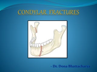
- Dr. Dona Bhattacharya
1. Introduction
2. Surgical anatomy
3. Classification
4. Etiology
5. Diagnosis
6. Management
7. Current concepts
8. Conclusion
9. References
Fractures of mandibular condyle
are common and account for 20-
30% of all mandibular fractures.
Forms imp component for TMJ
• Elliptical in shape, long axis
angled backwards between 15-
33 0 to frontal plane.
• Long axes of 2 condyle meet at
basion on anterior ligament of
foramen magnum forming an
angle 0f 145-160 degrees.
• Mediolateral width: 15-20 mm
• Anteroposterior width: 8-10mm
• Lateral pole: roughened, bluntly pointed,
projects from plane of ramus
• Medial pole: rounded, extends from
plane of ramus
• Fibrous layer thin on posterior aspect
and thick over convexity
Parameter Child Adult
Cortical bone Thin Thick
Condylar neck Broad Thin
Articular surface Thin Thick
Capsule Highly vascular Less vascular
Periosteum Highly active
osteogenic phase
Less active in latent
stage
Intracapsular fracture
& hemarthrosis
Very common Rare
Remodellin capacity
following trauma
Present Absent
Disturbance in growth Likely N.A.
Blood supply
TMJ area is highly vascular and innervated
Mainly from
• Superficial temporal artery
• Transverse facial artery
• Posterior tympanic artery
• Posterior deep temporal artery
Neural structures
• Facial nerve
• Auriculotemporal nerve
Al-Kayat& Bramley -> identifiable landmarks
Dingman & Grabb -> course
of marginal mandibular
nerve
• Posterior to facial artery it runs
above the inferior border of
mandible in 81%, in rest it coursed
in an arc with lowest border being
with in 1 cm from it.
• Anterior to the artery the nerve ran
above the inferior border in 100%
cases.
Incidence
∏ Assault
∏ RTA
∏ Sport injuries
∏ Falls
∏ Work-related incidents
Less obvious causes are:
Orotracheal intubation
Whiplash injury(Royd house,1973)
Childbirth
∏ Age
∏ Socioeconomic factors
∏ Geographic location
∏ Presence/absence of dentition
∏ Occlusion, mandibular position
∏ Dir & magnitude of force
∏ Muscle pull
Mechanism of injury
Given in 1915
Acc to location & direction of fracture:
• From above, downward & inward or reversed
• From above, backward & downward
Type I- The angle between the head and the long
axis of the ramus :10 to 45 degrees.
Type II- angle of 45 to 90 degrees, resulting in
tearing of the medial portion of the capsule.
Type III- the fragments are not in contact, and the
head is displaced mesially and forward owing to
traction of the lateral pterygoid muscle. confined
to within the glenoid fossa.
Type IV- fractures where the condylar head
articulates in an anterior position to the articular
eminence.
Type V- vertical or oblique fractures through the
head of the condyle.
Type I Non-displaced fracture
Type II Fracture deviation, where there is simple angulation of the condylar
process to the major fragment. (e.g. greenstick fracture)
Type III Fracture displacement, where there is simple overlap of the condylar
process and major mandibular fragments.
Type IV Fracture dislocation, where the head of the condyle is completely
disrupted from the articular fossa.
Intracapsular Fractures or High Condylar
i. Fractures involving the articular surface
ii. Fractures above or through the anatomical neck,
which do not involve the articular surface
Extracapsular or Low Condylar Fractures
Fractures associated with injury to the capsule, ligament and meniscus
Fractures involving adjacent bone
• Non-displaced fracture
• Low-neck fracture with
displacement, mostly with contact
between fragments
• High-neck fracture with
displacement, mostly without
contact between fragments
• Low-neck fracture with dislocation
• High-neck fracture with
dislocation
• Intracapsular fracture of condylar
head
Classification of condylar process fractures; M. Schneider, U. Eckelt; Journal of the
Canadian Dental Association December 2006, Vol. 68, No. 11
Type M: communited #; loss of vertical dimensions (Eckelt & Hlawitschaka)
Classification of condylar process fractures; M. Schneider, U. Eckelt; Journal of the
Canadian Dental Association December 2006, Vol. 68, No. 11
Anatomic location of the fracture
Condylar head
Condylar neck
Subcondylar
Relationship of condylar fragment to
mandible
Nondisplaced
Deviated
Displacement with medial or
lateral overlap
Displacement with anterior or
posterior overlap
No contact between fractured
segments
Relationship of condylar head & fossa
Nondisplaced
Displacement
Dislocation
Contusion of the TMJ
Fractures of the condylar process without displacement of the fragments
Fractures of the condyle
Transcapitular.
Subcapitular.
Fractures of the condylar neck
Basal fracture of the condylar process.
Fractures of the condylar process with displacement of the fragments.
Displacement of the small fragments
Ventrally
Dorsally
Medially
Laterally
Torsion of fragments
Sprains of the TMJ
Dislocation (subluxation) of the TMJ.
Dislocation of the condylar head (condyle).
Anteriorly
Posteriorly
Cranially (central dislocation)
Medially
Laterally
Classification of condylar process fractures; M. Schneider, U. Eckelt; Journal of the
Canadian Dental Association December 2006, Vol. 68, No. 11
Diacapitular fracture (through the head of
the condyle):
The fracture line starts in the articular surface
and may extend outside the capsule.
Fracture of the condylar neck:
The fracture line starts somewhere above line
A and in more than half runs above the line A
in the lateral view. Line A is the perpendicular
line through the sigmoid notch to the tangent
of the ramus.
Fracture of the condylar base:
The fracture line runs behind the mandibular
foramen and, in more than half, below line A
Classification of condylar process fractures;
M. Schneider, U. Eckelt; Journal of the
Canadian Dental Association December
2006, Vol. 68, No. 11
1. History
2. Clinical examination
3. Radiological
examination
Evidence of trauma.
Bleeding from external auditory canal.
Noticeable or palpable swelling – haemotoma / edema.
Facial asymmetry – foreshortening of ramus.
Pain & tenderness.
Crepitation over the joint.
Malocclusion
Deviation of mandibular condyle.
Muscle spasm.
Dentoalveloar injuries.
Management of Traumatic Dislocation of the Mandibular Condyle into the Middle
Cranial Fossa; Robert P. Barron, J Can Dent Assoc 2002; 68(11):676–80
A. Conventional Radiography
a. P A- View
b. Lateral Oblique
c. Towne's Projection
d. Panoramic view
e. TMJ views
B. CT
C. MRI
Aims for surgery:
1. Relief from pain
2. Stable occlusion
3. Restoration of inter- incisal opening
4. Full range of mandibular movements
5. To minimize deviation
6. Avoid growth disturbances
7. Avoid Ankylosis
2 schools of thought:
• Conservative-functional therapy
• Surgical treatment
Conservative-functional therapy
• Involves no surgical intervention of the fracture site
instead it reduces the fracture taking occlusion as a key
factor.
• Immobilization usually involves fixation with arch
bars, eyelet wires or splints.
• Period of immobilization varies from 7-17 days
Conservative management
• Exercise
• Increasing mouth opening
• Push the jaws laterally
• Diet: Soft diet
• Analgesics
• Anti-inflammatory
• Soft diet and mouth exercises-
• Teeth into normal occlusion
• Adequate ROM
• Elastic MMF for 2-3 weeks
• When occlusion is found to be altered
• Patient was unable to bring their teeth into normal occlusion presence
of pain or swelling
Physiotherapy
Elastic band – Class II light elastics
Review after 6
days
a) Normal occlusion: Remove when
brushing and replace immediately
b) Unable to achieve normal occlusion:
to be worn 24 hrs/day till next review
Review after
next 6 days
a) Occlusion maintainable: halt elastics
b) Occlusion difficult obtain: continue
elastics
Functional exercise:
• > 40 mm interincisal distance (adult)
• > 10 mm lateral excursion
• > 12 mm protrusion
Types of exercise:
• Maximal mouth opening
• Right lateral excursion
• Left lateral excursion
• Protrusive action
Indications:
• Condylar neck # with little or no displacement
• # occuring in child (10-12 yrs)
• Intracapsular #
Recently thermoforming plates is used for the same
Fixation strength is less than wiring so contraindicated in bilateral
fractures
Advantage:
• Smooth surface
• Transparent
Closed treatment of condylar fractures by intermaxillary fixation with
thermoforming plates; Haruhiko Terai et al, British Journal, 2004, Pg. 61-63
Advantage Disadvantage
• Relatively safe
• No injury of nerves and
blood vessels
• No postoperative
complications such as
infection or scar occurs.
• Fracture, loss, and
eruption delay of the
growing teeth can be
avoided in pediatric
patients as no tooth germ
injury occurs because of
no establishment of the
crown of the permanent
teeth
• Injury of the periodontal tissue and buccal
mucosa
• Poor oral hygiene,
• Pronunciation disorder
• Imbalanced nutrition
• Growth disorder and excessive growth of the
injured mandible may occur
• Facial asymmetry may occur in pediatric
patients aged 10 to 15 years due to growth disorder
or functional disorder, and that in particular, the
growth and functional disorders of the TMJ may
occur in 20% to 25% of pediatric patients aged 7
to 10 years
Closed reduction
New indications have slowly evolved with improvement in the surgical
techniques.
Zide’s 1989 indications
Absolute indications:
Fracture in to middle cranial fossa
Foreign body in to joint capsule
Lateral extracapsular deviation
Inability to open mouth or achieve occlusion after 1 week
Open fracture with potential for fibrosis
Possible indications:
Bilateral / unilateral fracture with crushed midface
Comminuted symphysis and condyle fracture with tooth loss
Displaced fracture with open bite or retrusion in mentally retarded or
medically compromised patients.
Displaced condyle in edentulous or partially edentulous mandible with
posterior bite collapse.
Blair’s Inverted
Hockey Stick
Incision
Thoma’s Angulated
Incision
Dingman’s
Incision
Popowich &
Crane Incision
Preauricular Incision
End aural Incision
Post auricular Incision
Submandibular Incision
Transparotid Incision
Retroparotid Incision
Rhytidectomy Incision
Intraoral Incision
e
e
Approach Advantage Disadvantage
Preauricular
Endaural
• Exposure of lateral and
anterior part of condyle
• Cosmetic (Endaural)
• Injury to facial nerve
• Injury to auriculotemporal
• Damage to middle ear
• Hemorrhage
• Parotid fistula
Postauricular • Esthetic
• Minimum risk of facial
nerve injury
• Permits harvest of conchal
cartilage for grafting
• Infection
• Hematoma
• Cartilage necrosis
Intraoral • No visible scar
• Adequate access to condylar
neck
• Injury to buccal, IAN
• Injury to lingual vessels
• Damage to maxillary
artery
Submandibular
(Risdon)
• Adequate access to condylar
neck & subcondyle
• Injury to MMB
Retromandibular • Adequate access to condyle
• Less risk of injury to MMB
• Scar
• Parotid fistula
The risks & benefits of surgery for temporomandibular joint internal
derangements; Simon Weinberg et al
Fixation of fracture
Fixation of medially displaced fracture
Eckelt technique of lag screw osteosynthesis
CONDYLAR
TRAUMA?
Clinical Sign
Malocclusion
Deviation
Range of motion
Negative clinical
exam
(-) Malocclusion
Minimal pain
Normal range of motion
No deviation on opening
Observation
Radiographs
Lateral obliques
Panorex
CT scan
No radiographic
evidence of condylar #
R/O hemathrosis
Joint effusion
(+) Condylr fractre
Normal occlusion Malocclusion
ORIF?
ROM
Pain
Deviation
Conservative IMF (7-21 days)
ORIF Other # ?
IMF (7-21 days) Reduction/fixation
of other #
Follow up
Yes
Yes
No
No
No
Yes
Ellis and Throckmorton conducted study with open or closed treatment
for fractures of the mandibular condylar process, in one hundred forty-six
patients, 81 treated by closed and 65 by open methods. The patients treated by
closed methods developed asymmetries characterized by shortening of the face
on the side of injury.
In the study of the Santler et al. two hundred 34 patients with fractures
of the mandibular condylar process were treated by open or closed methods. No
significant difference in mobility, joint problems, occlusion, muscle pain, or nerve
disorders were observed when the surgically and non-surgically treated patients
were compared. Surgically treated patients showed significantly more weather
sensitivity and pain on maximum mouth opening.
Renato VALIATI, 2008, The treatment of condylar fractures: to open or not to open? A
critical review of this controversy
To compare the occlusal relationships after open or closed treatment for
fractures of the mandibular condylar process, a total of 137 patients with unilateral
fractures of the mandibular condylar process (neck or subcondylar), 77 treated
closed and 65 treated open, were included in the study of Ellis, Simon and
Throckmorton. The patients treated by closed techniques had a significantly
greater percentage of malocclusion compared with patients treated by open
reduction, in spite of the initial displacement of the fractures being greater in
patients treated by open reduction.
Renato VALIATI, 2008, The treatment of condylar fractures: to open or not
to open? A critical review of this controversy
Mini-retromandibular approach to condylar fractures ; Federico BIGLIOLI,
Giacomo COLLETTI; Journal of Cranio-Maxillofacial Surgery (2008) 36, 378e383
No visible scar
Less complication rate
Intraoral approach for treatment of displaced Condylar fractures: case report;
valfrido pereira-filho et al; craniomaxillofacial trauma & reconstruction/volume 4,
number 2 2011
Condylar Fracture Repair: Use of the Endoscope to Advance Traditional Treatment
Philosophy ;Reid V. Mueller et al, Facial Plast Surg Clin N Am 14 (2006) 1–9
Resorbable triangular plate for osteosynthesis of fractures of the condylar neck;
Günter Lauer et al; British Journal of Oral and Maxillofacial Surgery 48 (2010) 532–535
Transmasseteric Anteroparotid Approach for Mandibular Condylar Fractures-
Merits and Demerits; AHMAD MAHROUS MOHAMAD, Egypt, J. Plast. Reconstr.
Surg., Vol. 35, No. 2, July: 227-232, 2011
Early complications:
1. Fracture of the tympanic plate
2. Fracture of the glenoid fossa with or without displacement of the
condylar segment into the middle cranial fossa
3. Damage to cranial nerves V & VII
4. Vascular injury
Late complications:
1. Malocclusion
2. Growth disturbance
3. Temporomandibular joint dysfunction
4. Ankylosis
5. Asymmetry
6. Frey’s syndrome
Fractures of the mandibular condyle constitute a significant
portion of mandibular fractures. A number of clinical signs and symptoms
are characteristic of injury to the condylar apparatus. The use of plain
radiographs in multiple view, or CT scans discloses most condylar
fractures and displacements, if any. A number of classification systems are
available to help in treatment planning and record keeping.
Non-surgical treatment is adequate for a majority of condylar
fractures. A period of immobilisation followed by active functional
therapy is indicated for most cases. Surgical management has specific
indications, and can be accomplished through a wide variety of
techniques. In general, complications are not common following condylar
trauma. Important among the possible complications are ankylosis,
growth disturbances and internal derangement.
1. Oral & maxillofacial trauma-Fonseca & walker vol 2
2. Oral & maxillofacial surgery-Fonseca vol 3
3. Oral & maxillofacial trauma-Rowe & Williams vol 2
4. Principles of Oral & maxillofacial surgery-Peterson
5. Fractures of middle third of face-Killey & Kay
6. Oral & maxillofacial surgery-Fragiskos
7. Maxillofacial trauma & facial reconstruction-Peter Ward Booth
8. Oral & maxillofacial surgery-Peter Ward Booth: vol 2
9. Chen Lee et al; Applications of the Endoscope in Facial fracture Management,
seminars in plastics surgery/volume 22, number 1 2008
10. Classification of condylar process fractures; M. Schneider, U. Eckelt; Journal of
the Canadian Dental Association December 2006, Vol. 68, No. 11
11. Management of Traumatic Dislocation of the Mandibular Condyle into the
Middle Cranial Fossa; Robert P. Barron, J Can Dent Assoc 2002; 68(11):676–80
12. Closed treatment of condylar fractures by intermaxillary fixation with
thermoforming plates; Haruhiko Terai et al, British Journal, 2004, Pg. 61-63
13. The risks & benefits of surgery for temporomandibular joint internal
derangements; Simon Weinberg et al
14. Resorbable triangular plate for osteosynthesis of fractures of the condylar neck;
Günter Lauer et al; British Journal of Oral and Maxillofacial Surgery 48 (2010) 532–
535
15. Condylar Fracture Repair: Use of the Endoscope to Advance Traditional Treatment
Philosophy ;Reid V. Mueller et al, Facial Plast Surg Clin N Am 14 (2006) 1–9
16. Intraoral approach for treatment of displaced Condylar fractures: case report;
valfrido pereira-filho et al; craniomaxillofacial trauma & reconstruction/volume 4,
number 2 2011
17. Mini-retromandibular approach to condylar fractures; Federico BIGLIOLI,
Giacomo COLLETTI; Journal of Cranio-Maxillofacial Surgery (2008) 36, 378e383
18. Renato VALIATI, 2008, The treatment of condylar fractures: to open or not to
open? A critical review of this controversy
19. Transmasseteric Anteroparotid Approach for Mandibular Condylar Fractures-
Merits and Demerits; AHMAD MAHROUS MOHAMAD, Egypt, J. Plast. Reconstr.
Surg., Vol. 35, No. 2, July: 227-232, 2011
Condylar #

Mais conteúdo relacionado

Mais procurados

zygomatic reduction,which comes when
 zygomatic reduction,which comes when zygomatic reduction,which comes when
zygomatic reduction,which comes whenJamil Kifayatullah
 
Management of condylar fractures
Management of condylar fracturesManagement of condylar fractures
Management of condylar fracturesdralimohammedhasan
 
ZYGOMATICO MAXILLARY COMPLEX FRACTURE
ZYGOMATICO MAXILLARY COMPLEX FRACTUREZYGOMATICO MAXILLARY COMPLEX FRACTURE
ZYGOMATICO MAXILLARY COMPLEX FRACTUREDr. Vishal Gohil
 
Apertognathia and its surgical management
Apertognathia and its surgical managementApertognathia and its surgical management
Apertognathia and its surgical managementHimanshu Soni
 
Sequencing in panfacial trauma
Sequencing in panfacial traumaSequencing in panfacial trauma
Sequencing in panfacial traumashivani gaba
 
Submandibular and retromandibular approach
Submandibular and retromandibular approachSubmandibular and retromandibular approach
Submandibular and retromandibular approachJamil Kifayatullah
 
Kaban protocol tmj ankylosis treatment new 2009
Kaban protocol tmj ankylosis treatment new 2009Kaban protocol tmj ankylosis treatment new 2009
Kaban protocol tmj ankylosis treatment new 2009Dr Pratiksha Malhotra
 
Surgical approaches to tmj
Surgical approaches to tmjSurgical approaches to tmj
Surgical approaches to tmjAditi Rajvanshi
 
6 maxillary osteotomies
6  maxillary osteotomies6  maxillary osteotomies
6 maxillary osteotomiesvasanramkumar
 
Approaches to maxillofacial skeleton
Approaches to maxillofacial skeletonApproaches to maxillofacial skeleton
Approaches to maxillofacial skeletonDr. SHEETAL KAPSE
 

Mais procurados (20)

zygomatic reduction,which comes when
 zygomatic reduction,which comes when zygomatic reduction,which comes when
zygomatic reduction,which comes when
 
Management of condylar fractures
Management of condylar fracturesManagement of condylar fractures
Management of condylar fractures
 
ZYGOMATICO MAXILLARY COMPLEX FRACTURE
ZYGOMATICO MAXILLARY COMPLEX FRACTUREZYGOMATICO MAXILLARY COMPLEX FRACTURE
ZYGOMATICO MAXILLARY COMPLEX FRACTURE
 
Genioplasty
 Genioplasty Genioplasty
Genioplasty
 
Condylar sag
Condylar sagCondylar sag
Condylar sag
 
Bad splits in bsso
Bad splits in bssoBad splits in bsso
Bad splits in bsso
 
Apertognathia and its surgical management
Apertognathia and its surgical managementApertognathia and its surgical management
Apertognathia and its surgical management
 
3 approaches to the tmj
3 approaches to the tmj3 approaches to the tmj
3 approaches to the tmj
 
Sequencing in panfacial trauma
Sequencing in panfacial traumaSequencing in panfacial trauma
Sequencing in panfacial trauma
 
NOE FRACTURE PPT
NOE FRACTURE PPTNOE FRACTURE PPT
NOE FRACTURE PPT
 
Genioplasty
GenioplastyGenioplasty
Genioplasty
 
Submandibular and retromandibular approach
Submandibular and retromandibular approachSubmandibular and retromandibular approach
Submandibular and retromandibular approach
 
Condylar fractures
Condylar fracturesCondylar fractures
Condylar fractures
 
Orthognathic surgery
Orthognathic surgeryOrthognathic surgery
Orthognathic surgery
 
Kaban protocol tmj ankylosis treatment new 2009
Kaban protocol tmj ankylosis treatment new 2009Kaban protocol tmj ankylosis treatment new 2009
Kaban protocol tmj ankylosis treatment new 2009
 
Surgical approaches to tmj
Surgical approaches to tmjSurgical approaches to tmj
Surgical approaches to tmj
 
6 maxillary osteotomies
6  maxillary osteotomies6  maxillary osteotomies
6 maxillary osteotomies
 
Approaches to maxillofacial skeleton
Approaches to maxillofacial skeletonApproaches to maxillofacial skeleton
Approaches to maxillofacial skeleton
 
Bsso
BssoBsso
Bsso
 
8. mandibular orthognathic procedures(113) Dr. RAHUL TIWARI
8. mandibular orthognathic procedures(113) Dr. RAHUL TIWARI8. mandibular orthognathic procedures(113) Dr. RAHUL TIWARI
8. mandibular orthognathic procedures(113) Dr. RAHUL TIWARI
 

Semelhante a Condylar #

Condylar fracture
Condylar fracture Condylar fracture
Condylar fracture sasa425020
 
Mandibular Condylar fractures & its Management
Mandibular Condylar fractures & its ManagementMandibular Condylar fractures & its Management
Mandibular Condylar fractures & its ManagementMehul Hirani
 
CONDYLAR FRACTURES management and survey
CONDYLAR FRACTURES management and surveyCONDYLAR FRACTURES management and survey
CONDYLAR FRACTURES management and surveyEUROUNDISA
 
MANDIBULAR FRACTURES MANAGEMENT PROTOCO
MANDIBULAR  FRACTURES MANAGEMENT PROTOCOMANDIBULAR  FRACTURES MANAGEMENT PROTOCO
MANDIBULAR FRACTURES MANAGEMENT PROTOCOEUROUNDISA
 
A34SD5FUGIHJOK.pptx
A34SD5FUGIHJOK.pptxA34SD5FUGIHJOK.pptx
A34SD5FUGIHJOK.pptxSPradhan10
 
Nailing it hip fractures short versus long; locked versus non locked
Nailing it hip fractures short versus long; locked versus non lockedNailing it hip fractures short versus long; locked versus non locked
Nailing it hip fractures short versus long; locked versus non lockedLove2jaipal
 
Proximal femoral fractures
Proximal femoral fracturesProximal femoral fractures
Proximal femoral fracturesDr Souvik Paul
 
Cranio facial fractures
Cranio facial fracturesCranio facial fractures
Cranio facial fracturesEhab Napih
 
Orthognathic Surgery
Orthognathic SurgeryOrthognathic Surgery
Orthognathic SurgeryHadi Munib
 
YGUVH BIJNOKM.pptx
YGUVH BIJNOKM.pptxYGUVH BIJNOKM.pptx
YGUVH BIJNOKM.pptxSPradhan10
 

Semelhante a Condylar # (20)

Condylar fracture
Condylar fracture Condylar fracture
Condylar fracture
 
Mandibular Condylar fractures & its Management
Mandibular Condylar fractures & its ManagementMandibular Condylar fractures & its Management
Mandibular Condylar fractures & its Management
 
CONDYLAR FRACTURES
CONDYLAR FRACTURESCONDYLAR FRACTURES
CONDYLAR FRACTURES
 
CONDYLAR FRACTURES management and survey
CONDYLAR FRACTURES management and surveyCONDYLAR FRACTURES management and survey
CONDYLAR FRACTURES management and survey
 
MANDIBULAR FRACTURES MANAGEMENT PROTOCO
MANDIBULAR  FRACTURES MANAGEMENT PROTOCOMANDIBULAR  FRACTURES MANAGEMENT PROTOCO
MANDIBULAR FRACTURES MANAGEMENT PROTOCO
 
CONDYLAR FRACTURE.pptx
CONDYLAR FRACTURE.pptxCONDYLAR FRACTURE.pptx
CONDYLAR FRACTURE.pptx
 
A34SD5FUGIHJOK.pptx
A34SD5FUGIHJOK.pptxA34SD5FUGIHJOK.pptx
A34SD5FUGIHJOK.pptx
 
Condylar fractures
Condylar fracturesCondylar fractures
Condylar fractures
 
Management of Mandibular Fractures
Management of Mandibular FracturesManagement of Mandibular Fractures
Management of Mandibular Fractures
 
Journal Club on Unstable trochanteric fractures
Journal Club on Unstable trochanteric fracturesJournal Club on Unstable trochanteric fractures
Journal Club on Unstable trochanteric fractures
 
iurygtrf.pptx
iurygtrf.pptxiurygtrf.pptx
iurygtrf.pptx
 
TMJ Ankylosis.pptx
TMJ Ankylosis.pptxTMJ Ankylosis.pptx
TMJ Ankylosis.pptx
 
Nailing it hip fractures short versus long; locked versus non locked
Nailing it hip fractures short versus long; locked versus non lockedNailing it hip fractures short versus long; locked versus non locked
Nailing it hip fractures short versus long; locked versus non locked
 
Proximal femoral fractures
Proximal femoral fracturesProximal femoral fractures
Proximal femoral fractures
 
age.pptx
age.pptxage.pptx
age.pptx
 
F67YOI.pptx
F67YOI.pptxF67YOI.pptx
F67YOI.pptx
 
Cranio facial fractures
Cranio facial fracturesCranio facial fractures
Cranio facial fractures
 
Orthognathic Surgery
Orthognathic SurgeryOrthognathic Surgery
Orthognathic Surgery
 
YGUVH BIJNOKM.pptx
YGUVH BIJNOKM.pptxYGUVH BIJNOKM.pptx
YGUVH BIJNOKM.pptx
 
EGRHG.pptx
EGRHG.pptxEGRHG.pptx
EGRHG.pptx
 

Último

world health day presentation ppt download
world health day presentation ppt downloadworld health day presentation ppt download
world health day presentation ppt downloadAnkitKumar311566
 
Hematology and Immunology - Leukocytes Functions
Hematology and Immunology - Leukocytes FunctionsHematology and Immunology - Leukocytes Functions
Hematology and Immunology - Leukocytes FunctionsMedicoseAcademics
 
Big Data Analysis Suggests COVID Vaccination Increases Excess Mortality Of ...
Big Data Analysis Suggests COVID  Vaccination Increases Excess Mortality Of  ...Big Data Analysis Suggests COVID  Vaccination Increases Excess Mortality Of  ...
Big Data Analysis Suggests COVID Vaccination Increases Excess Mortality Of ...sdateam0
 
Tans femoral Amputee : Prosthetics Knee Joints.pptx
Tans femoral Amputee : Prosthetics Knee Joints.pptxTans femoral Amputee : Prosthetics Knee Joints.pptx
Tans femoral Amputee : Prosthetics Knee Joints.pptxKezaiah S
 
Clinical Pharmacotherapy of Scabies Disease
Clinical Pharmacotherapy of Scabies DiseaseClinical Pharmacotherapy of Scabies Disease
Clinical Pharmacotherapy of Scabies DiseaseSreenivasa Reddy Thalla
 
Presentació "Real-Life VR Integration for Mild Cognitive Impairment Rehabilit...
Presentació "Real-Life VR Integration for Mild Cognitive Impairment Rehabilit...Presentació "Real-Life VR Integration for Mild Cognitive Impairment Rehabilit...
Presentació "Real-Life VR Integration for Mild Cognitive Impairment Rehabilit...Badalona Serveis Assistencials
 
History and Development of Pharmacovigilence.pdf
History and Development of Pharmacovigilence.pdfHistory and Development of Pharmacovigilence.pdf
History and Development of Pharmacovigilence.pdfSasikiranMarri
 
Case Report Peripartum Cardiomyopathy.pptx
Case Report Peripartum Cardiomyopathy.pptxCase Report Peripartum Cardiomyopathy.pptx
Case Report Peripartum Cardiomyopathy.pptxNiranjan Chavan
 
Glomerular Filtration and determinants of glomerular filtration .pptx
Glomerular Filtration and  determinants of glomerular filtration .pptxGlomerular Filtration and  determinants of glomerular filtration .pptx
Glomerular Filtration and determinants of glomerular filtration .pptxDr.Nusrat Tariq
 
ANTI-DIABETICS DRUGS - PTEROCARPUS AND GYMNEMA
ANTI-DIABETICS DRUGS - PTEROCARPUS AND GYMNEMAANTI-DIABETICS DRUGS - PTEROCARPUS AND GYMNEMA
ANTI-DIABETICS DRUGS - PTEROCARPUS AND GYMNEMADivya Kanojiya
 
Study on the Impact of FOCUS-PDCA Management Model on the Disinfection Qualit...
Study on the Impact of FOCUS-PDCA Management Model on the Disinfection Qualit...Study on the Impact of FOCUS-PDCA Management Model on the Disinfection Qualit...
Study on the Impact of FOCUS-PDCA Management Model on the Disinfection Qualit...MehranMouzam
 
low cost antibiotic cement nail for infected non union.pptx
low cost antibiotic cement nail for infected non union.pptxlow cost antibiotic cement nail for infected non union.pptx
low cost antibiotic cement nail for infected non union.pptxdrashraf369
 
SWD (Short wave diathermy)- Physiotherapy.ppt
SWD (Short wave diathermy)- Physiotherapy.pptSWD (Short wave diathermy)- Physiotherapy.ppt
SWD (Short wave diathermy)- Physiotherapy.pptMumux Mirani
 
Basic principles involved in the traditional systems of medicine PDF.pdf
Basic principles involved in the traditional systems of medicine PDF.pdfBasic principles involved in the traditional systems of medicine PDF.pdf
Basic principles involved in the traditional systems of medicine PDF.pdfDivya Kanojiya
 
VarSeq 2.6.0: Advancing Pharmacogenomics and Genomic Analysis
VarSeq 2.6.0: Advancing Pharmacogenomics and Genomic AnalysisVarSeq 2.6.0: Advancing Pharmacogenomics and Genomic Analysis
VarSeq 2.6.0: Advancing Pharmacogenomics and Genomic AnalysisGolden Helix
 
The next social challenge to public health: the information environment.pptx
The next social challenge to public health:  the information environment.pptxThe next social challenge to public health:  the information environment.pptx
The next social challenge to public health: the information environment.pptxTina Purnat
 
COVID-19 (NOVEL CORONA VIRUS DISEASE PANDEMIC ).pptx
COVID-19  (NOVEL CORONA  VIRUS DISEASE PANDEMIC ).pptxCOVID-19  (NOVEL CORONA  VIRUS DISEASE PANDEMIC ).pptx
COVID-19 (NOVEL CORONA VIRUS DISEASE PANDEMIC ).pptxBibekananda shah
 
Apiculture Chapter 1. Introduction 2.ppt
Apiculture Chapter 1. Introduction 2.pptApiculture Chapter 1. Introduction 2.ppt
Apiculture Chapter 1. Introduction 2.pptkedirjemalharun
 
epilepsy and status epilepticus for undergraduate.pptx
epilepsy and status epilepticus  for undergraduate.pptxepilepsy and status epilepticus  for undergraduate.pptx
epilepsy and status epilepticus for undergraduate.pptxMohamed Rizk Khodair
 
Informed Consent Empowering Healthcare Decision-Making.pptx
Informed Consent Empowering Healthcare Decision-Making.pptxInformed Consent Empowering Healthcare Decision-Making.pptx
Informed Consent Empowering Healthcare Decision-Making.pptxSasikiranMarri
 

Último (20)

world health day presentation ppt download
world health day presentation ppt downloadworld health day presentation ppt download
world health day presentation ppt download
 
Hematology and Immunology - Leukocytes Functions
Hematology and Immunology - Leukocytes FunctionsHematology and Immunology - Leukocytes Functions
Hematology and Immunology - Leukocytes Functions
 
Big Data Analysis Suggests COVID Vaccination Increases Excess Mortality Of ...
Big Data Analysis Suggests COVID  Vaccination Increases Excess Mortality Of  ...Big Data Analysis Suggests COVID  Vaccination Increases Excess Mortality Of  ...
Big Data Analysis Suggests COVID Vaccination Increases Excess Mortality Of ...
 
Tans femoral Amputee : Prosthetics Knee Joints.pptx
Tans femoral Amputee : Prosthetics Knee Joints.pptxTans femoral Amputee : Prosthetics Knee Joints.pptx
Tans femoral Amputee : Prosthetics Knee Joints.pptx
 
Clinical Pharmacotherapy of Scabies Disease
Clinical Pharmacotherapy of Scabies DiseaseClinical Pharmacotherapy of Scabies Disease
Clinical Pharmacotherapy of Scabies Disease
 
Presentació "Real-Life VR Integration for Mild Cognitive Impairment Rehabilit...
Presentació "Real-Life VR Integration for Mild Cognitive Impairment Rehabilit...Presentació "Real-Life VR Integration for Mild Cognitive Impairment Rehabilit...
Presentació "Real-Life VR Integration for Mild Cognitive Impairment Rehabilit...
 
History and Development of Pharmacovigilence.pdf
History and Development of Pharmacovigilence.pdfHistory and Development of Pharmacovigilence.pdf
History and Development of Pharmacovigilence.pdf
 
Case Report Peripartum Cardiomyopathy.pptx
Case Report Peripartum Cardiomyopathy.pptxCase Report Peripartum Cardiomyopathy.pptx
Case Report Peripartum Cardiomyopathy.pptx
 
Glomerular Filtration and determinants of glomerular filtration .pptx
Glomerular Filtration and  determinants of glomerular filtration .pptxGlomerular Filtration and  determinants of glomerular filtration .pptx
Glomerular Filtration and determinants of glomerular filtration .pptx
 
ANTI-DIABETICS DRUGS - PTEROCARPUS AND GYMNEMA
ANTI-DIABETICS DRUGS - PTEROCARPUS AND GYMNEMAANTI-DIABETICS DRUGS - PTEROCARPUS AND GYMNEMA
ANTI-DIABETICS DRUGS - PTEROCARPUS AND GYMNEMA
 
Study on the Impact of FOCUS-PDCA Management Model on the Disinfection Qualit...
Study on the Impact of FOCUS-PDCA Management Model on the Disinfection Qualit...Study on the Impact of FOCUS-PDCA Management Model on the Disinfection Qualit...
Study on the Impact of FOCUS-PDCA Management Model on the Disinfection Qualit...
 
low cost antibiotic cement nail for infected non union.pptx
low cost antibiotic cement nail for infected non union.pptxlow cost antibiotic cement nail for infected non union.pptx
low cost antibiotic cement nail for infected non union.pptx
 
SWD (Short wave diathermy)- Physiotherapy.ppt
SWD (Short wave diathermy)- Physiotherapy.pptSWD (Short wave diathermy)- Physiotherapy.ppt
SWD (Short wave diathermy)- Physiotherapy.ppt
 
Basic principles involved in the traditional systems of medicine PDF.pdf
Basic principles involved in the traditional systems of medicine PDF.pdfBasic principles involved in the traditional systems of medicine PDF.pdf
Basic principles involved in the traditional systems of medicine PDF.pdf
 
VarSeq 2.6.0: Advancing Pharmacogenomics and Genomic Analysis
VarSeq 2.6.0: Advancing Pharmacogenomics and Genomic AnalysisVarSeq 2.6.0: Advancing Pharmacogenomics and Genomic Analysis
VarSeq 2.6.0: Advancing Pharmacogenomics and Genomic Analysis
 
The next social challenge to public health: the information environment.pptx
The next social challenge to public health:  the information environment.pptxThe next social challenge to public health:  the information environment.pptx
The next social challenge to public health: the information environment.pptx
 
COVID-19 (NOVEL CORONA VIRUS DISEASE PANDEMIC ).pptx
COVID-19  (NOVEL CORONA  VIRUS DISEASE PANDEMIC ).pptxCOVID-19  (NOVEL CORONA  VIRUS DISEASE PANDEMIC ).pptx
COVID-19 (NOVEL CORONA VIRUS DISEASE PANDEMIC ).pptx
 
Apiculture Chapter 1. Introduction 2.ppt
Apiculture Chapter 1. Introduction 2.pptApiculture Chapter 1. Introduction 2.ppt
Apiculture Chapter 1. Introduction 2.ppt
 
epilepsy and status epilepticus for undergraduate.pptx
epilepsy and status epilepticus  for undergraduate.pptxepilepsy and status epilepticus  for undergraduate.pptx
epilepsy and status epilepticus for undergraduate.pptx
 
Informed Consent Empowering Healthcare Decision-Making.pptx
Informed Consent Empowering Healthcare Decision-Making.pptxInformed Consent Empowering Healthcare Decision-Making.pptx
Informed Consent Empowering Healthcare Decision-Making.pptx
 

Condylar #

  • 1. - Dr. Dona Bhattacharya
  • 2. 1. Introduction 2. Surgical anatomy 3. Classification 4. Etiology 5. Diagnosis 6. Management 7. Current concepts 8. Conclusion 9. References
  • 3. Fractures of mandibular condyle are common and account for 20- 30% of all mandibular fractures. Forms imp component for TMJ
  • 4. • Elliptical in shape, long axis angled backwards between 15- 33 0 to frontal plane. • Long axes of 2 condyle meet at basion on anterior ligament of foramen magnum forming an angle 0f 145-160 degrees.
  • 5. • Mediolateral width: 15-20 mm • Anteroposterior width: 8-10mm • Lateral pole: roughened, bluntly pointed, projects from plane of ramus • Medial pole: rounded, extends from plane of ramus • Fibrous layer thin on posterior aspect and thick over convexity
  • 6. Parameter Child Adult Cortical bone Thin Thick Condylar neck Broad Thin Articular surface Thin Thick Capsule Highly vascular Less vascular Periosteum Highly active osteogenic phase Less active in latent stage Intracapsular fracture & hemarthrosis Very common Rare Remodellin capacity following trauma Present Absent Disturbance in growth Likely N.A.
  • 7. Blood supply TMJ area is highly vascular and innervated Mainly from • Superficial temporal artery • Transverse facial artery • Posterior tympanic artery • Posterior deep temporal artery
  • 8. Neural structures • Facial nerve • Auriculotemporal nerve
  • 9. Al-Kayat& Bramley -> identifiable landmarks
  • 10. Dingman & Grabb -> course of marginal mandibular nerve • Posterior to facial artery it runs above the inferior border of mandible in 81%, in rest it coursed in an arc with lowest border being with in 1 cm from it. • Anterior to the artery the nerve ran above the inferior border in 100% cases.
  • 12.
  • 13. ∏ Assault ∏ RTA ∏ Sport injuries ∏ Falls ∏ Work-related incidents Less obvious causes are: Orotracheal intubation Whiplash injury(Royd house,1973) Childbirth
  • 14. ∏ Age ∏ Socioeconomic factors ∏ Geographic location ∏ Presence/absence of dentition ∏ Occlusion, mandibular position ∏ Dir & magnitude of force ∏ Muscle pull
  • 15.
  • 17.
  • 18. Given in 1915 Acc to location & direction of fracture: • From above, downward & inward or reversed • From above, backward & downward
  • 19. Type I- The angle between the head and the long axis of the ramus :10 to 45 degrees. Type II- angle of 45 to 90 degrees, resulting in tearing of the medial portion of the capsule. Type III- the fragments are not in contact, and the head is displaced mesially and forward owing to traction of the lateral pterygoid muscle. confined to within the glenoid fossa. Type IV- fractures where the condylar head articulates in an anterior position to the articular eminence. Type V- vertical or oblique fractures through the head of the condyle.
  • 20. Type I Non-displaced fracture Type II Fracture deviation, where there is simple angulation of the condylar process to the major fragment. (e.g. greenstick fracture) Type III Fracture displacement, where there is simple overlap of the condylar process and major mandibular fragments. Type IV Fracture dislocation, where the head of the condyle is completely disrupted from the articular fossa.
  • 21. Intracapsular Fractures or High Condylar i. Fractures involving the articular surface ii. Fractures above or through the anatomical neck, which do not involve the articular surface Extracapsular or Low Condylar Fractures Fractures associated with injury to the capsule, ligament and meniscus Fractures involving adjacent bone
  • 22. • Non-displaced fracture • Low-neck fracture with displacement, mostly with contact between fragments • High-neck fracture with displacement, mostly without contact between fragments • Low-neck fracture with dislocation • High-neck fracture with dislocation • Intracapsular fracture of condylar head Classification of condylar process fractures; M. Schneider, U. Eckelt; Journal of the Canadian Dental Association December 2006, Vol. 68, No. 11
  • 23. Type M: communited #; loss of vertical dimensions (Eckelt & Hlawitschaka) Classification of condylar process fractures; M. Schneider, U. Eckelt; Journal of the Canadian Dental Association December 2006, Vol. 68, No. 11
  • 24. Anatomic location of the fracture Condylar head Condylar neck Subcondylar Relationship of condylar fragment to mandible Nondisplaced Deviated Displacement with medial or lateral overlap Displacement with anterior or posterior overlap No contact between fractured segments Relationship of condylar head & fossa Nondisplaced Displacement Dislocation
  • 25. Contusion of the TMJ Fractures of the condylar process without displacement of the fragments Fractures of the condyle Transcapitular. Subcapitular. Fractures of the condylar neck Basal fracture of the condylar process. Fractures of the condylar process with displacement of the fragments. Displacement of the small fragments Ventrally Dorsally Medially Laterally Torsion of fragments
  • 26. Sprains of the TMJ Dislocation (subluxation) of the TMJ. Dislocation of the condylar head (condyle). Anteriorly Posteriorly Cranially (central dislocation) Medially Laterally Classification of condylar process fractures; M. Schneider, U. Eckelt; Journal of the Canadian Dental Association December 2006, Vol. 68, No. 11
  • 27. Diacapitular fracture (through the head of the condyle): The fracture line starts in the articular surface and may extend outside the capsule. Fracture of the condylar neck: The fracture line starts somewhere above line A and in more than half runs above the line A in the lateral view. Line A is the perpendicular line through the sigmoid notch to the tangent of the ramus. Fracture of the condylar base: The fracture line runs behind the mandibular foramen and, in more than half, below line A Classification of condylar process fractures; M. Schneider, U. Eckelt; Journal of the Canadian Dental Association December 2006, Vol. 68, No. 11
  • 28. 1. History 2. Clinical examination 3. Radiological examination
  • 29. Evidence of trauma. Bleeding from external auditory canal. Noticeable or palpable swelling – haemotoma / edema. Facial asymmetry – foreshortening of ramus. Pain & tenderness. Crepitation over the joint. Malocclusion Deviation of mandibular condyle. Muscle spasm. Dentoalveloar injuries.
  • 30. Management of Traumatic Dislocation of the Mandibular Condyle into the Middle Cranial Fossa; Robert P. Barron, J Can Dent Assoc 2002; 68(11):676–80
  • 31. A. Conventional Radiography a. P A- View b. Lateral Oblique c. Towne's Projection d. Panoramic view e. TMJ views B. CT C. MRI
  • 32. Aims for surgery: 1. Relief from pain 2. Stable occlusion 3. Restoration of inter- incisal opening 4. Full range of mandibular movements 5. To minimize deviation 6. Avoid growth disturbances 7. Avoid Ankylosis
  • 33. 2 schools of thought: • Conservative-functional therapy • Surgical treatment
  • 34. Conservative-functional therapy • Involves no surgical intervention of the fracture site instead it reduces the fracture taking occlusion as a key factor. • Immobilization usually involves fixation with arch bars, eyelet wires or splints. • Period of immobilization varies from 7-17 days
  • 35. Conservative management • Exercise • Increasing mouth opening • Push the jaws laterally • Diet: Soft diet • Analgesics • Anti-inflammatory • Soft diet and mouth exercises- • Teeth into normal occlusion • Adequate ROM • Elastic MMF for 2-3 weeks • When occlusion is found to be altered • Patient was unable to bring their teeth into normal occlusion presence of pain or swelling
  • 36. Physiotherapy Elastic band – Class II light elastics Review after 6 days a) Normal occlusion: Remove when brushing and replace immediately b) Unable to achieve normal occlusion: to be worn 24 hrs/day till next review Review after next 6 days a) Occlusion maintainable: halt elastics b) Occlusion difficult obtain: continue elastics
  • 37. Functional exercise: • > 40 mm interincisal distance (adult) • > 10 mm lateral excursion • > 12 mm protrusion Types of exercise: • Maximal mouth opening • Right lateral excursion • Left lateral excursion • Protrusive action
  • 38. Indications: • Condylar neck # with little or no displacement • # occuring in child (10-12 yrs) • Intracapsular #
  • 39. Recently thermoforming plates is used for the same Fixation strength is less than wiring so contraindicated in bilateral fractures Advantage: • Smooth surface • Transparent Closed treatment of condylar fractures by intermaxillary fixation with thermoforming plates; Haruhiko Terai et al, British Journal, 2004, Pg. 61-63
  • 40. Advantage Disadvantage • Relatively safe • No injury of nerves and blood vessels • No postoperative complications such as infection or scar occurs. • Fracture, loss, and eruption delay of the growing teeth can be avoided in pediatric patients as no tooth germ injury occurs because of no establishment of the crown of the permanent teeth • Injury of the periodontal tissue and buccal mucosa • Poor oral hygiene, • Pronunciation disorder • Imbalanced nutrition • Growth disorder and excessive growth of the injured mandible may occur • Facial asymmetry may occur in pediatric patients aged 10 to 15 years due to growth disorder or functional disorder, and that in particular, the growth and functional disorders of the TMJ may occur in 20% to 25% of pediatric patients aged 7 to 10 years Closed reduction
  • 41.
  • 42.
  • 43. New indications have slowly evolved with improvement in the surgical techniques. Zide’s 1989 indications Absolute indications: Fracture in to middle cranial fossa Foreign body in to joint capsule Lateral extracapsular deviation Inability to open mouth or achieve occlusion after 1 week Open fracture with potential for fibrosis Possible indications: Bilateral / unilateral fracture with crushed midface Comminuted symphysis and condyle fracture with tooth loss Displaced fracture with open bite or retrusion in mentally retarded or medically compromised patients. Displaced condyle in edentulous or partially edentulous mandible with posterior bite collapse.
  • 44.
  • 45.
  • 46.
  • 47. Blair’s Inverted Hockey Stick Incision Thoma’s Angulated Incision Dingman’s Incision Popowich & Crane Incision Preauricular Incision
  • 56. Approach Advantage Disadvantage Preauricular Endaural • Exposure of lateral and anterior part of condyle • Cosmetic (Endaural) • Injury to facial nerve • Injury to auriculotemporal • Damage to middle ear • Hemorrhage • Parotid fistula Postauricular • Esthetic • Minimum risk of facial nerve injury • Permits harvest of conchal cartilage for grafting • Infection • Hematoma • Cartilage necrosis Intraoral • No visible scar • Adequate access to condylar neck • Injury to buccal, IAN • Injury to lingual vessels • Damage to maxillary artery Submandibular (Risdon) • Adequate access to condylar neck & subcondyle • Injury to MMB Retromandibular • Adequate access to condyle • Less risk of injury to MMB • Scar • Parotid fistula The risks & benefits of surgery for temporomandibular joint internal derangements; Simon Weinberg et al
  • 57.
  • 59. Fixation of medially displaced fracture
  • 60. Eckelt technique of lag screw osteosynthesis
  • 61. CONDYLAR TRAUMA? Clinical Sign Malocclusion Deviation Range of motion Negative clinical exam (-) Malocclusion Minimal pain Normal range of motion No deviation on opening Observation Radiographs Lateral obliques Panorex CT scan No radiographic evidence of condylar # R/O hemathrosis Joint effusion (+) Condylr fractre Normal occlusion Malocclusion ORIF? ROM Pain Deviation Conservative IMF (7-21 days) ORIF Other # ? IMF (7-21 days) Reduction/fixation of other # Follow up Yes Yes No No No Yes
  • 62.
  • 63. Ellis and Throckmorton conducted study with open or closed treatment for fractures of the mandibular condylar process, in one hundred forty-six patients, 81 treated by closed and 65 by open methods. The patients treated by closed methods developed asymmetries characterized by shortening of the face on the side of injury. In the study of the Santler et al. two hundred 34 patients with fractures of the mandibular condylar process were treated by open or closed methods. No significant difference in mobility, joint problems, occlusion, muscle pain, or nerve disorders were observed when the surgically and non-surgically treated patients were compared. Surgically treated patients showed significantly more weather sensitivity and pain on maximum mouth opening. Renato VALIATI, 2008, The treatment of condylar fractures: to open or not to open? A critical review of this controversy
  • 64. To compare the occlusal relationships after open or closed treatment for fractures of the mandibular condylar process, a total of 137 patients with unilateral fractures of the mandibular condylar process (neck or subcondylar), 77 treated closed and 65 treated open, were included in the study of Ellis, Simon and Throckmorton. The patients treated by closed techniques had a significantly greater percentage of malocclusion compared with patients treated by open reduction, in spite of the initial displacement of the fractures being greater in patients treated by open reduction. Renato VALIATI, 2008, The treatment of condylar fractures: to open or not to open? A critical review of this controversy
  • 65. Mini-retromandibular approach to condylar fractures ; Federico BIGLIOLI, Giacomo COLLETTI; Journal of Cranio-Maxillofacial Surgery (2008) 36, 378e383 No visible scar Less complication rate
  • 66. Intraoral approach for treatment of displaced Condylar fractures: case report; valfrido pereira-filho et al; craniomaxillofacial trauma & reconstruction/volume 4, number 2 2011
  • 67. Condylar Fracture Repair: Use of the Endoscope to Advance Traditional Treatment Philosophy ;Reid V. Mueller et al, Facial Plast Surg Clin N Am 14 (2006) 1–9
  • 68. Resorbable triangular plate for osteosynthesis of fractures of the condylar neck; Günter Lauer et al; British Journal of Oral and Maxillofacial Surgery 48 (2010) 532–535
  • 69. Transmasseteric Anteroparotid Approach for Mandibular Condylar Fractures- Merits and Demerits; AHMAD MAHROUS MOHAMAD, Egypt, J. Plast. Reconstr. Surg., Vol. 35, No. 2, July: 227-232, 2011
  • 70. Early complications: 1. Fracture of the tympanic plate 2. Fracture of the glenoid fossa with or without displacement of the condylar segment into the middle cranial fossa 3. Damage to cranial nerves V & VII 4. Vascular injury Late complications: 1. Malocclusion 2. Growth disturbance 3. Temporomandibular joint dysfunction 4. Ankylosis 5. Asymmetry 6. Frey’s syndrome
  • 71. Fractures of the mandibular condyle constitute a significant portion of mandibular fractures. A number of clinical signs and symptoms are characteristic of injury to the condylar apparatus. The use of plain radiographs in multiple view, or CT scans discloses most condylar fractures and displacements, if any. A number of classification systems are available to help in treatment planning and record keeping. Non-surgical treatment is adequate for a majority of condylar fractures. A period of immobilisation followed by active functional therapy is indicated for most cases. Surgical management has specific indications, and can be accomplished through a wide variety of techniques. In general, complications are not common following condylar trauma. Important among the possible complications are ankylosis, growth disturbances and internal derangement.
  • 72. 1. Oral & maxillofacial trauma-Fonseca & walker vol 2 2. Oral & maxillofacial surgery-Fonseca vol 3 3. Oral & maxillofacial trauma-Rowe & Williams vol 2 4. Principles of Oral & maxillofacial surgery-Peterson 5. Fractures of middle third of face-Killey & Kay 6. Oral & maxillofacial surgery-Fragiskos 7. Maxillofacial trauma & facial reconstruction-Peter Ward Booth 8. Oral & maxillofacial surgery-Peter Ward Booth: vol 2 9. Chen Lee et al; Applications of the Endoscope in Facial fracture Management, seminars in plastics surgery/volume 22, number 1 2008 10. Classification of condylar process fractures; M. Schneider, U. Eckelt; Journal of the Canadian Dental Association December 2006, Vol. 68, No. 11 11. Management of Traumatic Dislocation of the Mandibular Condyle into the Middle Cranial Fossa; Robert P. Barron, J Can Dent Assoc 2002; 68(11):676–80
  • 73. 12. Closed treatment of condylar fractures by intermaxillary fixation with thermoforming plates; Haruhiko Terai et al, British Journal, 2004, Pg. 61-63 13. The risks & benefits of surgery for temporomandibular joint internal derangements; Simon Weinberg et al 14. Resorbable triangular plate for osteosynthesis of fractures of the condylar neck; Günter Lauer et al; British Journal of Oral and Maxillofacial Surgery 48 (2010) 532– 535 15. Condylar Fracture Repair: Use of the Endoscope to Advance Traditional Treatment Philosophy ;Reid V. Mueller et al, Facial Plast Surg Clin N Am 14 (2006) 1–9 16. Intraoral approach for treatment of displaced Condylar fractures: case report; valfrido pereira-filho et al; craniomaxillofacial trauma & reconstruction/volume 4, number 2 2011 17. Mini-retromandibular approach to condylar fractures; Federico BIGLIOLI, Giacomo COLLETTI; Journal of Cranio-Maxillofacial Surgery (2008) 36, 378e383 18. Renato VALIATI, 2008, The treatment of condylar fractures: to open or not to open? A critical review of this controversy 19. Transmasseteric Anteroparotid Approach for Mandibular Condylar Fractures- Merits and Demerits; AHMAD MAHROUS MOHAMAD, Egypt, J. Plast. Reconstr. Surg., Vol. 35, No. 2, July: 227-232, 2011