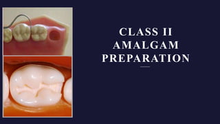
Class 2 cavity amalgam
•Download as PPTX, PDF•
13 likes•1,815 views

This document provides details on preparing Class II amalgam restorations involving only one proximal surface. It describes the initial and final tooth preparation stages, including entering the pit nearest the involved surface, establishing target depths, and visualizing the final locations of facial and lingual walls. It also discusses creating a proximal ditch cut, modifying preparations based on factors like gingiva location and condition, and designing resistance and retention forms to protect the restoration and remaining tooth structure.

Recommended
More Related Content
What's hot
What's hot (20)
Similar to Class 2 cavity amalgam
Similar to Class 2 cavity amalgam (20)
Recently uploaded
Recently uploaded (20)
Class 2 cavity amalgam
- 2. DEFINITION Preparations involving the proximal surfaces of posterior teeth are termed Class II. Involves the proximal, proximo – facial ( lingual ) , proximo – occlusal preparation on molars and premolars.
- 5. C L A S S I I A M A LG A M R E S TO R AT I O N S I N VO LV I N G O N LY O N E P ROX I M A L S U R FAC E Carious lesion on one proximal surface. STEPS IN PREPRARATION - 1. Initial tooth preparation stage 2. Final tooth preparation stage
- 6. 1. Initial tooth preparation stage - A. Outline form - Enter the pit nearest the involved proximal surface with a punch cut using a No. 245 bur . Entering the pit nearest the involved proximal surface allows the mesial pit (in this case) not to be included if it is sound. Viewed from the proximal and lingual (facial) aspects, the long axis of the bur and the long axis of the tooth crown should remain parallel during the cutting procedures.
- 8. As the bur enters the pit, a target depth of 0.1–0.2 mm into dentin should be established (i.e., one-half to two-thirds the length of the cutting portion of a No. 245 bur); 1.5 mm as measured at the central fissure, and approximately 2 mm on the prepared external walls such that the DEJ is identified
- 9. While maintaining the same depth and bur orientation, the bur is moved to extend the outline to include the central fissure and the opposite pit (the distal pit, in this example), if necessary
- 10. Isthmus width should be as narrow as possible, preferably no wider than one-quarter the intercuspal distance.
- 11. The amount of remaining tooth structure is more important to restoration longevity than is the restorative material used. Occlusal convergence - provides favorable amalgam angles at the margins. The facial, lingual, and distal walls should be extended until a sound DEJ is reached.
- 12. It may be necessary to tilt the bur to diverge occlusally at the distal wall if further distal extension would undermine the marginal ridge of its dentinal support.
- 13. The dovetail feature is not required in the occlusal step of a single proximal surface preparation, unless a fissure emanating from an occlusal pit indicates it. Without a dovetail, however, the occlusal step should not be in a straight direction, which may reduce the retention form. This type of retention form also is provided by any extension of the central fissure preparation that is not in a straight direction from pit to pit.
- 14. Before extending into the involved proximal marginal ridge (the mesial ridge, in this example), the final locations of the facial and lingual walls of the proximal box are visualized. This action prevents overextension of the occlusal outline form (i.e., occlusal step) where it joins the proximal outline form (i.e., proximal box).
- 15. The preparation is extended mesially, stopping approximately 0.8 mm short of cutting through the marginal ridge into the contact area. The occlusal step in this region is made slightly wider faciolingually than in the Class I preparation because additional width is necessary for the proximal box.
- 16. Proximal Outline Form (Proximal Box) The desired final location of the facial and lingual walls of the proximal box or the proximal outline form relative to the contact area is visualized. The objectives for the extension of the proximal margins are as follows: 1. Include all caries, defects, or existing restorative material 2.Create 90-degree cavosurface margins (i.e., butt-joint margins) 3. Establish (ideally) not more than 0.5 mm clearance with the adjacent proximal surface facially, lingually, and gingivally
- 17. 1. PROXIMAL DITCH CUT (two-thirds at the expense of enamel and one-third at the expense of dentin )
- 19. . G, The faciolingual dimension of the proximal ditch is greater at the gingival level than at the occlusal level. H, To isolate and weaken the proximal enamel further, the bur is moved toward and perpendicular to the proximal surface. I, The side of the bur may emerge slightly through the proximal surface at the level of the gingival floor.
- 20. Clearance of the proximal margins (i.e., mesiofacial, mesiolingual, gingival) greater than 0.5 mm is excessive, unless indicated to include any caries, undermined enamel, or existing restorative material. Extending gingival margins into the gingival sulcus should be avoided, where possible, because subgingival margins are more difficult to restore and may be a contributing factor to periodontal disease.
- 21. If the proximal ditch cut is entirely in dentin, the axial wall usually is too deep. The ideal dentinal depth of the axial wall of the proximal boxes of premolars and molars should be the same (two-thirds to three-fourths the diameter of the No. 245 bur [or 0.5–0.6 mm]). The proximal ditch cut may diverge gingivally to ensure that the faciolingual dimension at the gingival aspect is greater than at the occlusal aspect .
- 22. The gingival divergence contributes to the retention form and provides for the desirable extension of the facial and lingual proximal margins to include defective tooth structure or old restorative material at the gingival level, while conserving the marginal ridge and providing for 90-degree amalgam at the margins on this ridge.
- 27. A P P L I C AT I O N S O F T H E G E N E R A L P R I N C I P L E S O F C AV I T Y P R E PA R AT I O N TO C L A S S I I L E S I O N S 1. Outline form - o Extension for convenience or access – For most molars and premolars with proximal lesions , the occlusal surface should be included for access purposes . o Few cases in which proximal cavity can be prepared from the facial or lingual embrasure.
- 28. Location and condition of the gingiva - A –Young teeth and high cariogenic activity B – Crest of free gingiva C- Recession – Supragingival margins
- 29. Condition of the marginal ridge - o If the proximal lesion can be instrumented from the buccal or lingual embrasure , the integrity of the ridge occlusally and proximally evaluated. o If it is not undermined , a proximal cavity can be prepared or occlusal and proximal cavity prepared separately with the marginal ridge separating them.
- 30. Location and extent of the contact areas - o Purpose of class 2 restoration – to replace the diseased tissue and correct the local conditions which initiated caries. o Proper relationship b/w 4 anatomic landmarks – Marginal ridge , contact areas, embrasures, gingiva – modify the outline form accordingly by overextension or underextension .
- 31. Modifying factors influencing the outline form - 1. Masticatory loads – inversely proportional to bucco lingual extent of cavity 2. Generalized plaque index - directly proportional to extent of cavity 3. Localized cariogenic factors – greater activity – greater extent of cavity into self cleansable areas 4. Esthetics – minimize the facial extent of cavity esp. In mesiofacial margins of premolars and first molars. 5. Tooth position – malaligned tooth , rotation
- 32. 2. RESISTANCE FORM - Concept is based on the reaction between the restoration and the remaining tooth structure. Nature of this reaction - development of internal stresses – can lead to structural failure. So tooth and restoration must resist failure by designing the cavity which is best able to withstand the distribution and magnitude of stresses on tooth and restoration.
- 33. Occlusal loading – Tensile stresses at axiopulpal line angle ( isthmus) and compressive stress in the underlying dentin and shear stress at junction of bulk of proximal part and its self retained part.
- 34. Premature contacts by the amalgam will exaggerate the stresses normally induced in that area. Can lead to mechanical and biological failure. Eliminate pre existing premature contacts before treatment.
- 35. A. Design features to protect the mechanical integrity of the restoration - 1. Slanting the axial wall towards the pulpal floor. 2. Rounding of axio pulpal line angles 3. Flat pulpal and gingival floor 4. Every part of cavity self retentive 5. Avoid marginal excess – can fracture because of low tensile Strength of amalgam – avoid feather edge margins – butt joint
- 36. B. Design features to protect the physio mechanical integrity of remaining tooth structure - 1. ¼ to 1/5 of intercuspal distance – width at isthmus – avoid excess width 2. Occlusal cavity – rounded line angles , perpendicular facial , lingual walls at isthmus 3. Ideal length to width ratio in cuspal wall surrounding a cavity – 1:1 ( flat table )
- 37. When facial or lingual wall from occlusal surface meet the proximal surface , should form right angles . These walls must terminate past the contact areas in the embrasures. Reverse S curve design - involvement of contact area, 90 degrees cavosurface margin and termination of facial and lingual walls in embrasure , preservation of tooth structure.
- 38. Reverse S curve – mainly on facial wall, minimal lingually.
- 39. C . R E T E N T I O N F O R M - 1. Primary , secondary retentive features